
Dzumacoccus baylaci Hodgson, Germain & Matile-Ferrero, 2018
|
publication ID |
https://doi.org/ 10.11646/zootaxa.4387.2.8 |
|
publication LSID |
lsid:zoobank.org:pub:58200C75-C2D7-4A3B-8DB7-4F531548EF0D |
|
DOI |
https://doi.org/10.5281/zenodo.5976915 |
|
persistent identifier |
https://treatment.plazi.org/id/EC62184F-E918-FFFC-5E8A-FE4CFA55FBC2 |
|
treatment provided by |
Plazi |
|
scientific name |
Dzumacoccus baylaci Hodgson, Germain & Matile-Ferrero |
| status |
sp. nov. |
Dzumacoccus baylaci Hodgson, Germain & Matile-Ferrero spec. n.
Material studied. Holotype: NEW CALEDONIA, Mount Dzumac, piste Ouiné, 700 m alt., on Gymnostoma poissonianum (Casuarinaceae) , 18/xi/1990, coll. Michel Baylac (MNHN 14917-1): 1 slide with 1 adult female; paratypes: same data as for holotype (MNHN 14917-2): 1 slide with 1 first-instar nymph. Also: same data as holotype (MNHN 14918): 1 slide with 3 first-instar nymphs.
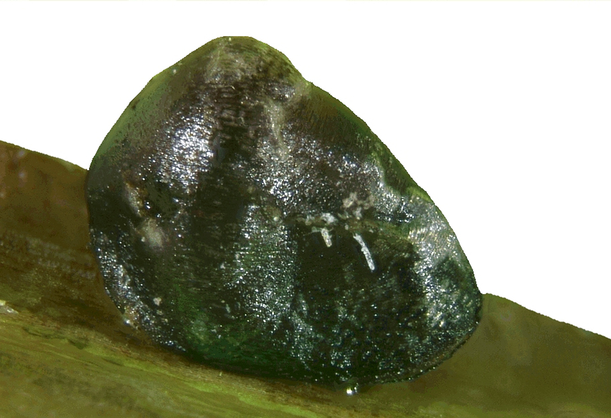
Adult female. Unmounted specimen ( Fig. 2 View FIGURE 2 ): adult female, preserved in alcohol, entirely covered by a tapered thick waxy coat, black-brown to greenish black, trilobite-like.
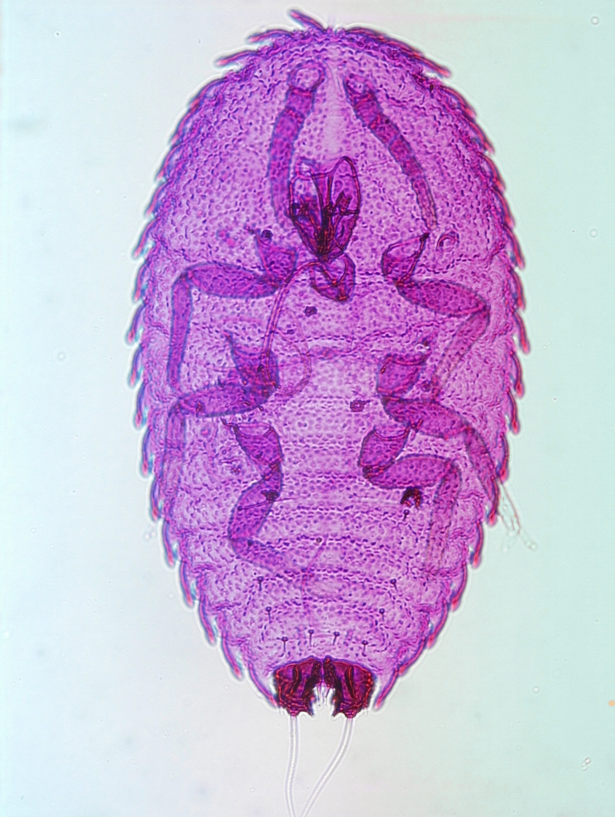
Slide-mounted specimen ( Figs 3 View FIGURE 3 and 4 View FIGURE 4 ): body elongate oval, broadest across abdomen, with a slight indentation in margin medially on head. Anal cleft and stigmatic clefts absent. Entire body margin with a row of strongly sclerotized, triangular spines that all point posteriorly. Length 1.1 mm, width 0.85 mm.
Dorsum. Derm lightly sclerotized, with an abundant series of shallow ridges, rather similar to finger prints in appearance. Dorsal setae, each 6–7 µm long and mostly slightly curved, sparse, arranged in a submarginal line and in a broad medial band. Dorsal microducts small, each with a long inner ductule and a small glandular inner end, those near margin with inner ductule 12–14 µm long but ductules situated more medially appearing to become shorter, down to about 10 µm long. No other pores or ducts present. Anal lobes modified into heavily sclerotized anal plates, each about 40 µm long and 65 µm wide; each anal plate with 4 setae, each seta situated on a sclerotized lobe, as follows: outermost seta on posterior margin spinose, about 8 µm long; middle seta long and setose, about 40 µm long; innermost seta on posterior margin finely spinose and about 5 µm long; and with a spinose seta on ventral inner margin, also about 5 µm long. Anal tube short, anal ring lying more or less beneath anterior margins of anal plates; structure of anal ring unknown but probably with six fairly short setae. Suranal setae: present ventral to anal ring, each 23–25 µm long.
Margin. Margin with a line of heavily sclerotized, triangular, strongly spinose setae around entire circumference, all pointing posteriorly (with spines facing in opposite directions medially on head); each spine 12– 13 µm long and 10–11 µm wide across base, each attached to side of a fleshy marginal lobe; with 85–87 setae on each side, giving a saw-toothed appearance.
Venter. Derm membranous apart from some unusual markings between pro- and mesothoracic coxae, consisting of mildly sclerotized spots and lines. Loculate pores, each 4–5 µm wide with 5 loculi, present in a broad, sparse submarginal band on head and thorax and anteriorly on abdomen on about 1 or 2 segments; band broadest on about prothorax, where the pores extend medially to mouthparts; pores fewest on abdomen; also with a single pore in each spiracular atrium and with 0-2 along margin of each posterior spiracular atrium. Ventral setae rather few and mainly setose, with 3 pairs in a longitudinal line between antennae; a small group present laterad to each anterior spiracle; 4 pairs submedially on thorax; sparse lines of setae present across all abdominal segments, with perhaps 4–6 setae across each anterior segment and about 12–14 across each of segments V and VI; also around vulva. No preantennal pores or cruciform pores observed. Eyespot situated on venter between each antenna and margin, each small, lightly sclerotized, about 4 µm wide. Antennae situated quite close to anterior margin, each perhaps three segmented, with terminal segment composed of apical three segments fused; total length 75–78 µm; setal distribution: scape 4; pedicel 2 + a campaniform pore; apical segment with about 7 long fleshy setae, a stiff long apical seta and 2 or 3 hair-like setae. Clypeolabral shield about 110 µm long; number of labial segments unclear. Spiracles each situated within a sclerotized atrium 25–27 µm wide, total length with apodeme about 45 µm; each with a single loculate pore within atrium. Legs well developed, without a tibio-tarsal articulation and with tibia shorter than tarsus; metacoxa with a sclerotized cavity on proximal dorsal surface, making metacoxa slightly shorter that other coxae; other coxal pores absent; lengths in µm: mesocoxa 73–77, metacoxa 66–70; metatrochanter + femur 100; metatibia 40–42; metatarsus 78–81; claw about 13; setal distribution: coxa 4?; trochanter 1; femur 1; tibia 2; tarsus 3 or 4; tarsus with a campaniform pore. Tarsal digitules dissimilar, one larger than other and both slightly longer than claw. Claw without an obvious denticle; claw digitules dissimilar, one much narrower and shorter than other but both with capitate apices. Vulva not visible, probably situated between abdominal segments VII and VIII.
Comments. The structure of the marginal spinose setae in this genus appears to be unique within the Eriococcidae . In addition, the presence of the odd sclerotizations on the venter of the thorax are also unusual. Another eriococcid species also recently described from New Caledonia, Choneochiton casuarinae Hodgson, Mille & Cazères (Hodgson et al. 2014) , also has odd sclerotizations in this position, although otherwise it is very different. Another feature of interest is the deep sclerotized “pores” on the dorsal surface of each metacoxa. Most eriococcids have spinulae or pores of various sizes on the metacoxae, mostly situated on the lateral surface and much smaller than those on D. baylaci . Other features considered to be of interest are: (i) the complete absence of macrotubular ducts from both dorsal and ventral surfaces; (ii) the absence of spinose setae from the dorsum; (iii) the presence of very long, narrow ductules in each dorsal microtubular duct; (iv) the structure of the anal lobes, which have been modified into structures resembling anal plates; (v) the absence of cruciform pores on the venter; (vi) dissimilar digitules on both tarsus and claw, and (vii) possible fusion of the apical antennal segments. The structure of the anal plates is somewhat similar to that in Montanococcus sp. ( Henderson 2007) but D. baylaci is otherwise very different.
First-instar nymph ( Figs 5 View FIGURE 5 and 6 View FIGURE6 ). Slide-mounted specimen: body elongate oval, broadest across thorax. Entire body margin with a row of strong, sclerotized lobe-like extensions. Length 500 µm, width 285 µm.
Dorsum. Derm lightly sclerotized, completely covered in small nodulations. Dorsal setae of 2 sizes: (i) larger setae, each about 10 µm long with a basal socket about 5 µm wide, present as follows: a pair submedially on abdominal segment VII and also with 1 submarginally on each of abdominal segments III–VII (occasionally with 1 replaced by a smaller seta), and (ii) smaller setae, each about 2.5–3.0 µm long with a basal socket about 2 µm wide, present in (a) a submarginal line of 5 setae on each thoracic segment and abdominal segments I and II; (b) a complete submedial line of about 11 setae extending from abdominal segment VI anteriorly to head; and (c) also with 1 seta more laterally on head. Dorsal microducts small, each with a long inner ductule 8–9 µm long and a small glandular end, with 1 associated with each marginal spinose extension; also smaller ducts randomly distributed elsewhere on dorsum (perhaps segmentally arranged on abdomen but not easy to locate). Anal lobes modified into heavily sclerotized anal plates, each plate about 45 µm long, both plates together 75 µm wide; each plate with 4 setae, each seta situated on a sclerotized lobe, as follows: outermost seta on posterior margin finely spinose, about 5 µm long; middle seta long and setose, 90–95 µm long; innermost seta on posterior margin spinose, about 3 µm long; and a spinose seta on ventral inner margin 6.5–7.0 µm long. Margin of each plate drawn out into a spine-like point medially on posterior margin. Suranal setae each about 25 µm long. Anal tube short, anal ring lying more or less beneath anterior margins of anal plates; structure of anal ring unknown but probably bearing 6 setae.
Margin. Margin with a line of membranous lobes, each leaning posteriorly with outer margin heavily sclerotized, probably slightly flattened and twisted towards apex; each sclerotized part of lobe 38–40 µm long and about 8 µm wide, numbering 19 on each side: with 1 on each side of each abdominal segment, perhaps 4 along thoracic margin and 8 pairs on head. Eyespot perhaps situated on margin between 3rd and 4th marginal lobes, each associated with seta about 10 µm long.
Venter. Derm membranous. Loculate pores, each about 3 µm wide with probably 5 very small loculi, present as follows: 2 between each spiracle and margin, with one very close to margin and other nearer spiracle; also with single pores mediolaterally on abdominal segments II–VII. Ventral setae all setose, as follows: with 1 seta just anterior to each scape and 2 further pairs of interantennal setae situated more posteriorly; with a long seta (about 13 µm long) just mesad to each mesocoxa; with short setae (each about 5 µm long) just mesad to each metacoxa and also mediolaterally in abdominal segments II-VI; also with similar setae forming an inner submarginal line, 1 per abdominal segment; seta on segment VII very long, about 50 µm long; submarginal setae present on each abdominal segment plus 2 on each side of thorax, and with a pair of long submarginal setae medially on head, each about 20 µm long. Antennae situated quite close to anterior margin, close together, each 6 segmented and 130–135 µm long, with apical segment as long as previous 2 segments; setal distribution: scape 4; pedicel 2 + a campaniform pore; III with 1 hair-like seta; IV & V each with 1 fleshy seta and 1 hair-like seta, apical segment with perhaps 3 long fleshy setae, a long stiff apical seta and 2 or 3 hair-like setae. Clypeolabral shield about 75 µm long; number of labial segments unclear but with 4 pairs of setae. Spiracles each small, with peritreme about 8 µm wide. Legs well developed, without tibio-tarsal articulation and with tibia shorter than tarsus; coxal pores absent; lengths of metathoracic legs in µm: coxa 48–50; trochanter + femur 69–71; tibia about 35; tarsus about 55; claw about 15; setal distribution: coxa 4; trochanter 1; femur 1; tibia 1; tarsus 3 or 4; tarsus with a campaniform pore; tarsal digitules dissimilar, one larger than other and slightly longer than claw; claw without an obvious denticle; claw digitules dissimilar, one very narrow and other broad but both with capitate apices and subequal in length to tarsal digitules.
Comments. Like the adult, the first-instar nymphs of Dzumacoccus baylaci can be immediately identified by the unusual structure of the marginal spines. The structure of the anal lobes is rather similar to those of Montanococcus sp. ( Henderson 2007), currently only known from New Zealand, but it is otherwise different. Other characters worthy of note are: (i) the 2 sizes of dorsal setae; (ii) the structure of the dorsal microducts; (iii) the heavily sclerotized anal lobes; (iv) the dissimilar digitules on the tarsus and claw, and (v) the line of ventral loculate pores submedially on abdomen.
Male: not observed.
Etymology. The genus is named after the geographical locality Mount Dzumac. The species is named after our colleague Michel Baylac, dipterist, Muséum national d’Histoire naturelle, Paris, France, who collected this material during an expedition in November 1990.
Discussion. Dzumacoccus is the fourth endemic eriococcid genus now known from New Caledonia but differs significantly from the other genera, namely Rhopalotococcus , Chazeauana and Choneochiton (see key below). Dzumacoccus is the second endemic eriococcid genus to be found on Casuarinaceae endemic to New Caledonia, the other being Choneochiton casuarinae Hodgson, Mille & Cazères, 2014 .
According the website ScaleNet ( García Morales et al. 2016), 110 species of scale insects have been reported on species of Casuarinaceae , of which 53 are either polyphagous and/or cosmopolitan; the remainder (57 species belonging to 7 families) appear to be monophagous on Casuarinaceae , as follows: Diaspididae 22, Eriococcidae 17, Pseudococcidae 11, Asterolecanidae 2, Monophlebidae 2, Coccidae 1, Kerriidae 1 and Lecanodiaspididae 1. Most of these species are found in Australia (44), of which the Diaspididae and Eriococcidae are much the largest families with 19 and 17 species respectively. In New Caledonia, in addition to the 2 endemic eriococcid species, 2 other scale insect species appear to be restricted to Casuarinaceae , namely the diaspidid Diaspis casuarinae Williams & Watson and the monophlebid Tessarobelus ordinarius Bhatti. Ten other scale insect species are currently recorded as having been found only on Casuarinaceae , namely the coccid Houardia mozambiquensis Hodgson from Mozambique; the diaspidids Melanaspis casuarinae Mamet from Madagascar and Pseudaulacaspis papulosa Williams & Watson from Indonesia; the pseudococcids Dysmicoccus senegalensis Balachowsky from Senegal, Fijicoccus casuarinae Williams & Watson from Fiji, Papuacoccus barymelus Williams & Watson from Papua New Guinea, and Pseudococcus casuarinae (Takahashi) from Micronesia; the lecanodiaspid Lecanodiaspis casuarinae Williams & Watson and the kerriid Paratachardina morobensis Williams & Watson , both from Papua New Guinea, and the monophlebid Walkeriana tosariensis Reyne from Indonesia.
Key to adult female Eriococcidae View in CoL from New Caledonia (modified after Williams 2007 and Hodgson et al. 2014).
1. Anal lobes absent.................................................................................... 2
- Anal lobes strongly developed and prominent............................................................... 3
2. Hind leg with tibia + tarsus conspicuously longer than those of more anterior legs. Antenna 7 segmented. Microducts absent. Anal ring with 2 setae only. Suranal setae absent............................... Rhopalotococcus metrosideri Williams
- Hind legs with tibia + tarsus about same size as more anterior legs. Antennae six segmented. Microducts present. Anal ring with four setae. Suranal setae present......................................... Rhopalotococcus dugdalei Williams
3. Legs vestigial............................................................. Chazeauana gahniae Matile-Ferrero View in CoL
- Legs well developed................................................................................... 4
4. Marginal enlarged setae absent................................ Choneochiton casuarinae Hodgson, Mille & Cazères
- Marginal enlarged setae present.......................................................................... 5
5. Marginal setae flattened and triangular, as wide as long, robust, forming a continuous row with a saw-toothed appearance................................................................................ Dzumacoccus baylaci spec. n.
- Marginal setae longer than wide, conical................................................................... 6
6. Dorsal setae short, truncate..................................................... Eriococcus araucariae Maskell View in CoL
- Dorsal setae pointed, same shape as marginal setae..................................... Eriococcus millei Williams


No known copyright restrictions apply. See Agosti, D., Egloff, W., 2009. Taxonomic information exchange and copyright: the Plazi approach. BMC Research Notes 2009, 2:53 for further explanation.