
Macrohyliota militaris (Erichson, 1842)
|
publication ID |
https://doi.org/ 10.11606/1807-0205/2020.60.special-issue.05 |
|
DOI |
https://doi.org/10.5281/zenodo.4985471 |
|
persistent identifier |
https://treatment.plazi.org/id/EC1EBD5B-FFD1-8A3C-FF7C-F326FF5DED5A |
|
treatment provided by |
Carolina |
|
scientific name |
Macrohyliota militaris (Erichson, 1842) |
| status |
|
Macrohyliota militaris (Erichson, 1842)
Descriptions of Larvae and Pupae Morphologies
Larva (last instar) ( Figs. 1-2 View Figure 1 View Figure 2 )
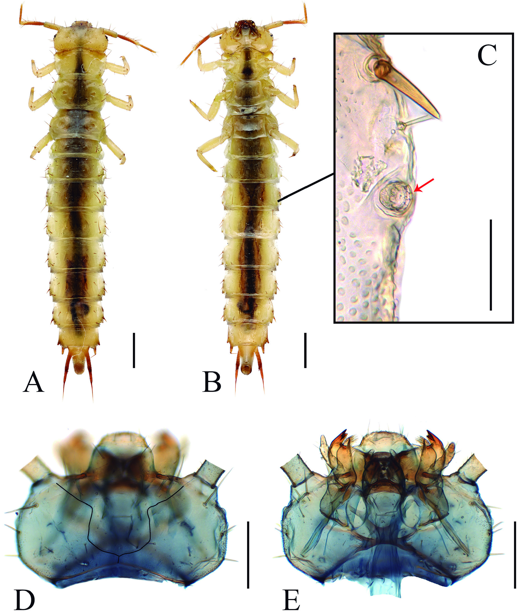
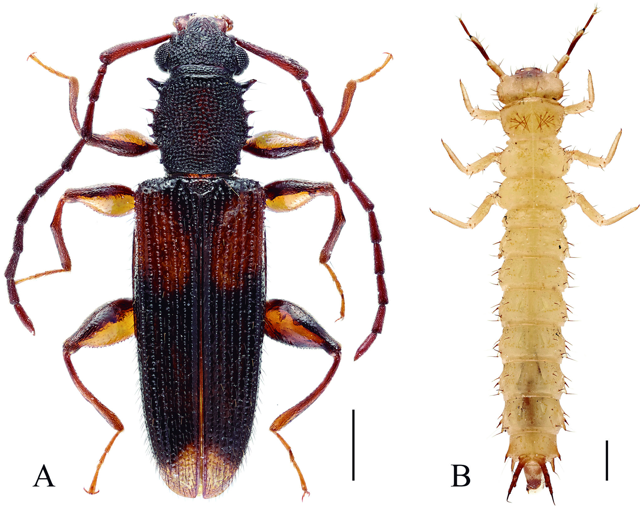
Length 10-11 mm, head width about 1.6 mm. Body distinctly flattened and slender; lateral margins nearly parallel ( Figs. 1A, 1B View Figure 1 ).
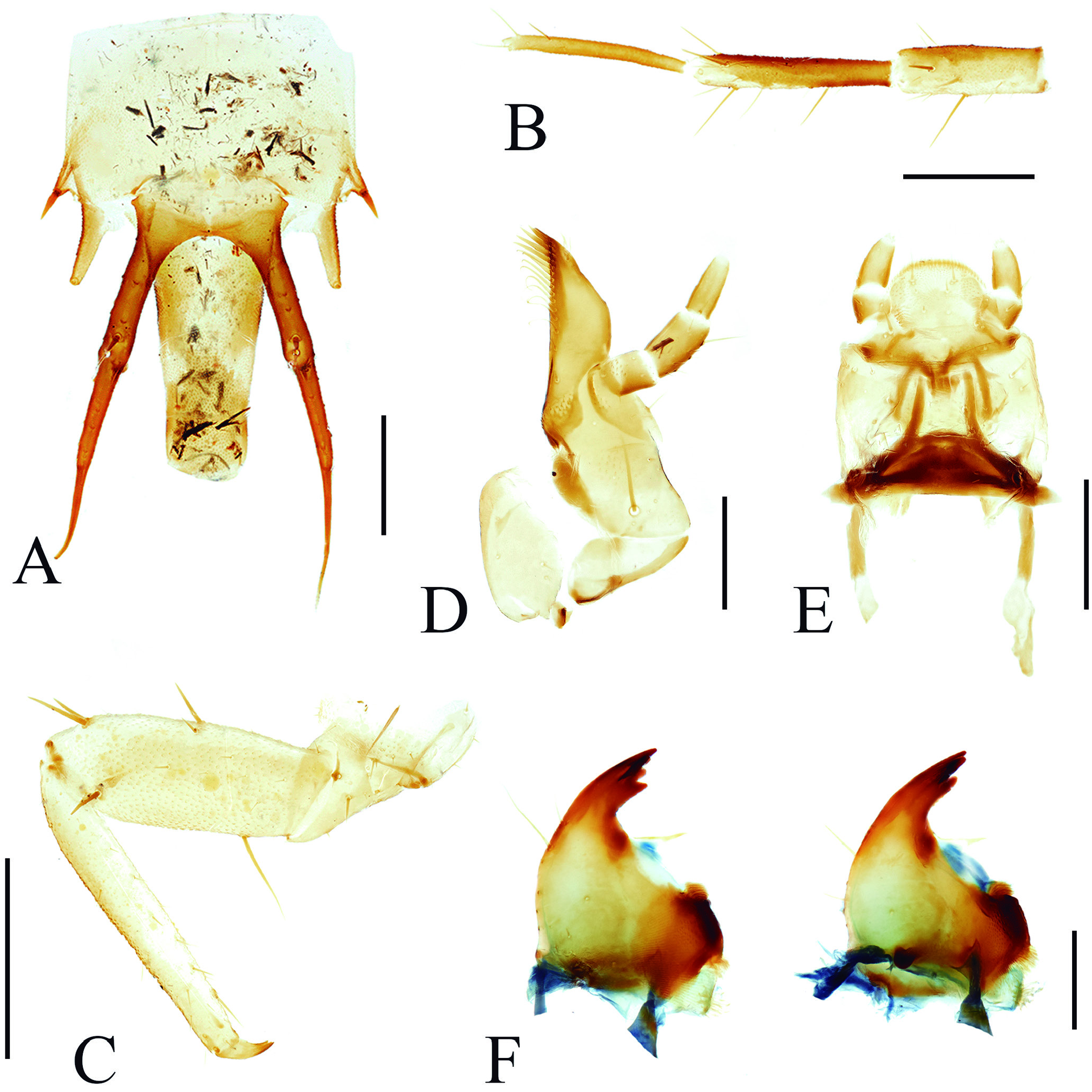
Head transverse with 5 stemmata in one group on each side; frontal arms U-shaped and complete, epicranial stem very short ( Fig. 1D View Figure 1 ). Dorsal surface with two moderately long setae near each antennal insertion, a pair of long lateral setae on anterior area and three pairs near posterolateral edge, otherwise surfaces covered with several pairs of shorter setae. Ventral side ( Fig. 1E View Figure 1 ): gular sutures widely separated, divergent both anteriorly and posteriorly; hypostomal rods distinct and long, diverging posteriorly; ventral surface with pair of long setae below antennal insertion and several pairs of short setae. Clypeus trapezoid, fused with frons, bearing a pair of moderately long setae on anterior area and several pairs of short setae near apex. Labrum transverse with rounded apical margin, covered with short setae. Antenna ( Fig. 2B View Figure 2 ) elongate, slender, about 1.1 times as long as head width; scape relatively stout,with a long seta at mid length and some shorter and very short setae; pedicel about 1.4 times as long as scape, with short, conical sensorium and several long setae near apex and one near mid length, flagellum distinctly narrower than the basal segments. Mandible ( Fig. 2F View Figure 2 ) triangular with broad basal area, apex bidentate, incisor area with two teeth on left mandible and one on right mandible; prostheca membranous, triangular and long, almost transparent; mola with asperities or small tubercles transversely arranged with a posterior and an anterior penicillus; ventral process sub-triangular with pointed apex. Ventral mouthparts deeply retracted. Maxilla ( Fig. 2D View Figure 2 ) with well-developed articulating area; cardo sub-triangular and transverse;stipes large with several distinct setae; mala falcate with inner edge acute, bearing 2-3 sclerotized unci and dense setae apically; palpifer distinct. Maxillary palps 3-segmented, covered with sparse setae: first palpomere stout, the second longer than first, terminal palpomere fusiform with rounded apex. Labium with submentum trapezoid, fused with gula at middle; mentum nearly quadrate, prementum transverse with distinct palpigers laterally; ligula broad with rounded apical margin, covered with dense short setae apically and several longer setae on surface. Labial palps ( Fig. 2E View Figure 2 ) two-segmented with the first segment stout, the second palpomere nearly 1.7 times as long as the first one, apex rounded. Hypopharyngeal sclerome forming transverse bar.
Tergites of thorax simple, without distinct sclerites or tubercles; integument with very fine asperities laterally, visible under high magnification only. Prothorax sub-trapezoid, widest apically, dorsum with two pairs of setae anterolaterally and one posterolaterally; venter with pair of relatively long setae anteriorly adjacent to procoxae and several short setae. Meso- and metathorax sub-rectangular with lateral margins slightly curved, widest at about middle. Mesothorax with three pairs of distinct lateral setae; metathorax with two pairs of setae on dorsum; ventral side of each segment with pair of long setae anterolaterally and several shorter setae. Thoracic spiracle annular, located between pro- and mesothorax.Coxae separated by more than two coxal diameters ( Fig. 1B View Figure 1 ). Legs ( Fig. 2C View Figure 2 ) elongate; trochanter subtriangular and stout with several short setae; femur about 2.4 times as long as wide, covered with several distinct setae and some sparse and short setae; tibiotarsus slender, about 6.2 times as long as wide and 1.3 times as long as femur, covered with short setae along inner edge; pretarsal claw simple. Abdomen 10 segmented; ratio of segments lengths I-VIII as 1.0: 1.3: 1.5: 1.6: 1.6: 1.5: 1.3: 0.9. The first segment short, trapezoid, widest posteriorly, bearing pair of long setae posterolaterally; abdominal segments II-VIII each with a pair of long lateral hairs that are exfoliate. Segments II-VII with lateral margin slightly convex, with 3 pairs of distinct lateral setae; mediotergites with pair of long setae posterolaterally and pair of short setae anteriorly; each sternite with pair of long setae posterolaterally. Segment VIII distinctly narrower and shorter than VII, lateral margins almost parallel with pair of distinct posterolateral projections, extending posteriorly and bearing a long seta. Urogomphi ( Fig. 2A View Figure 2 ) long and U-shaped, strongly bifurcate with sharp apex, gradually diverging posteriorly, each side with two setae underneath at about middle. Terminal segment posteriorly oriented, forming sub-cylindrical pygopod, with anal opening apically. Spiracles annular with distinctly crenulate peritreme ( Fig. 1C View Figure 1 ) on abdominal segments I-VIII, located on very short spiracular tubes.
Remarks: The larva in a prepupa stage ( Fig. 3B View Figure 3 ) is almost identical to the last instar larva, but with the stemmata almost invisible, and the setae on the pronotum almost fused with the pronotum.
Pupa: ( Fig. 4 View Figure 4 )
Length 8.3 mm. Colour of integument changing from initial khaki to rufous in latter stages preceding eclosion; colour changes are especially apparent on head, pronotum, antennae, elytra and legs. Abdomen with terminal segments surrounded by skin cast from last instar larva.
Head ( Fig. 4A View Figure 4 ) bent to ventral side, with a pair of protrusions around anterior margin in front of eyes, bearing distinct setae; frons with paired setae between eyes near antennal insertions. Eyes moderately large, with distinct facets. Antenna with scape slender and transverse, antennomeres 2-11 longer than wide, extending posteriorly along the lateral margin of abdomen, reaching to the ventral side of fourth abdominal ventrite; each segment with several distinct spine-shaped protrusions around the apex and several short setae near middle.
Prothorax ( Fig. 4B View Figure 4 ) sub-quadrate with paired apical protrusions that are expanded apically, bearing a long seta on each side. Anterolateral area with pair of protrusions that are distinctly bifurcate apically,bearing two setae on each side. Lateral margins with two pairs of small and triangular protrusions anteriorly and two posteriorly, each protrusion bearing a long seta. Posterior area with pair of setae on dorsal side. Mesothorax transverse with long wing pads on dorsal side, sterna with moderately broad sternal process. Metathorax relatively large, also with long wing pads, sterna with distinct discrimen. Elytron on ventral side, covering parts of pterothorax, abdominal ventrites and hind legs; surface with weak grooves. Legs relatively long, with compact femora and tibiae transversely located, tarsi longitudinal; fore and mid femora with three distinct lateral setae while hind femora only have one on each side.
Abdomen with eight exposed tergites, tergite I-VII from transverse to sub-triangular, the last two segments concealed by skin of last instar larva; segments II-IV with short lateral protrusions bearing distinct setae,segments V-VII with longer transversely lateral setigerous protrusions, tergite VI also with pair of posterolateral setae. Ventral side with five exposed ventrites, ventrite 5 from transverse to sub-triangular as matured; the first two sternites absent, the last three sternites and urogomphi are concealed by the exuvium of last instar larva.
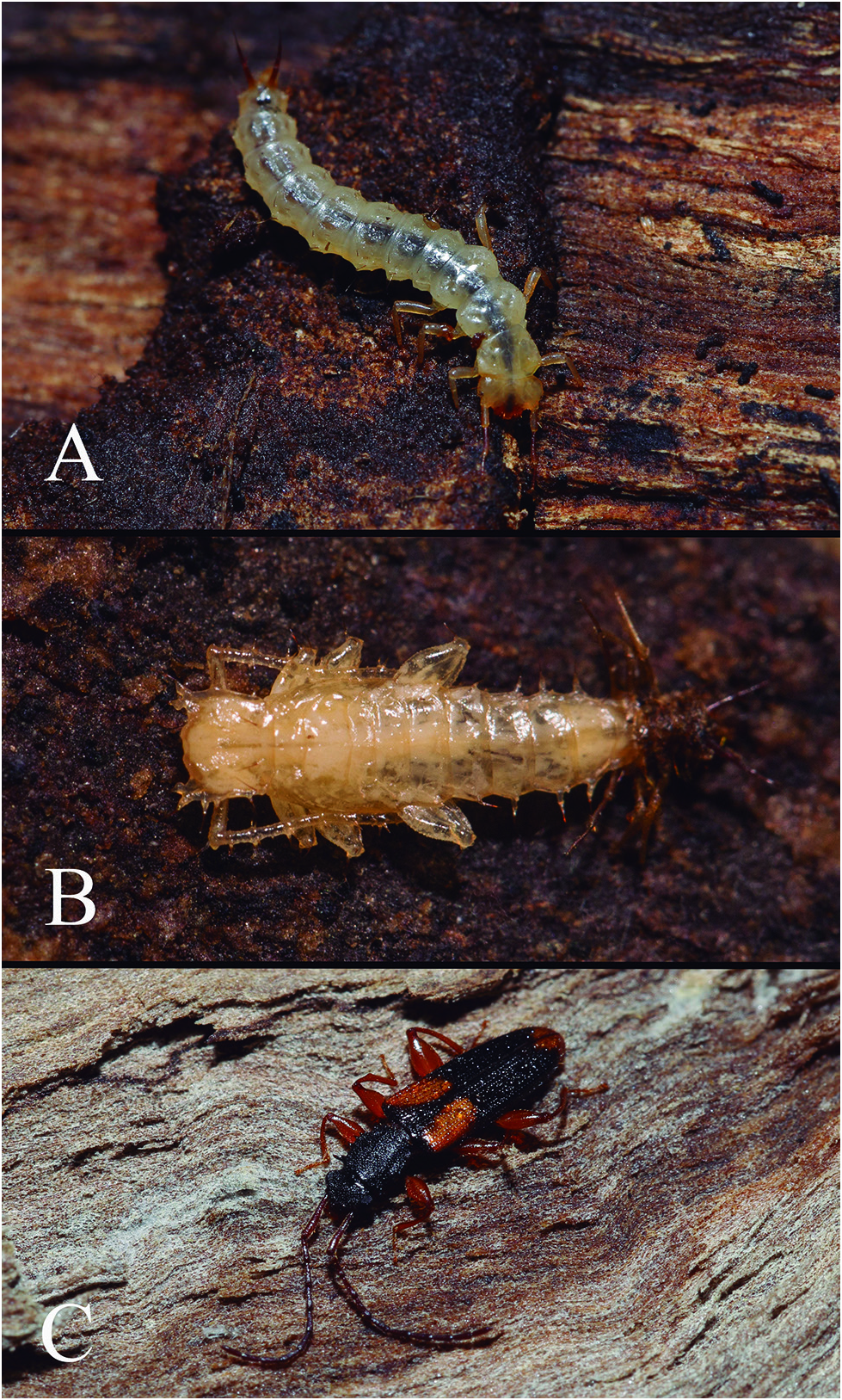
Biology: Larvae and adults specimens were collected under bark of a dead Eucalyptus tree ( Figs. 5A, 5C View Figure 5 ), moving quickly after being exposed. Larvae were observed in the lab grazing on spores and decaying bark. Dark tissues and spores were clearly visible in the esophagus and gut ( Fig. 5A View Figure 5 ), confirming that the larvae feed on the rotten tissues of dead Eucalyptus and the fungi growing on it. Pupae were attached to the wood or bark substrate at the apex of abdomen ( Fig. 5B View Figure 5 ).
Specimens examined: Australia, ACT: 35.27°S 149.11°E Canberra,Black Mt. 20/05/2017 Under bark of Eucalyptus, Zhenhua Liu (2 larvae dissected; 1 larva mounted on card; 1 larva, 2 adults and 1 pupa preserved in ethanol; ANIC).

No known copyright restrictions apply. See Agosti, D., Egloff, W., 2009. Taxonomic information exchange and copyright: the Plazi approach. BMC Research Notes 2009, 2:53 for further explanation.
|
Kingdom |
|
|
Phylum |
|
|
Class |
|
|
Order |
|
|
Family |
|
|
SubFamily |
Brontinae |
|
Tribe |
Brontini |
|
Genus |