
Rhyacophila aphrodite Malicky 1975
|
publication ID |
https://doi.org/ 10.11646/zootaxa.4623.3.8 |
|
publication LSID |
lsid:zoobank.org:pub:6755FA6F-B5CE-4686-A317-1CBFEE0A19B0 |
|
DOI |
https://doi.org/10.5281/zenodo.5941113 |
|
persistent identifier |
https://treatment.plazi.org/id/E262D664-F47D-FFC0-8691-FF16FC8DFE0F |
|
treatment provided by |
Plazi |
|
scientific name |
Rhyacophila aphrodite Malicky 1975 |
| status |
|
Description of the final instar larva of Rhyacophila aphrodite Malicky 1975 View in CoL
Biometry. Body length of final instar larva 18.2 to 22.0 mm, head width 1.24 to 1.34 mm (n = 2). Chaetotaxonomy according to Williams & Wiggins (1981) and Friedrich et al. (2015), anal proleg terminology following Nielsen (1942).
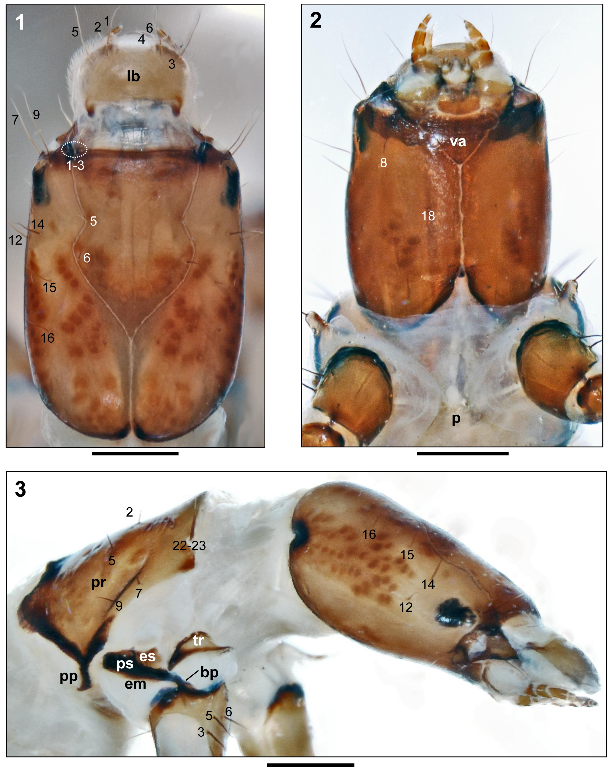
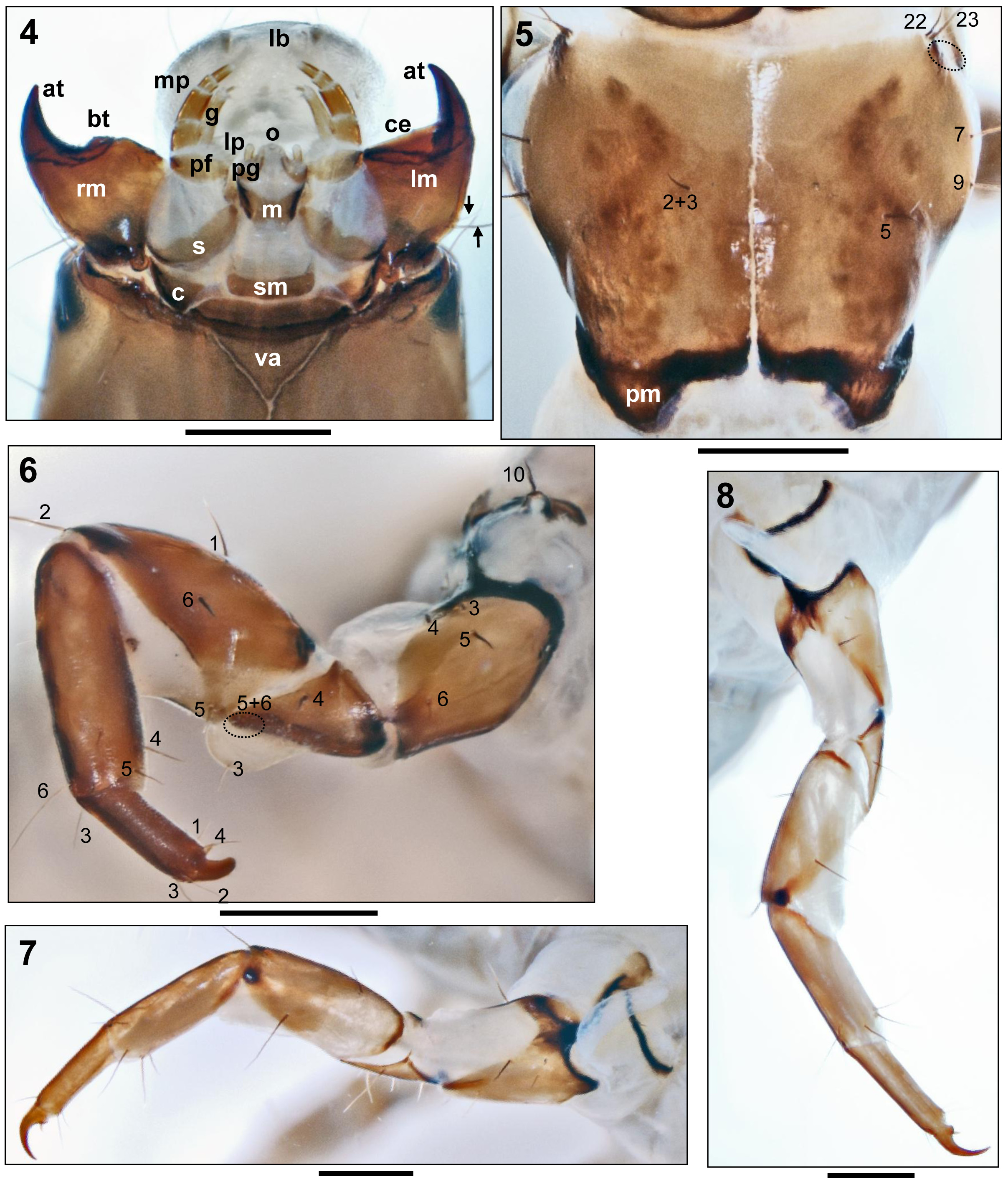
Head. Head capsule elongate, mean length: width ratio 1.33, lateral margins slightly convex with maximum width at height of posterior tip of frontoclypeal apotome ( Fig. 1 View FIGURES 1–3 ); surface finely wrinkled. Base coloration yellow, with conspicuous reddish brown, oval muscle attachment spots on dorsal, ventral, and lateral sides of parietalia, on frontoclypeal apotome, and along coronal suture ( Figs. 1–3 View FIGURES 1–3 ). Additional brown pigmentation on parietalia, dorsally from posterior frontoclypeal suture along posterior coronal suture to foramen occipitale, laterally from setae #12 and #14 to apophysis of foramen occipitale and ventrally, from foramen occipitale to center of parietalia ( Figs. 1–3 View FIGURES 1–3 ). On each parietal, setae #7, #9, #12, #14, #15, and #16 long and conspicuous ( Fig. 1 View FIGURES 1–3 ). Frontoclypeal apotome with deep central constriction, 5 pairs of primary setae, dark brown anterior margin, and U-shaped light brown pigmented area posterior of central constriction including 4–5 light muscle attachment spots ( Fig. 1 View FIGURES 1–3 ). Antennae inconspicuous, close to anterior parietal border, with short flagellum (not visible in Fig. 1 View FIGURES 1–3 ). Yellowish labrum with 6 pairs of primary setae ( Fig. 1 View FIGURES 1–3 ). Ventral apotome ( Fig. 2 View FIGURES 1–3 va) shaped like isosceles triangle, dark brown, with thickened anterior border ( Figs. 2 View FIGURES 1–3 , 4 View FIGURES 4–8 ). Sclerites of maxillolabium yellowish brown; lateral borders of cardines, stipites, and mentum dark brown to black ( Fig. 4 View FIGURES 4–8 ). Mandibles medium brown, almost black apically, and asymmetrical ( Fig. 4 View FIGURES 4–8 ): right mandible with short, stout basal tooth ( Fig. 4 View FIGURES 4–8 bt), lacking on left mandible and replaced there by straight cutting edge ( Fig. 4 View FIGURES 4–8 ce). Each mandible with apical tooth ( Fig. 4 View FIGURES 4–8 at) and 2 lateral setae ( Fig. 4 View FIGURES 4–8 , arrows).
Thorax. Pronotum fully covered by 2 large sclerites with their maximum widths at insertions of setae # 7 and 9; sclerites tapering anteriorly and posteriorly; lateral borders slightly concave ( Fig. 5 View FIGURES 4–8 ). Pronotal sclerites yellowish, each with reddish brown muscle attachment spots over light brown pigmentation creating arrow-shaped pattern pointing mesad ( Fig. 5 View FIGURES 4–8 ). In addition, oval concentrations of muscle attachment spots in posterolateral sections of sclerites, each with seta #5 near its anterior border. Additional muscles attachment spots and narrow stripes of pigment bordering posterior half of median suture ( Fig. 5 View FIGURES 4–8 ). Posterior margin thick and black, with pair of semicircular posterior submesal bulges each with yellow center ( Fig. 5 View FIGURES 4–8 pm) and posterolateral prolongations ( Fig. 3 View FIGURES 1–3 pp). Posterolateral border of pronotal sclerites with black stripe fading laterally, absent anterior of insertion of seta #9; with thin black stripe above insertions of setae #9 and #7 ( Fig. 3 View FIGURES 1–3 ). Pronotal notch at anterolateral corner semicircular, with 2 long setae ( Figs. 3 View FIGURES 1–3 and 5 View FIGURES 4–8 , #22, #23) and 3 tiny setae ( Fig. 5 View FIGURES 4–8 , dotted oval). Prosternal horn lacking, prosternite ill-defined, quadrangular, yellowish white ( Fig. 2 p View FIGURES 1–3 ). Meso- and metathoraces totally unsclerotized, with pale purplish-blue coloured areas subdivided by whitish longitudinal and transversal stripes; each with single pair of anterior (setal area 1 = sa 1) setae and pair of groups of one long and two tiny posterior (setal area 2 = sa 2) setae.
Procoxae each with triangular conical basal process ( Fig. 3 View FIGURES 1–3 bp) touching ventral extension of black pleural suture ( Fig. 3 View FIGURES 1–3 ps). Dorsal apex of pleural suture close to posterolateral prolongations of pronotum ( Fig. 3 View FIGURES 1–3 pp). Proepisternum small, quadrangular, yellowish brown; pro-epimeron very narrow, creating yellowish ventral plate below pleural suture ( Fig. 3 View FIGURES 1–3 em). Protrochantin triangular, yellow, with dark posterior tip and dark dorsal and ventral borders ( Fig. 3 View FIGURES 1–3 tr); its anterior process finger-shaped, bearing seta #10 ( Fig. 6 View FIGURES 4–8 ). Episterna and epimera of meso- and metathoraces almost completely reduced to black vertical pleural sutures ( Fig. 9 View FIGURES 9–12 ps), fused with horizontal trochantin bearing seta #10 ( Lepneva 1964; seta #10 hidden by thoracic gill in Fig. 9 View FIGURES 9–12 ). Legs yellowish brown, with forefemora distinctly wider than mid- and hind femora, and sclerite borders dark brown to black ( Figs. 6–9 View FIGURES 4–8 View FIGURES 9–12 ). Counts of long setae on each leg as follows: coxa 5 (#1, #3, #4, #5, #6; Figs. 6 View FIGURES 4–8 , 9 View FIGURES 9–12 ), trochanter 5 (#3–#6 visible in anterior view), femur 6 (#1, #2, #5, and #6 visible in anterior view), tibia 6 (#3–#6 and one secondary seta visible in anterior view), tarsus 4 (#1–#4; Fig. 6 View FIGURES 4–8 ). Tarsal claws sickle-shaped, pointed (in Fig. 6 View FIGURES 4–8 , tarsal claw heavily worn), with basal spur originating from conical base ( Figs. 6–8 View FIGURES 4–8 ).
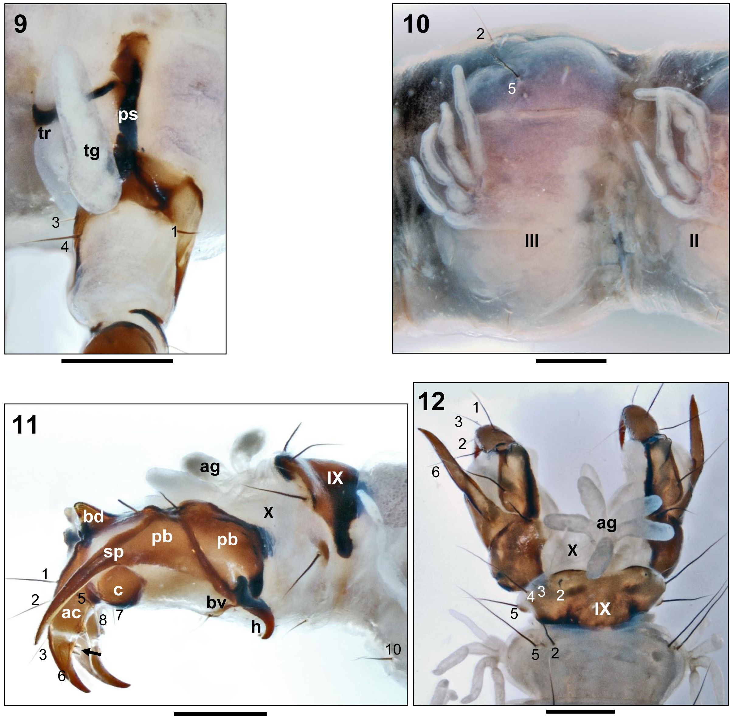
Abdomen. Abdomen slightly depressed (flattened dorsoventrally), with purplish–blue dorsal colouration subdivided by whitish lines. Laterally and ventrally pale cream coloured. Each segment with 2 pairs of long dorsal primary setae ( Fig. 10 View FIGURES 9–12 , setae #2, #5), one pair of tetrafilament lateral gills ( Fig. 10 View FIGURES 9–12 ), and pair of ventral setae #10 ( Fig. 11 View FIGURES 9–12 ). Lateral fringe lacking.
Abdominal dorsum IX covered by large, yellow quadrangular sclerite with black anterior and posterior borders and dark brown markings and pair of lateral black projections; setae #2 and #3 inserted on sclerite, seta #4 on posterior edge and seta #5 inserted on small isolated posterolateral sclerite ( Figs. 11, 12 View FIGURES 9–12 ). With tufted, white anal gills ( Figs. 11, 12 View FIGURES 9–12 ag). Anal proleg sclerites yellowish brown, with dark brown borders and dark brown suture between anterior and posterior parts of proximal sclerite ’b’ ( Fig. 11 View FIGURES 9–12 ). Each proleg having proximal sclerite ’b’ ( Fig. 11 View FIGURES 9–12 pb) with oblique dark bar of suture extending onto strong basoventral hook ( Fig. 11 h View FIGURES 9–12 ) anteriorly and with long terminal sword process ( Fig. 11 View FIGURES 9–12 sp) posteriorly; distal sclerite ’b’ with dorsal hornlike process with blunt tip ( Fig. 11 View FIGURES 9–12 bd). Ventral sclerite ‘c’ nearly round ( Fig. 11 c View FIGURES 9–12 ). Ventral sclerite ( Fig. 11 View FIGURES 9–12 bv) with short median seta. Anal claw (ac) partially divided by ventral membrane into proximal and distal sections, latter fitted with two short basoventral teeth ( Fig. 11 View FIGURES 9–12 , arrow). Anal claw teeth perpendicular to longitudinal axis of anal claw; distal anal claw tooth shorter than half anal claw width at distal tooth insertion ( Fig. 11 View FIGURES 9–12 arrow).
No known copyright restrictions apply. See Agosti, D., Egloff, W., 2009. Taxonomic information exchange and copyright: the Plazi approach. BMC Research Notes 2009, 2:53 for further explanation.