
Hatschekia sumireyakko, Uyeno, Daisuke & Nagasawa, Kazuya, 2012
|
publication ID |
https://doi.org/ 10.5281/zenodo.208870 |
|
DOI |
https://doi.org/10.5281/zenodo.5658715 |
|
persistent identifier |
https://treatment.plazi.org/id/E23F9A16-7767-A629-FF3F-FF78EFB83455 |
|
treatment provided by |
Plazi |
|
scientific name |
Hatschekia sumireyakko |
| status |
sp. nov. |
Hatschekia sumireyakko n. sp.
( Figs. 15–36 View FIGURES 15 – 22 View FIGURES 23 – 28 View FIGURES 29 – 36 )
Material examined. Holotype, female ( NSMT –Cr 21662), ex Centropyge venusta (Yasuda & Tominaga) ( Perciformes : Pomacanthidae ), off Torishima Islet (26°19ʹN, 126°49ʹE), Kumejima Island, the Ryukyu Islands, East China Sea, Japan. 19 November, 2009. Allotype: a male ( NSMT –Cr 21663) and Paratypes: 3 females and 3 males ( NSMT –Cr 21664); 1 female and 2 males ( RUMF –ZC–1503), Collection data of allotype and paratypes same as that of holotype.
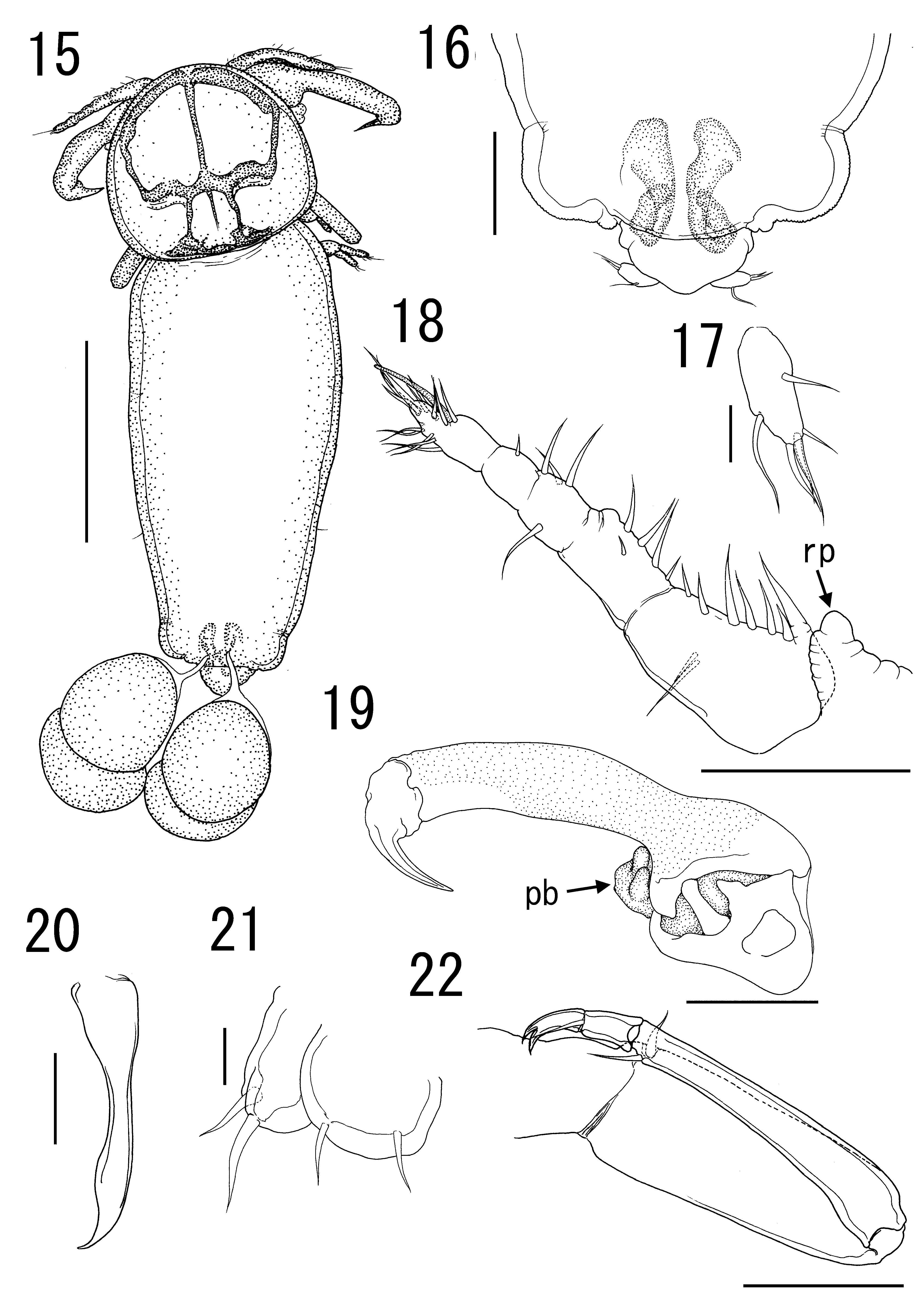
Description of female holotype. Body ( Fig. 15 View FIGURES 15 – 22 ) 624 long, excluding caudal rami, on holotype. Cephalothorax round, slightly shorter than wide (190 × 210); dorsal chitinous frame with double semicircle with posterior ring and lateral bar along posterior margin of cephalothorax. Trunk fusiform, longer than wide (466 × 208), widest anteriorly, gradually narrowed posteriorly. Urosome ( Fig. 16 View FIGURES 15 – 22 ), excluding caudal rami, shorter than wide 23 × 52. Caudal ramus ( Fig. 17 View FIGURES 15 – 22 ) slightly longer than wide 22 × 9, bearing 6 naked setae.
Rostrum with 1 round process on posterolateral corners ( Fig. 18 View FIGURES 15 – 22 ). Antennule ( Fig. 18 View FIGURES 15 – 22 ) indistinctly 5- segmented, 124 long; armature formula: 9, 5, 4, 1, 13 + 1 aesthetasc. Antenna ( Fig. 19 View FIGURES 15 – 22 ) 3-segmented; proximal segment (coxa) unarmed; middle segment (basis) ornamented with surface pits; terminal claw unarmed; proximal segment length 53; middle segment length 163; terminal claw length 22; total length 238. Parabasal papilla ( Fig. 19 View FIGURES 15 – 22 ) well developed. Oral cone robust. Mandible ( Fig. 20 View FIGURES 15 – 22 ) slender, with sharp apex. Maxillule ( Fig. 21 View FIGURES 15 – 22 ) bilobate; both lobes armed with 2 tapering elements; 2 small elements on inner lobe. Maxilla ( Fig. 22 View FIGURES 15 – 22 ) 4-segmented; proximal segment unarmed; second segment rod-like, with 1 basal seta; third segment elongate, with 1 distal seta; terminal segment small, with 1 small seta and bifid claw. Maxilliped absent.
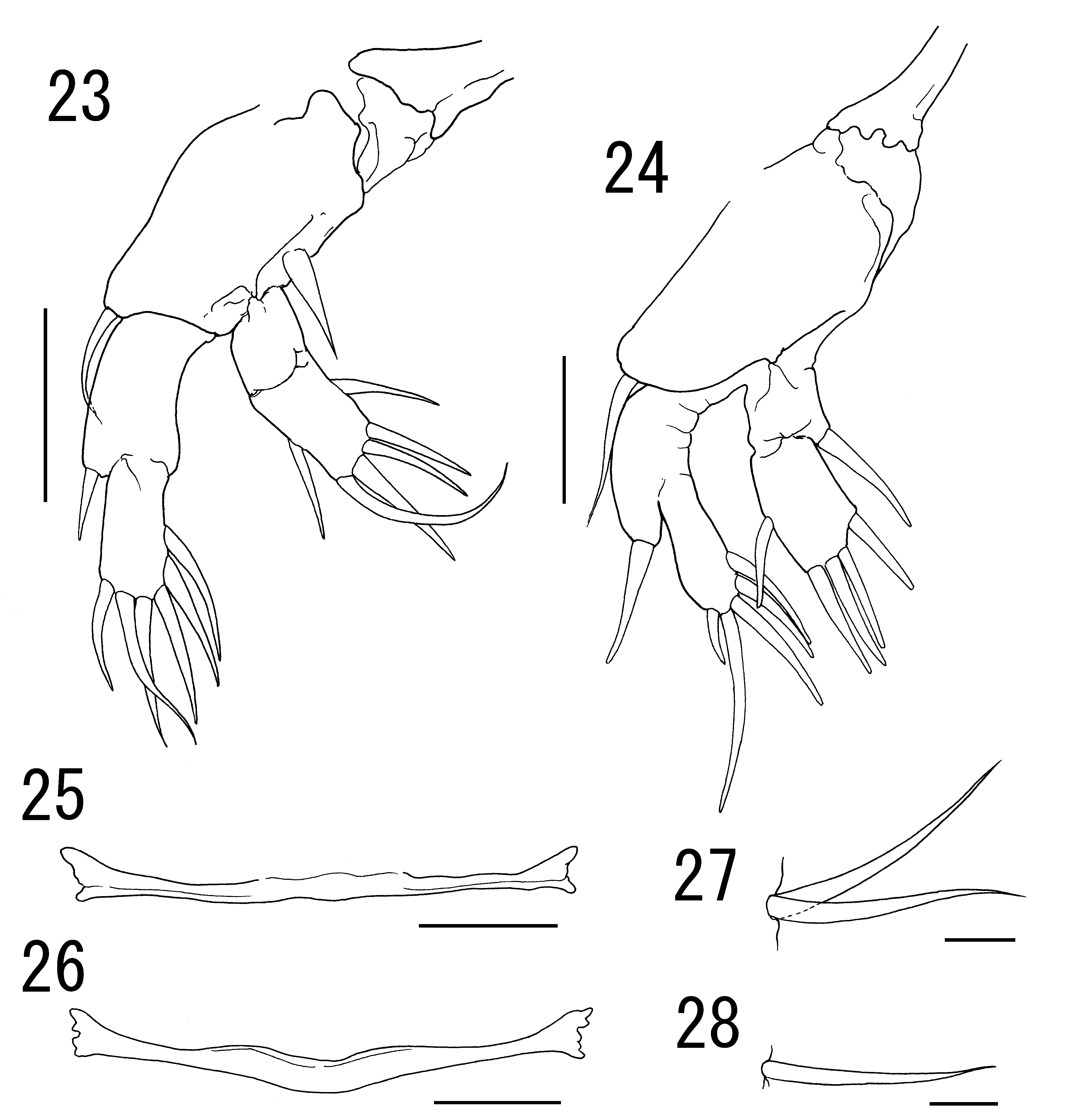
Legs 1 and 2 ( Figs 23–24 View FIGURES 23 – 28 ) biramous, bearing 2-segmented rami; leg armature formula as follows:
Leg 1 ( Fig. 23 View FIGURES 23 – 28 ) 83 long; protopod length 32; exopod length 30 exceeding endopod length 21. Leg 2 ( Fig. 24 View FIGURES 23 – 28 ) length 80; protopod length 47; exopod length 32; endopod length 28.
Intercoxal sclerite of legs 1 and 2 ( Figs 25–26 View FIGURES 23 – 28 ) rod-like, unarmed.
Leg 3 ( Fig. 27 View FIGURES 23 – 28 ) represented by 2 simple setae on anterior 1/3 surface of trunk. Leg 4 ( Fig. 28 View FIGURES 23 – 28 ) represented by 1 simple lateral seta on posterior 2/3 of trunk.
Variability of measurement. Paratype females share all important morphological characters with the holotype. Measurement range of their body parts and appendages of type series (n = 5) was as follows: body length (excluding caudal rami) 541–657 (589 ± 49), cephalothorax length 175–197 (187 ± 8), cephalothorax width 187–210 (196 ± 9), trunk length 382–489 (425 ± 49), trunk width 184–233 (205 ± 21), urosome (excluding caudal rami) length 22–26 (23 ± 2), urosome width (excluding caudal rami) 40–55 (50 ± 6), caudal ramus length 17–22 (18 ± 2) ×, caudal ramus width 6–9 (8 ± 1), antennule length 108–124 (112 ±7), antenna proximal segment length 28–53 (40 ± 10), antenna middle segment length 117–165 (148 ± 20), antenna terminal segment length 20–34 (26 ± 5), antenna total length 174–245 (214 ± 29), leg 1 length 73–83 (78 ± 4), leg 1 protopod length 32–36 (34 ± 1), leg 1 exopod length 24–30 (26 ± 2), leg 1 endopod length 15–21 (18 ± 2), leg 2 length 63–80 (67 ± 8), leg 2 protopod length 33–47 (38 ± 6), leg 2 exopod length 26–32 (29 ± 2), leg 2 endopod length 21–28 (25 ± 3).
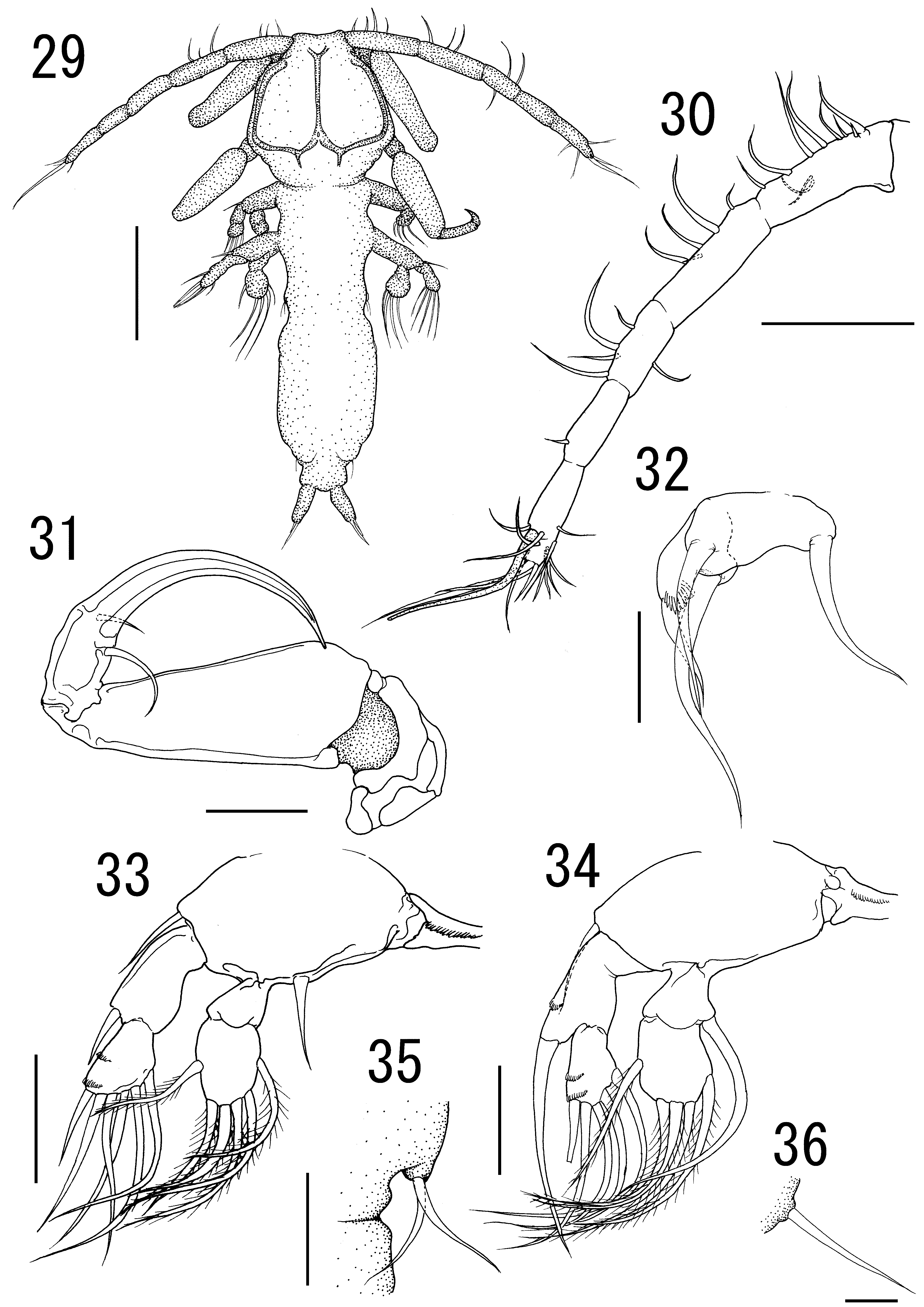
Description of male allotype. Body ( Fig. 29 View FIGURES 29 – 36 ) 240 long, excluding caudal rami, on allotype. Sexual dimorphism present in cephalothorax, proportion of trunk and caudal ramus, rostrum, antennule, antenna, absence of parabasal papilla, maxillule, legs 1 to 3, and presence of leg 5. Cephalothorax round with frontal part prominent 81 × 78; dorsal chitinous frame with 3 vertical bars connecting each other by posterior, horizontal bar. Trunk fusiform, longer than wide 160 × 52, widest anteriorly, gradually narrowed posteriorly. Urosome excluding caudal rami shorter than wide 16 × 26. Caudal ramus distinctly longer than wide 21 × 9.
Rostrum without distinct process. Antennule ( Fig. 30 View FIGURES 29 – 36 ) indistinctly 5-segmented, 187 long; armature formula: 10, 5, 5, 1, 13 + 1 aesthetasc. Antenna ( Fig. 31 View FIGURES 29 – 36 ) 3-segmented; proximal and middle segments (coxa and basis) unarmed; terminal claw with 2 setae near basis; proximal segment length 27; middle segment length 66; terminal claw length 26; total length 120. Maxillule ( Fig. 32 View FIGURES 29 – 36 ) bilobate; both lobes armed with 2 tapering elements; inner lobe with row of blunt spinules.
Legs 1 and 2 ( Figs 33–34 View FIGURES 29 – 36 ) biramous, bearing 2-segmented rami; terminal segment of endopods bearing plumose setae; leg armature formula as follows:
Leg 1 ( Fig. 33 View FIGURES 29 – 36 ) 90 long; protopod length 38; exopod length 32 exceeding endopod length 20. Leg 2 ( Fig. 24 View FIGURES 23 – 28 ) length 77; protopod length 45; exopod length 32; endopod length 26.
Intercoxal sclerite of legs 1 and 2 ( Figs 33–34 View FIGURES 29 – 36 ) with 1 row of blunt spinules.
Leg 3 ( Fig. 35 View FIGURES 29 – 36 ) represented by 2 simple setae on conical process located at slightly anterior to midlength of lateral margin of trunk. Leg 5 ( Fig. 36 View FIGURES 29 – 36 ) represented by 1 simple lateral seta at posterolateral corner of trunk.
Variability of measurement. Paratype males share all important morphological characters with the allotype. Measurement range of their body parts and appendages of type series (n = 6) was as follows: body (excluding caudal rami) length 240–320 (293 ± 31), cephalothorax length 81–93 (88 ± 4), cephalothorax width 78–96 (91 ± 7), trunk length 160–230 (205 ± 27), trunk width 52–68 (61 ± 6), urosome (excluding caudal rami) length 16–28 (23 ± 5), urosome (excluding caudal rami) width 26–34 (31 ± 3), caudal ramus length 21–29 (26 ± 3), caudal ramus width 9–11 (10 ± 1), antennule length 161–187 (177 ±11), antenna proximal segment length 23–38 (30 ± 5), antenna middle segment length 63–73 (79 ± 3), antenna terminal segment length 17–26 (22 ± 4), antenna total length 114–128 (122 ± 4), leg 1 length 75–90 (81 ± 6), leg 1 protopod length 34–38 (36 ± 2), leg 1 exopod length 22–32 (27 ± 3), leg 1 endopod length 17–21 (19 ± 2), leg 2 length 71–79 (76 ± 4), leg 2 protopod length 43–48 (46 ± 2), leg 2 exopod length 28–32 (30 ± 2), leg 2 endopod length 24–28 (25 ± 1).
Attachment site. Gill filaments.
Remarks. Based on the female specimens, Hatschekia sumireyakko n. sp. shares the dorsal frame of the cephalothorax, which forms into the posterior annulus, with H. khahajya , H. monacanthi and H. triannuli n. sp. Hatschekia triannuli n. sp. differs from H. sumireyakko n. sp. by the presence of 3 protrusions along the anterior margin of the rostrum; the posterolateral lobe being extending near the posterior end of the abdomen; a 1- segmented endopod of leg 2; a considerably lesser cephalothorax length/body length ratio [0.15 ± 0.01 vs. 0.32 ± 0.02 (U-test; p <0.001), Table 1 View TABLE 1 ]; and a greater width/length ratio of the horizontal cephalothorax [1.64 ± 0.07 vs. 1.05 ± 0.05 (U-test; p <0.001), Table 1 View TABLE 1 ]. Hatschekia sumireyakko n. sp. is separable from former 2 species by the absence of process on the posterior margin of the intercoxal sclerites of legs 1 and 2 (vs. 4 processes present in H. khahajya and H. monacanthi ). Hatschekia sumireyakko n. sp. can be distinguished from 4 species, H. crenata , H. nohu , H. pacifica , and H. pagellibogneravi that have been insufficiently described for the dorsal frame on the cephalothorax by the characters that distinguish these four species from H. triannuli n. sp. (see Remarks of H. triannuli n. sp.).
Male morphology of Hatschekia species have only been described in nine out of 111 species, H. conifera Yamaguti, 1939 , H. hippoglossi (Guérin-Méneville, 1837) , H. harkema Pearse, 1948 , H. iridescens , H. monacanthi Yamaguti, 1939 , H. petiti Nuñes-Ruivo, 1954 , H. pinguis Wilson, 1908 , and H. prionoti Pearse, 1947 , and H. siganicola El-Rashidy & Boxshall, 2011 (see El-Rashidy & Boxshall 2011; Schram & Aspholm 1997; Uyeno & Nagasawa 2009a). In H. sumireyakko n. sp., sexual dimorphism is saliently shown on the characters of the cephalothorax, trunk, rostrum, antennule, antenna, parabasal papilla, maxillule, and leg 5. Hatschekia monacanthi also show sexual dimorphism in the above characters (see Uyeno & Nagasawa 2009a).
Etymology. The specific name of the new species, sumireyakko , refers to the Japanese common name of the host. The name is used as a noun in apposition.
| NSMT |
National Science Museum (Natural History) |
No known copyright restrictions apply. See Agosti, D., Egloff, W., 2009. Taxonomic information exchange and copyright: the Plazi approach. BMC Research Notes 2009, 2:53 for further explanation.