
Attheyella (Delachauxiella) freyi Löffler, 1963
|
publication ID |
https://doi.org/ 10.5281/zenodo.209217 |
|
DOI |
https://doi.org/10.5281/zenodo.5659229 |
|
persistent identifier |
https://treatment.plazi.org/id/DB104374-FFB5-FFBB-2B93-8989FD047236 |
|
treatment provided by |
Plazi |
|
scientific name |
Attheyella (Delachauxiella) freyi Löffler, 1963 |
| status |
|
Attheyella (Delachauxiella) freyi Löffler, 1963
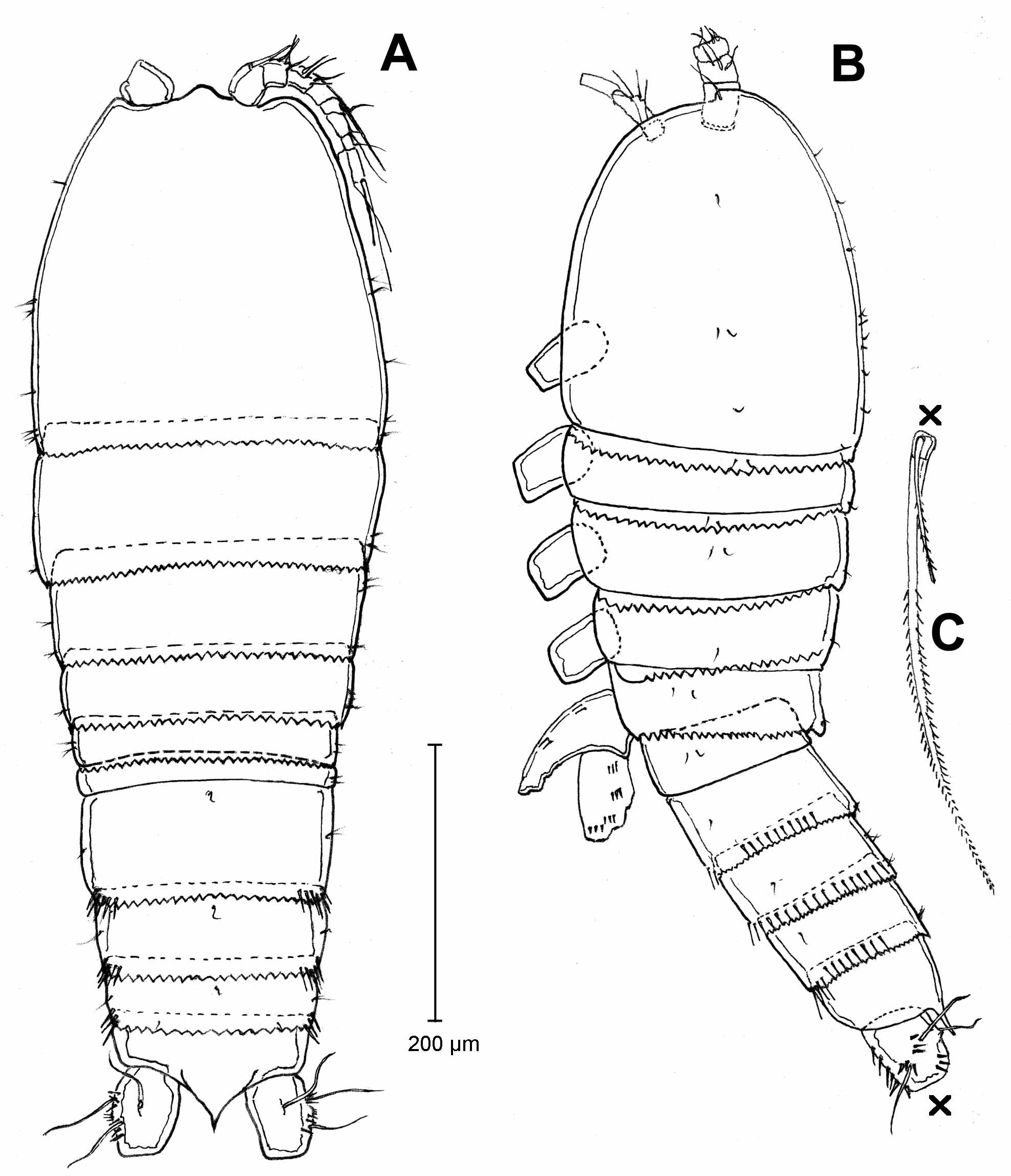
( Figs. 14–25 View FIGURE 14 View FIGURE 15 View FIGURE 16 View FIGURE 17 View FIGURE 18 View FIGURE 19 View FIGURE 20 View FIGURE 21 View FIGURE 22 View FIGURE 23 View FIGURE 24 View FIGURE 25 )
Material examined. Colombia, Cauca State, Puracé, pond near Laguna de San Rafael, coll. S. Gaviria, 14.12.1991: MNHN-Cp2301, 1 female dissected on 1 slide; 1 MNHN-Cp2302, 1 male dissected on 1 slide; NHMW 25218, 1 female undissected mounted on slide; NHMW 25219 – 25220 and NHMW 25222, 3 females dissected on slides; NHMW 25223, 1 male undissected, mounted on slide; NHMW 25221, NHMW 25224 – 25227, 5 males dissected on slides. MNHN-Cp2303, 4 females and 1 male, undissected, ethanol preserved; NHWM-25228, 5 females and 5 males, undissected, ethanol preserved. Additionally, 1 female and 1 male were used for SEM analysis. Ecuador (syntypes, coll. Prescott, January-May 1958). Locality: El Angel, NHMW 22290, 23594 and 23596, each one with 1 female, dissected on slide; NHMW 23589, 23590, 23592 and 23593, each one with 2 females, dissected on 1 slide; NHMW 22291 and 23596, each one with 1 male, dissected on slide. Locality: Antisama, NHMW 22333 and 22334, each with 1 female, dissected on slide; NHMW 23595, 1 male, dissected on slide.
Diagnosis. Female. Frills of posterior edge of body somites (except anal somite) serrated. Genital doublesomite not fused externally, genital tube long, genital pore located at 1/3 of the second urosomite. Genital doublesomite as well as urosomites 3 and 4 with row of spines ventrally and laterally near posterior edge, ventrally always with middle or lateral gaps. Row of spinules dorsally very variable. Ventral surface laterally with double row of spinules in genital double-somite, urosomites 3 and 4. Anal operculum rounded with long posterior arrow-head extention, margin fine serrated, extends to 2/3 of length of caudal rami. Leg 1 with 3-segmented endopod, legs 2-4 with 2-segmented endopod. Leg 1: endopod 1 extends to proximal half of exopod 3. Endopod last segment of leg 2, with 4 (in exceptional cases 5), of leg 3 with 5, of leg 4 with 5 (in exceptional cases with 6) spines or setae. Leg 5: baseoendopod with 6 setae, seta I (inner most), V and VI approximately with equal length and shorter than II-IV, exopod 2.5 times longer than width, with 4 setae, ventral surface of both segments with rows of spinules. Male. Thoracic somites and urosomites with posterior edge dorsally serrated. Urosomites with posterior edge ventrally serrated or smooth. Urosomites with row of spinules dorsally and ventrally very variable. Anal operculum strongly extended into a narrow arrow-head shape, edge finely serrated, extended beyond posterior edge of caudal rami. Legs 1 and 3 with 3- segmented endopod, legs 2 and 4 with 2-segmented endopod. Leg 1: endopod 1 extends to proximal half of exopod 3. Endopod 1 of leg 1, 2 and 4 with 1 seta on inner margin, of leg 3 without seta. Leg 3 with apophysis on second segment, with two terminal barbs. Apical segment of endopods of leg 2 with 3 (rarely 4), of leg 3 with 2, of leg 4 with 5 (2 small elements of outer margin are considered setae). Baseoendopod of leg 5 with 2 inner spines, exopod with 4 setae.
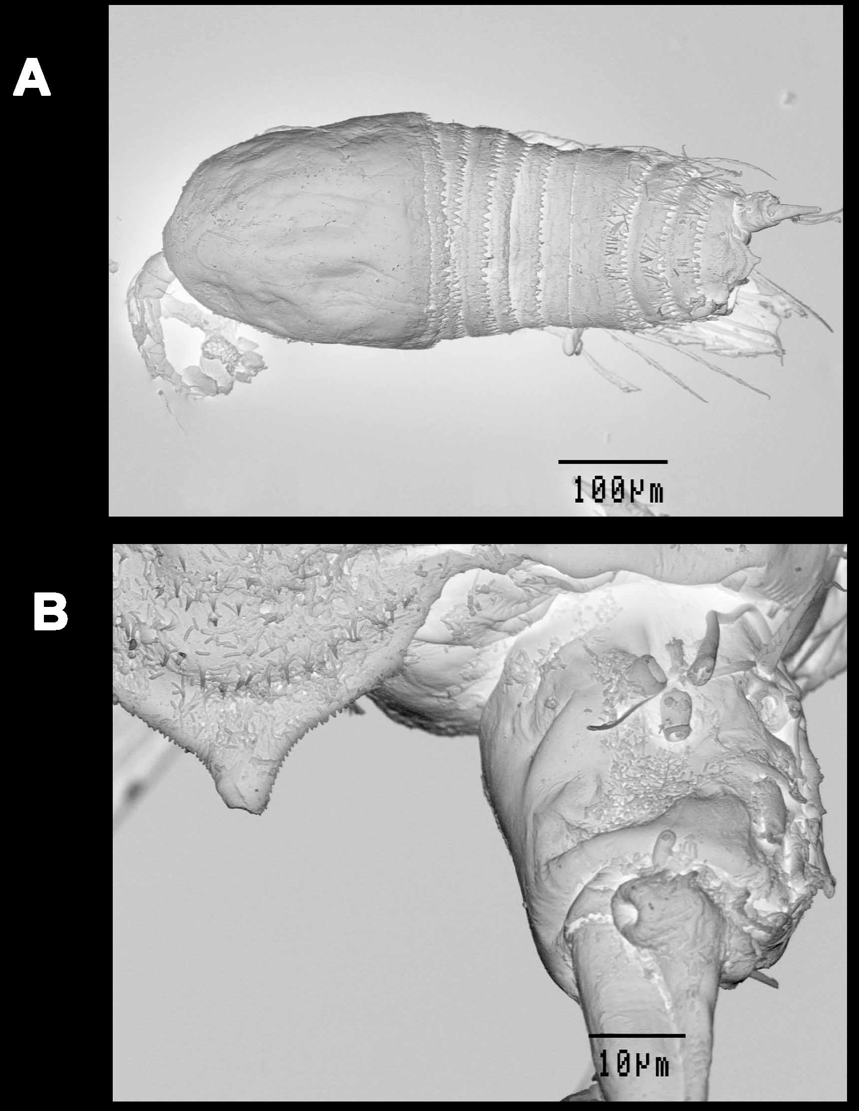
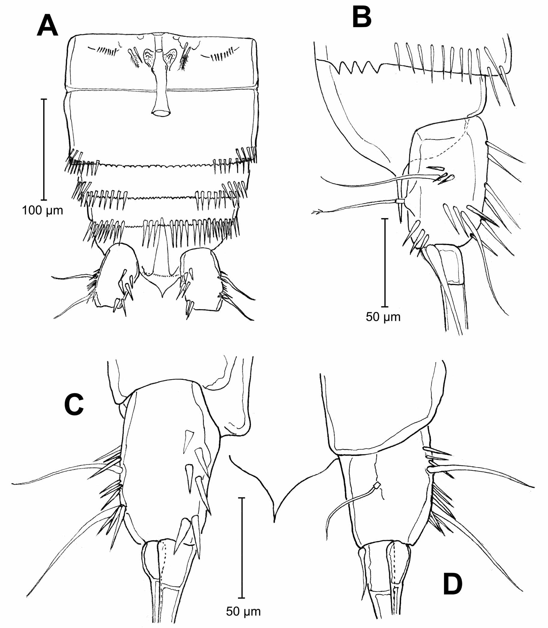
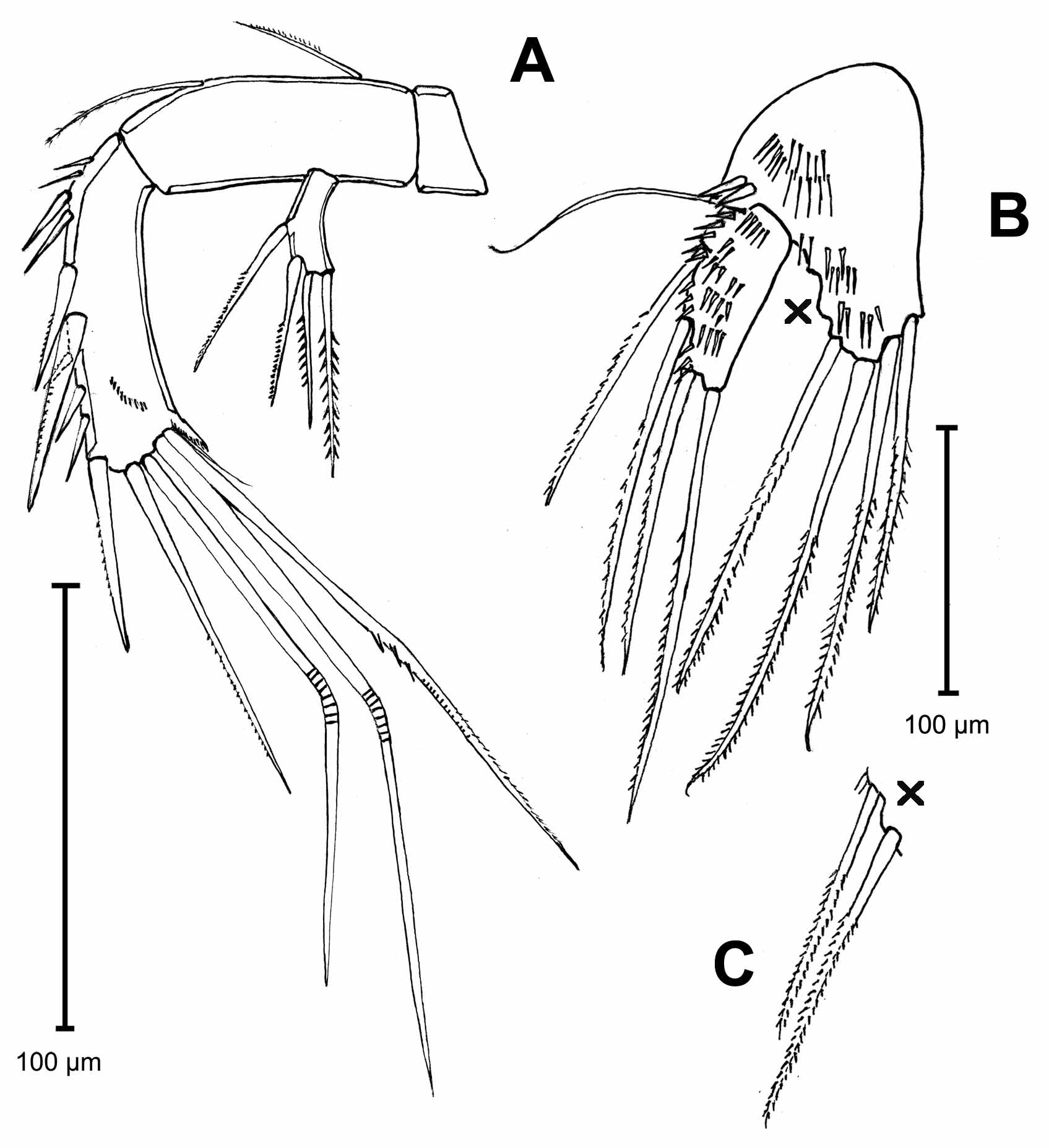
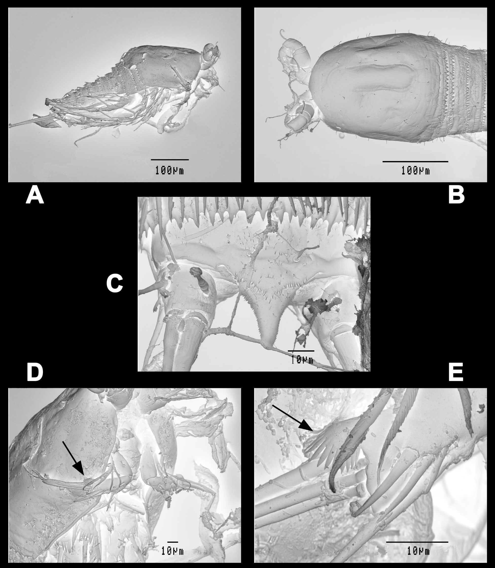
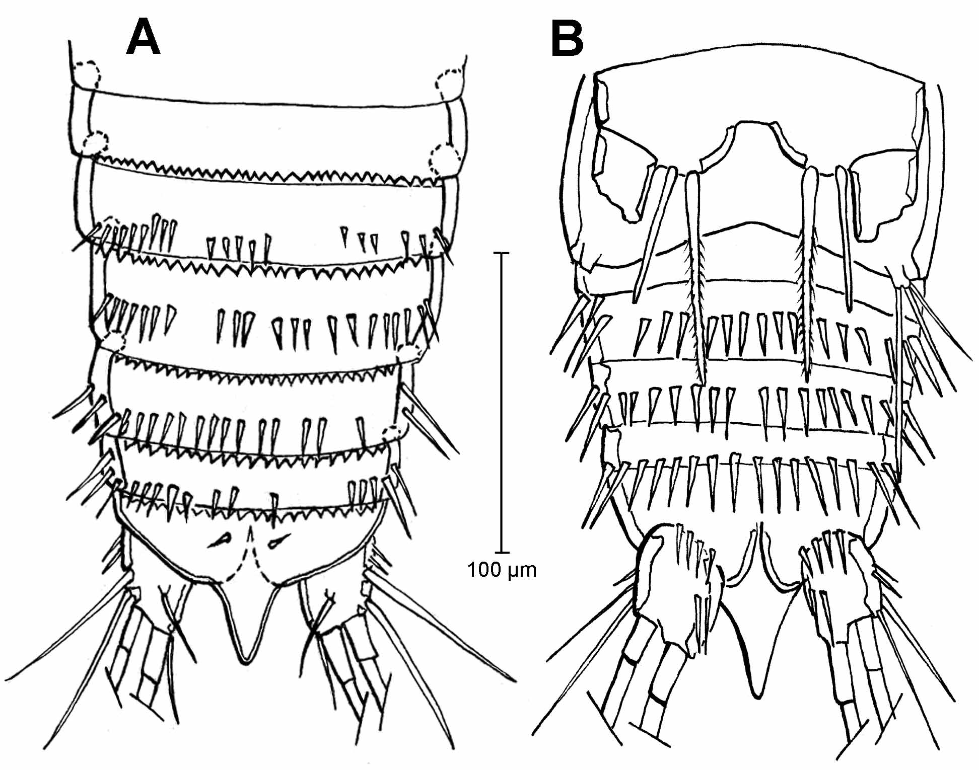
Supplementary description of female. Body cylindrical ( Figs. 14 View FIGURE 14 and 15 View FIGURE 15 A), cephalosome slightly broader than thorax and abdomen, broadest section at its posterior margin. Cephalosome and body somites dorsally and laterally with sensilla. Integument strongly sclerotized. Frills of posterior edge of body somites (except anal somite) serrated. Dorsal serration consists of fine sharp teeth. In contrast, ventral serration is weak and blunt. Body surface including anal operculum, dorsally with additional rows of tiny spinules (visible only in SEM photographs, Fig. 15 View FIGURE 15 ). Abdominal somites with row of spinules near posterior edge, dorsally limited to lateral margins (extension of spinules rows dorsally very variable: see variability), ventrally incomplete in the middle of somites. Ventral surface laterally with double row of spinules on genital double-somite, third and fourth urosomites ( Fig. 16 View FIGURE 16 A). Genital double-somite externally not fused, copulatory pore located at one-third of the second urosomite; genital field laterally with row of tiny spinules. Anal urosomite without spinules. Anal operculum rounded, with central, long posterior extension pointed at tip ( Figs. 14 View FIGURE 14 , 16 View FIGURE 16 A, 16B and 16D) and border finely serrated (visible only in SEM photographs, Fig. 15 View FIGURE 15 B). Anal operculum extends to 2/3 the length of caudal rami. Caudal rami subquadrate, 1.7 times longer than wide (ventral view), with 2 lateral setae and 1 dorsal inserted in distal half of the ramus; with 3 terminal setae; inner seta very tiny, its length less than ½ of width of caudal rami; median seta longer than urosome; outer seta tiny, its length 1/3 of middle seta ( Fig. 14 View FIGURE 14 B); outer margin of caudal ramus with 3 groups of 3 slender spines, ventral surface with 3 groups of paired robust spines ( Fig. 16 View FIGURE 16 ) inserted near inner margin.
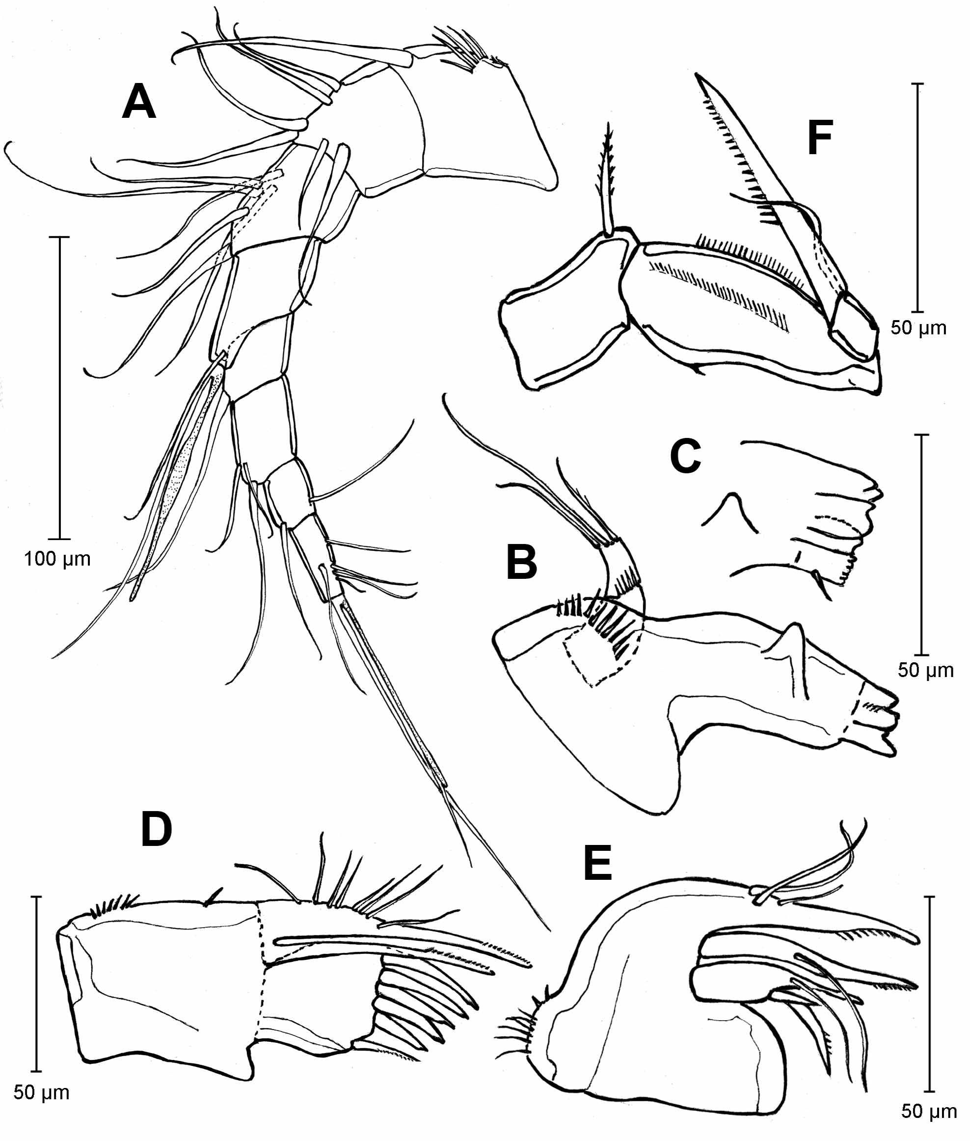
Antennule ( Fig. 17 View FIGURE 17 A) 8-segmented, segment 1 with 4 spinules. Setation formula: 1, 6, 6, 2 + aesthetasc (one seta and aesthetasc with conjoined bases), 1, 3, 2 and 6 + aesthetasc (one seta and aesthetasc with conjoined bases). Setae conjoined with aesthetask of fourth segment longer than 4 apical segments together; setae conjoined with aesthetask of segment 8 longer than 5 apical segments together.
Antenna ( Fig. 18 View FIGURE 18 A): Coxa unarmed, allobasis with 2 setae on inner margin; endopod 1-segmented with row of spinules on dorsal surface near apical margin, inner margin of endopod with 4 proximal spinules followed by 2 strong spines, 3 spinules and 1 spine inserted almost apically; apical margin with 2 spines and 3 geniculate setae, outer seta with inner margin carrying 3 secondary spines near geniculation point. Outer corner of endopod with fan-like spine with similar morphology as in male (see male Fig. 21 View FIGURE 21 E). Exopod 1-segmented, with 2 lateral and 2 apical setae.
Mandible ( Figs. 17 View FIGURE 17 B and 17C): coxal gnathobase with cutting edge composed of 3 main large and coarse teeth, a row of fringed teeth and one lateral seta; lateral surface of gnathobase proximally with row of 11 spinules, a triangular process located on the 2/3 of the distance between the palp and the teeth, distally with strong medial teeth; palp 2-segmented, first segment with row of spinules at apical margin, second segment with 3 apical setae, middle seta longer than lateral setae.
Maxillule ( Fig. 17 View FIGURE 17 D): inner margin of precoxa with proximal row of 7 spinules, distally with 1 spinule; arthrite of the praecoxa with 5 apical fringed spines and 1 subapical seta; coxal endite composed by one long spine; basal endite with one spine and 8 lateral setae.
Maxilla ( Fig. 17 View FIGURE 17 E): composed of syncoxa with 2 endites and basis; proximal endite of syncoxa distally very narrow, with 2 lateral spines of different sizes, stronger spine distally with simple pinnate row of spinules, and 1 lateral seta; distal endite composed by 1 spine with 1 lateral seta, distal section with simple row of spinules; basis distally with simple row of spinules, ending in a narrow tip, with 3 lateral setae. Outer margin of maxilla with proximal row of setulae.
Maxilliped ( Fig. 17 View FIGURE 17 F): coxa bearing 1 seta on inner margin; basis ornamented with two long rows of tiny spinules on anterior margin and dorsal surface, respectively, posterior margin with 1 spinule; endopod 1-segmented, bearing long claw and 1 long seta, claw with distal row of spinules.
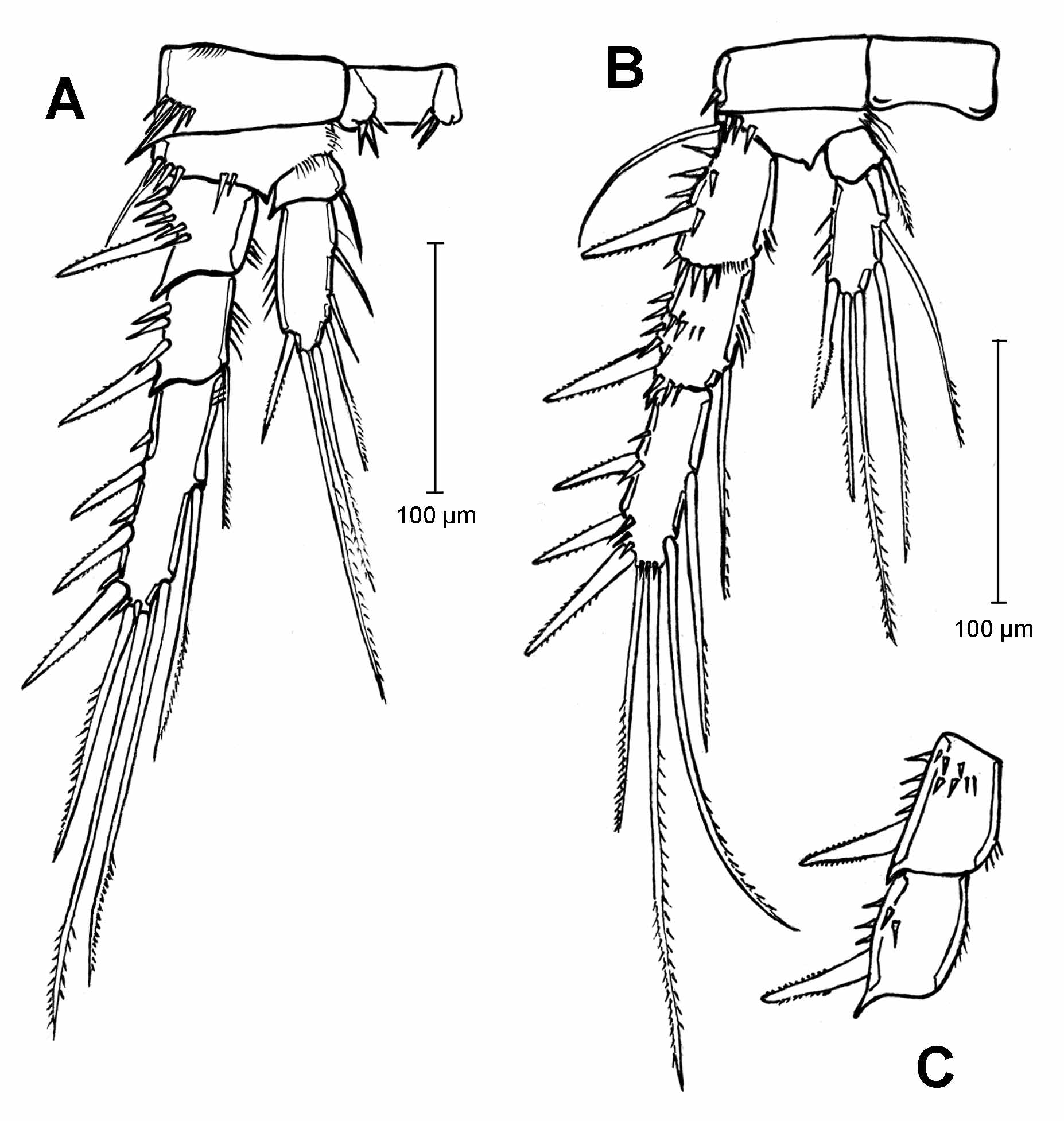
Leg 1 ( Figs. 19 View FIGURE 19 A, 19B and 19C): intercoxal plate with 2 groups of 5 spinules on anterior surface. Coxa, anterior surface with 2 spinules near outer margin and row of 5 spinules on outer apical corner; basis with one strong bipinnate outer and one strong naked inner spine, anterior surface with 2 rows of 3 spinules each, inserted near base of outer spine, row of 7 spinules and row of 3 spinules on apical margin, hairs on inner margin and near insertion point of inner spine. Exopod 3-segmented; first segment with 1 bipinnate spine on outer margin, row of spinules near base of spine and near apical margin; second segment with expanded outer distal corner and hairs on inner margin, 1 bipinnate spine on outer margin and other ornaments as first segment; third segment with 1 bipinnate spine and a row of spinules near outer margin, apically with 1 long unipinnate spine and 2 geniculate setae ( Fig. 19 View FIGURE 19 B). Endopod 3-segmented, longer than exopod; first segment longer than first two segments of exopod, with 1 short inner bipinnate spine and 2 small setae inserted distally and a row of spinules on outer margin; second segment with 1 bipinnate spinule on inner margin inserted distally and row of spinules distally on outer margin; third segment with row of spinules on outer margin, with 1 outer unipinnate spine, 1 median geniculate seta and 1 short inner unipinnate seta on apical margin ( Fig. 19 View FIGURE 19 C).
Leg 2 ( Fig. 19 View FIGURE 19 D): anterior surface of intercoxal plate with one group of 3 and one group of 2 spinules near each distal corner. Coxa with 1 small spine on outer margin. Basis with 1 bipinnate outer spine, 1 row of spinules near insertion point of exopod and rows of hairs near insertion point of endopod and on inner margin. Exopod 3-segmented; outer distal corner of segment strongly extended, outer margin of first segment with 1 bipinnate spine and spinules, row of spinules near distal margin and row of hairs on inner margin; second segment with outer distal corner similarly extended as in first segment, outer margin with 1 bipinnate spine and spinules, inner margin with 1 long seta inserted distally unipinnate at its distal part, and bearing 4 spinules near its base on outer margin; third segment about 1.5 times longer than first and second segments together, outer margin with 3 bipinnate spines and spinules, inner margin with 1 long median bipinnate seta, apical margin with 1 outer unipinnate spine and 1 inner bipinnate seta. Endopod 2-segmented; first segment short, with 1 bipinnate spine on inner margin and hairs on outer margin; second segment with 1 distal unipinnate seta and spinules on outer margin, 2 bipinnate setae on apical margin and 1 median seta unipinnate at its end on inner margin. Both endopods of one specimen with 1 additional seta on inner margin, two specimens with this additional seta in one of both endopods (see variability).
Leg 3 ( Fig. 20 View FIGURE 20 A): intercoxal plate as in leg 2. Coxa with expanded outer apical corner, outer apical surface with a row of spinules. Basis with 1 short outer naked seta, with two rows of spinules near insertion point of exopod, with row of hairs near insertion point of endopod and on inner margin. Exopod 3-segmented; first segment with expanded outer apical corner, outer margin with 1 bipinnate spine and spinules, inner margin with row of hairs; second segment with armature as in first segment, additionally with a long seta inserted on distal inner corner, unipinnate at its distal part and bearing 3 spinules on outer margin near its base; outer margin of third segment with 3 bipinnate spines and spinules, inner margin with 2 setae unipinnate in their distal part, apical margin with 1 unipinnate spine and 1 seta, bipinnate at its extremity. Endopod 2-segmented, first segment short, with spine on inner margin; outer margin of second segment with 1 bipinnate distal spine and spinules, inner margin with 3 setae, apical margin with 2 setae, the inner bipinnate, the outer unipinnate.
Leg 4 ( Figs. 20 View FIGURE 20 B and 20C): intercoxal plate without ornaments. Coxa with spinule on outer margin. Basis with 1 long outer naked seta and a row of spinules on anterior surface near base of exopod, inner margin with row of hairs. Exopod 3-segmented; first segment with 1 bipinnate spine and row of spinules on outer margin, inner margin with hairs; anterior ( Fig. 20 View FIGURE 20 B) and posterior ( Fig. 20 View FIGURE 20 C) surfaces of segment with spinules, distal margin of anterior surface with row of spinules of two different sizes; outer margin of second segment ornamented as in first segment, inner margin with long unipinnate seta and row of hairs; anterior ( Fig. 20 View FIGURE 20 B) and posterior ( Fig. 20 View FIGURE 20 C) surfaces with spinules, distal margin of anterior surface with row of spinules; third segment with 3 bipinnate spines and spinules on outer margin, inner margin with 2 setae unipinnate in their distal half, apical margin with 1 unipinnate spine, 1 unipinnate seta and row of spinules near base of armature. Endopod 2-segmented, first segment short, with bipinnate spine on inner margin; inner margin of second segment with 2 long setae, unipinnate at their distal part, outer margin with 1 bipinnate spine and spinules, apical margin with 2 setae, the outer unipinnate, the inner bipinnate. One specimen bears an additional short seta on proximal inner margin of third segment (see variability).
Leg 5 ( Figs. 18 View FIGURE 18 B and 18C): baseoendopods separated at base, bearing 1 thin naked seta on outer margin and 6 bippinate setae on distal margin, seta I (innermost), V and VI approximately equal in length and shorter than II-IV; exopod 2.5 times longer than wide, bearing 4 unipinnate setae; row of spinules on anterior surfaces of baseoendopod and exopod as in Figure 18 View FIGURE 18 B.
Legs 1–5 with following formula of spines (Roman numerals) and setae (Arabic numerals). Spinules are not included in the formula.
Leg 6 ( Fig. 16 View FIGURE 16 A): reduced to a small plate and located posterior to seminal receptacle, bearing 1 uniserially setulated seta and 1 naked seta.
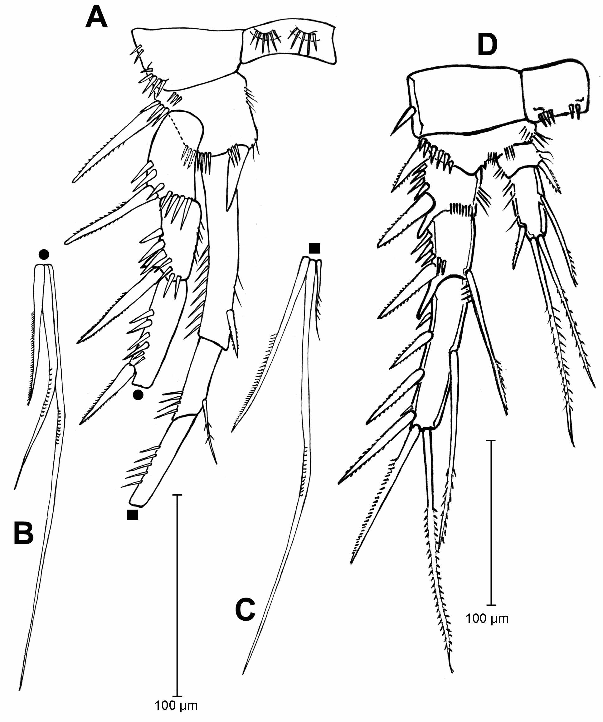
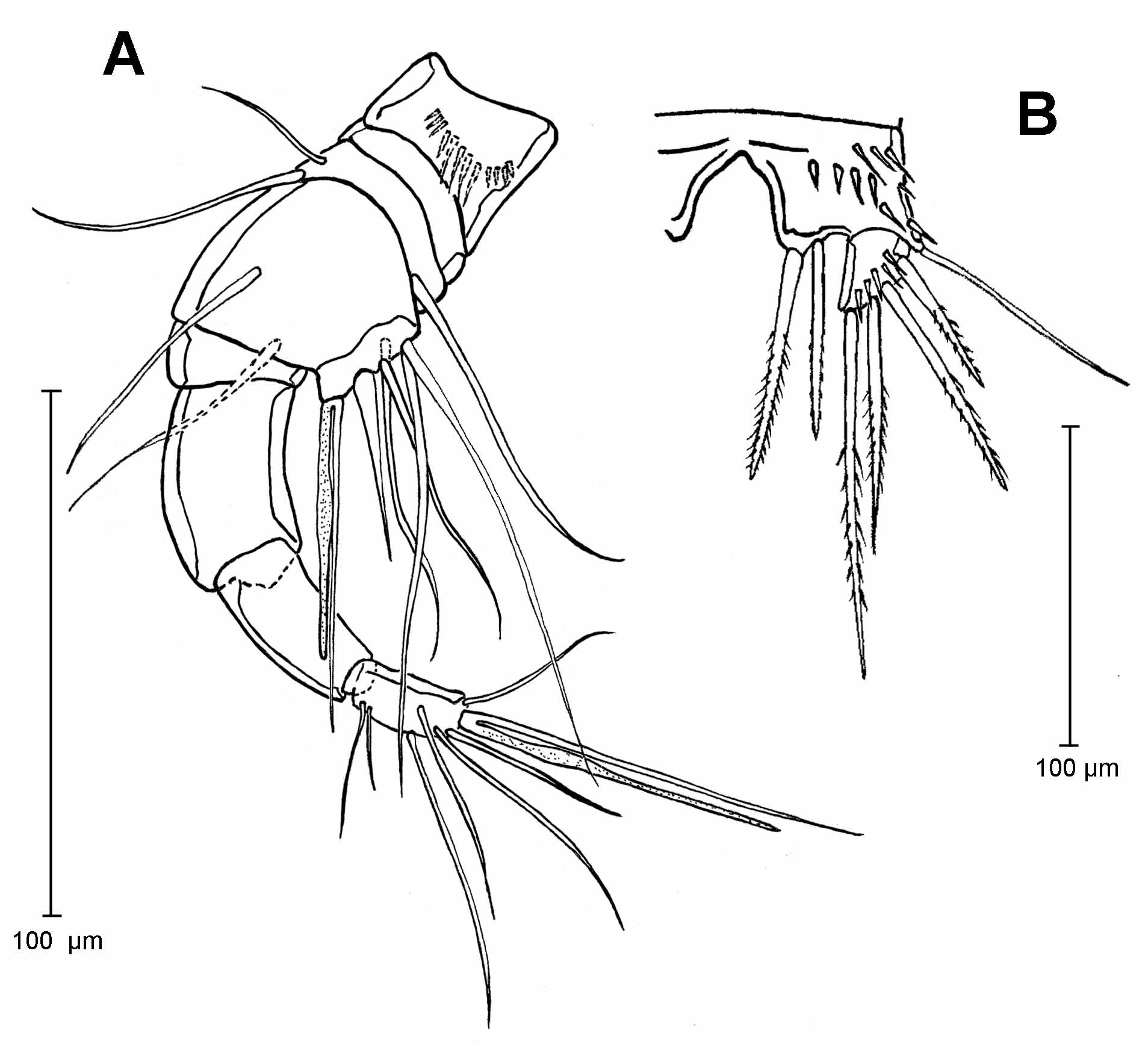
Supplementary description of male. Body with cephalothorax wider and considerably higher than urosome ( Figs. 21 View FIGURE 21 A and 21B). Broadest body section at the posterior margin of the integumentary window. Integumentary window long, rectangular with rounded corners, with slightly narrow waistline at posterior third ( Fig. 21 View FIGURE 21 B). Cephalosome and body somites dorsally and laterally with sensilla. All body somites with frill of posterior edge dorsally serrated. Posterior margin of thoracic somites and urosomites dorsally serrated ( Figs. 21 View FIGURE 21 A, 21B and 22A). Urosomite 1 with posterior margin ventrally smooth, urosomites 2 to 4 with posterior margin ventrally serrated or smooth; urosomites 1 to 4 dorsally with discontinuous rows of spinules, urosomites 2 to 4 ventrally with continuous row of spinules near the posterior margin ( Figs. 22 View FIGURE 22 A and 22B) (see morphological variability). Body somites and anal operculum ornamented on the surface with rows of spinules (visible only in SEM photographs, Figs. 21 View FIGURE 21 B and 21 C). Anal somite with two large sensillae near base of operculum; distal margin ventrally with 4 spines on each side. Anal operculum posteriorly strongly extended into a narrow arrowhead-shape, border finely serrated. Caudal rami outer margin with 2 proximal spines and 2 lateral setae, dorsal surface with one seta, apical margin with 3 setae, the meddle one tiny; ventral surface with 2 spines near inner margin.
Antennula geniculate ( Figs. 23 View FIGURE 23 A and 21B), with 8 segments, first segment with row of spinules on dorsal surface, setation formula ( Fig. 23 View FIGURE 23 A): 0, 0, 3, 7 + aesthetasc and seta with conjoined bases, 0, 0, 0, 7 + aesthetasc and seta with conjoined bases.
Antenna as in female. Endopod with spine on outer apical corner transformed into a fan-like structure, clearly visible in SEM photograph ( Figs. 21 View FIGURE 21 D and 21E). This structure is also present in female.
Mandible, maxillule, maxilla and maxilliped not examined.
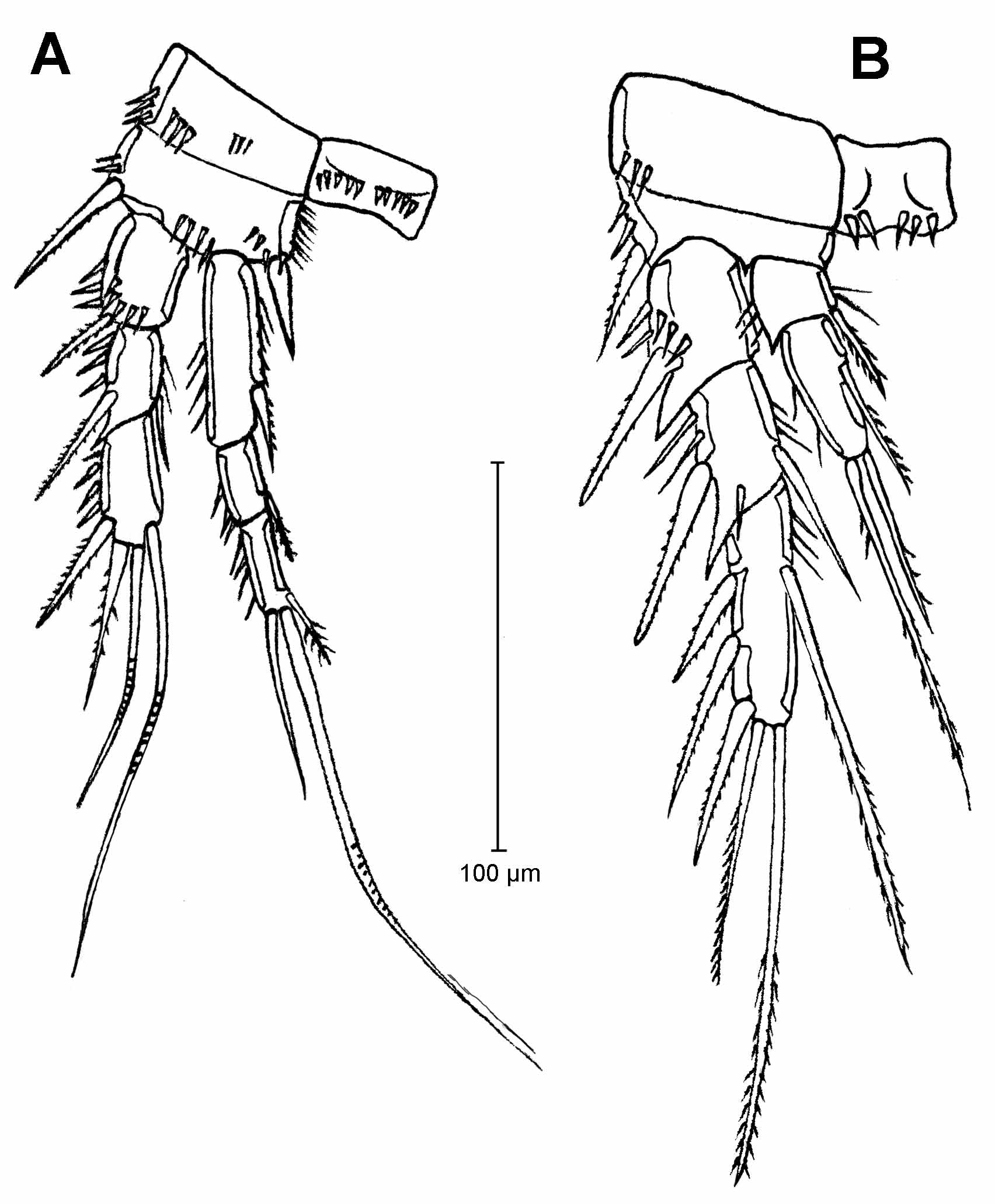
Leg 1 ( Fig. 24 View FIGURE 24 A): intercoxal plate with 2 groups of 5 spinules on anterior surface. Coxa, anterior surface with 2 groups of 3 spinules on anterior surface and a group of 3 spinules on outer apical corner; basis with one large outer bipinnate spine and one large inner naked spine, anterior surface with two rows of 4 and 3 spinules each, inserted near distal margin, hairs on inner margin. Exopod 3-segmented; first segment with 1 bipinnate spine and spinules on outer margin, anterior surface with row of spinules near outer and distal margins; second segment with one bipinnate spine and spinules on outer margin, with one naked seta and 2 spinules on inner margin; third segment with 1 bipinnate spine and a row of spinules on outer margin, apically with 1 bipinnate spine and 1 geniculate seta, outer margin with 1 geniculate seta inserted subapically. Endopod 3-segmented; first and second segments with armature like in female, third segment with apical margin with 1 short outer simple seta and a geniculate inner seta, inner margin with a distal bipinnate seta.
Leg 2 ( Fig. 24 View FIGURE 24 B): anterior surface of intercoxal plate with 1 group of 2 and 3 spinules near each distal corner. Coxa with 3 spinules on outer distal corner. Basis with 1 bipinnate spine and spinules on outer margin. Exopod 3- segmented; first segment with outer distal corner strongly extended, outer margin with 1 bipinnate spine and spinules, anterior surface with row of spinules near insertion point of spine; second segment with outer distal corner similarly extended as in first segment, outer margin with 1 bipinnate spine and spinules, anterior surface with 1 spinule near distal margin, inner margin with spinules and 1 seta inserted distally (shorter than in female), seta naked; third segment as long as first and second segments together, armature as in female but without spinule on outer margin and with spinules on inner margin. Endopod 2-segmented; first segment as in female, inner spine shorter than in female; second segment with spinules and without seta on outer margin, with 2 bipinnate setae apically and 1 median unipinnate seta on inner margin. One specimen carries an additional seta distally on inner margin of the right leg (see variability).
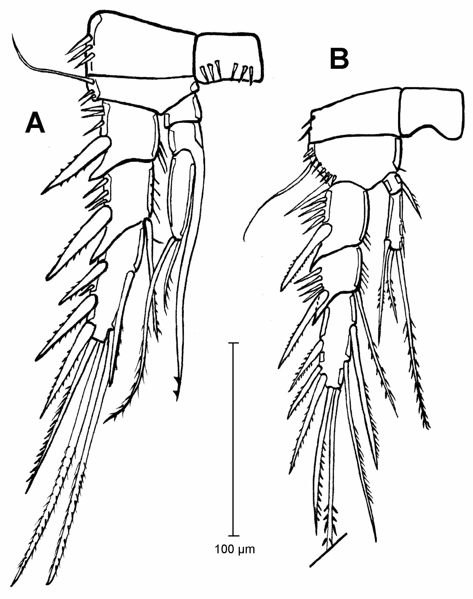
Leg 3 ( Fig. 25 View FIGURE 25 A): intercoxal plate with group of 3 spinules near each distal corner. Coxa with row of 3 spinules near outer margin. Basis with 1 long naked seta and 2 spinules on outer margin. Exopod 3-segmented; first segment as in female; second segment as in female but seta on inner margin much longer, seta unipinnate in its distal part and bearing 1 spinule on inner margin near its base; third segment shorter than in female, armature as in female but distal seta on inner margin of third segment is bipinnate. Endopod 3-segmented, first segment short, unarmed; second segment with long inner apophysis ending in 2 barbs, third segment with 2 setae of different size.
Leg 4 ( Fig. 25 View FIGURE 25 B): intercoxal plate without ornaments. Coxa with spinules on outer margin. Basis with 1 long outer naked seta; anterior surface with a row of spinules near outer margin, inner margin with 1 hair. Exopod 3-segmented; first segment with outer distal corner extended, with 1 bipinnate spine and row of spinules on outer margin, inner margin distally with hairs; second segment with outer margin ornamented as in first segment, inner margin with long bipinnate seta and row of hairs; third segment with 3 bipinnate spines on outer margin, 2 unipinnate setae on inner margin and 1 unipinnate spine and 1 bipinnate seta on apical margin. Endopod 2-segmented, first segment short, with spine on inner margin; inner margin of second segment with 2 small setae, outer margin with 1 spine inserted distally, apical margin with 2 unequal bipinnate setae. All Colombian males carry 5 elements on the last segment of the endopod. Both small elements on the inner margin are definite setae because they are set in notches on the edge. That condition was also noted by Wells (2007) regarding the illustrations of animals from Ecuador described by Löffler (1963).
Leg 5 ( Fig. 23 View FIGURE 23 B): baseoendopods fused, anterior surface with row of spinules inserted as in figure, with 1 long outer seta and 2 inner bipinnate apical spines; exopod with 4 bipinnate setae, anterior surface with 5 spinules. Legs 1–4 with following formula of spines and setae:
Leg 6 ( Fig. 22 View FIGURE 22 B): represented by a small segment, bearing 3 setae, the innermost longer than the others and reaching the anal urosomite.
Variability of females. Females of the species from Ecuador and Colombia show more morphological variability than males, particularly in the ornamentation of body somites and in the chaetotaxy of legs. In all specimens the ventral rows of spinules at the genital double-somite and at the urosomites 3 and 4 have a midventral small or large gap. The rows of spinules on the ventral surface of somites are generally double (but with different extension), although in one specimen from Ecuador (NHMW 23592) the rows are simple. Genital double-somite and urosomite 3 and 4 always bear lateral row of spinules.
The dorsal rows of spinules show greater variability than the ventral rows. In general, the animals (4 specimens from Colombia, 9 from El Angel, 2 from Antisama) bear only a dorsolateral row of spinules, as illustrated in Figure 14 View FIGURE 14 A. Only two specimens from El Angel (NHMW 23592 and 22290) show dorsally complete rows of spinules on the genital double-somite and urosomite 4. Urosomite 3 shows a complete row in one of the latter specimens (NHMW 23592) and shows a middle gap in the other one. Only one specimen from Colombia (animal used for SEM-photograph, Figure 15 View FIGURE 15 ) bears dorsally row of spinules on the genital double-somite and on urosomites 3 and 4, all rows showing several gaps.
The chaetotaxy of legs is constant on the first, third and fifth legs, but variable on endopods of the second and fourth legs. Of the 4 Colombian specimens dissected, two bear 4 setae (NHMW 2518 and 25220), one bears 5 setae (MNHN Cp-2301), and one (NHMW 25222) shows asymmetry, with 4 setae on the left endopod of leg 2 and 5 on the right one. Ten of eleven specimens of El Angel ( Ecuador) bear 4 setae on the endopods of the second leg and one specimen (NHWW 23590) bears 5 setae on one leg and 4 on the other. Both specimens of Antisama ( Ecuador) bear 4 setae on endopods of the same leg. The chaetotaxy of the fourth legs is less variable: only one specimen from El Angel shows 5 setae on the left endopod and 6 setae on the right one (NHMW 23589), all remaining specimens from Ecuador and all from Colombia bear 5 setae on these endopods.
Leg 5: there is no variability in the chaetotaxy of leg 5. The baseoendopod of Colombian females always bears 6 setae on its apical margin. Although the drawing of the female from Ecuador shows 5 setae ( Löffler 1963), all studied syntypes of the locality El Angel bear 6 setae. The animal used by Löffler for his illustration probably has lost seta III. The place of insertion of the lost seta is clearly visible on the distal edge of the leg.
Length of Colombian females 588 – 756 µm (n=15). Length of Ecuadorian females 800 – 980 µm (n=13) ( Löffler 1963).
Variability of males. Ornamentation of body somites and chaetotaxy of second swimming legs of Colombian and Ecuadorian specimens show morphological variability.
Urosomite 1, posterior margin smooth ventrally, whereas urosomites 2 to 4 are serrated (e.g. NHMW 25226) or smooth ( Fig. 22 View FIGURE 22 B) (NHMW 25221). The same two types of ornamentation were observed in the Ecuadorian specimens ( Löffler 1963).
Dorsal rows of spinules show great variability, as follows: 1) animals with urosomites 1 to 4 with discontinuous rows of spinules ( Fig. 22 View FIGURE 22 A) (MNHN 2302), 2) animals with complete rows on urosomites 2 to 4 and no spines on urosomite 1 (NHMW 25226), 3) animals without spines on urosomites 1 to 3 and with a middle gap in the row on urosomite 4 (NHMW 25227) and 4) animals without spines on urosomites (NHMW 25223). Ecuadorian specimens show any combination between complete dorsal rows (NHMW 23595, 23596) and a condition without spines ( Löffler 1963). The ventral row of spines on urosomites also show variability: 1) animals with continuous rows on all urosomites (MNHN 2301, NHMW 25221), 2) animals with complete rows on urosomites 3 and 4 and two ventrolateral gaps in the row on urosomite 2 (NHMW 25226) or a middle gap on the same urosomite (NHMW 25225) and 3) animals with urosomite 3 and 4 with ventrolateral gaps (NHMW 25226). In Ecuadorian specimens, most of those rows are complete (e.g. NHMW 22291, 23596), but they can be also incomplete with ventrolateral gaps (e.g. NHMW 23595).
The number of ventral spines on the posterior margin of urosomite 5 varies in Colombian specimens from 4 (MNHN 2302, NHMW 25223, 25226) to 2 (NHMW 25221, 25225, 25227) on each side. In Ecuador, the illustrated specimen (NHMW 23596) shows 3 spines on each side ( Löffler, 1963).
Leg 2: the second endopod of most of the animals shows 3 setae on the last segment, but few specimens carry 4 setae: one specimen (NHMW 25226) bears an additional small seta inserted distally on the inner margin, and one specimen (NHMW 25225) bears 3 seta on the left endopod and 4 on the right one. Within the three syntype males from Ecuador, two bear 3 setae and one animal bears 4 setae ( Löffler 1963).
Length of Colombian males 493 – 588 µm (n=13); length of Ecuadorian males 540 – 700 µm (n=3) ( Löffler 1963).
Distribution. Known only from Ecuador ( Löffler 1963) and Colombia, inhabiting benthic habitats of water bodies in the “páramo” region, above 3000 m altitude.
| NHMW |
Naturhistorisches Museum, Wien |
No known copyright restrictions apply. See Agosti, D., Egloff, W., 2009. Taxonomic information exchange and copyright: the Plazi approach. BMC Research Notes 2009, 2:53 for further explanation.