
Siamitoides, Jałoszyński, 2020
|
publication ID |
https://doi.org/ 10.11646/zootaxa.4868.2.5 |
|
publication LSID |
lsid:zoobank.org:pub:B32CFC79-57D5-49D1-93C3-EE7A692DFFF5 |
|
DOI |
https://doi.org/10.5281/zenodo.4436428 |
|
persistent identifier |
https://treatment.plazi.org/id/D752EF5A-8E3C-7C3A-FF04-F94BFEAA5EF8 |
|
treatment provided by |
Plazi |
|
scientific name |
Siamitoides |
| status |
gen. nov. |
Siamitoides View in CoL gen. n.
Type species: Siamitoides carinatus , here designated.
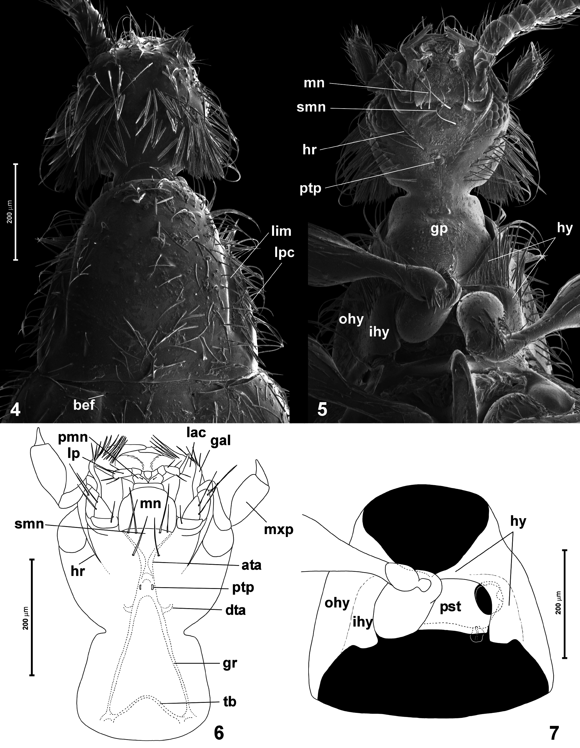
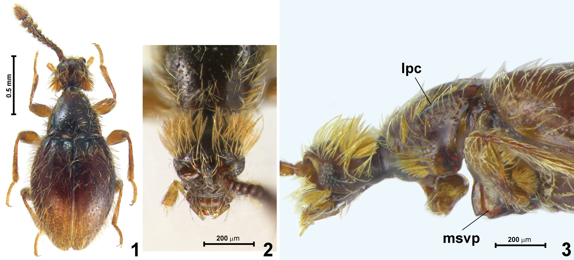
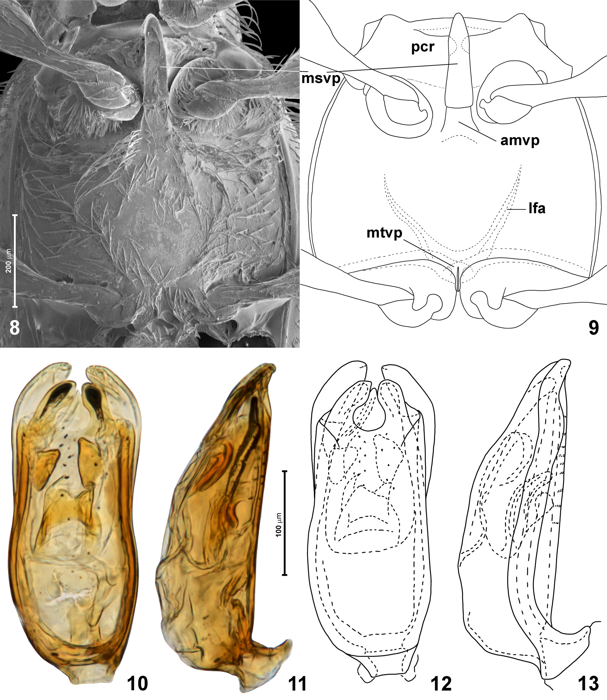
Diagnosis. Siamitoides is unique among world Stenichnini in having two apomorphies: (1) each lateral pronotal carina ( Fig. 4 View FIGURES 4–7 ; lpc) that forms lateral margin of pronotum accompanied mesally by a broad and deep, elongate lateral impression ( Fig. 4 View FIGURES 4–7 ; lim) with sharply marked mesal margin; and (2) hypomera ( Figs 5, 7 View FIGURES 4–7 ; hy) in front of procoxal cavities expanding mesad and meeting at middle, so that basisternal region of prosternum is indiscernible. Additionally, the following combination of characters is not found in any other genus (but separately each of them is shared with other Stenichnini): body with deep constriction between head and prothorax but shallow between prothorax and elytra ( Fig. 1 View FIGURES 1–3 ); antennae gradually thickened ( Fig. 1 View FIGURES 1–3 ); head short, with eyes closer to mandibular bases than to occipital constriction ( Figs 2–3 View FIGURES 1–3 ), in lateral view head capsule ( Fig. 3 View FIGURES 1–3 ) subtriangular, flattened ventrally and with the highest point above antennal fossae, steeply declining toward clypeus and less so toward occiput, with flat vertex; thick bristles present on head (vertex and tempora) and on anteromedian regions of hypomera ( Figs 2–5 View FIGURES 1–3 View FIGURES 4–7 ); occipital constriction ( Figs 4–5 View FIGURES 4–7 ) slightly narrower than half width of head at eyes, constriction on neck region abrupt, step-wise; frontoclypeal groove absent; posterior tentorial pits ( Figs 5–6 View FIGURES 4–7 ) exposed, small, situated far in front of occipital constriction; pre-tentorial gular sutures (= lateral sutures of submentum; see Jałoszyński (2020a)) lacking; hypostomal ridges present ( Figs 5–6 View FIGURES 4–7 ), incomplete, extending from lateral margins of cardines to middle between anterior submental margin and posterior tentorial pits; mentum ( Figs 5–6 View FIGURES 4–7 ) subtrapezoidal; labial palpomere 2 ( Fig. 6 View FIGURES 4–7 ) subcylindrical; maxillary palpomere 3 ( Fig. 6 View FIGURES 4–7 ) not swollen, but only slightly more than twice as long as broad; maxillary palpomere 4 ( Fig. 6 View FIGURES 4–7 ) subconical; pronotum ( Fig. 4 View FIGURES 4–7 ) subconical, broadest at base and narrowing anterad, lacking antebasal pits, groove and sublateral carinae, but with nearly complete, sharply marked lateral carinae; prosternum ( Fig. 7 View FIGURES 4–7 ) with posterior margin weakly concave; procoxal cavities ( Fig. 7 View FIGURES 4–7 ) closed by posterior lobe of coxosternum fused with hypomeron; hypomeron ( Fig. 7 View FIGURES 4–7 ) divided into asetose and elevated inner portion and broad outer portion covered with bristles and setae, border between these areas not marked by hypomeral ridges but by nearly step-wise elevation of inner hypomeral region; mesoventrite ( Figs 8–9 View FIGURES 8–13 ) with anteromedian impressions functioning as procoxal rests lacking defined posterior and lateral margins and with anterior margins indicated only near mesoventral intercoxal process; mesoventral process ( Figs 8–9 View FIGURES 8–13 ) massive, broadly carinate, strongly elevated anteriorly, with flattened ventral surface, slightly broadening posterad and anteriorly reaching anterior mesoventral margin; lateral foveae between meso- and metaventrite lacking; mesocoxal projections prominent; anterior metaventral process ( Figs 8–9 View FIGURES 8–13 ) present, subrectangular, anteriorly touching posterior margin of mesoventral process, demarcated only by shallow transverse fissure; metaventral intercoxal process ( Figs 8–9 View FIGURES 8–13 ) composed of pair of long and slender spines, barely separating metacoxae; metafurca ( Fig. 9 View FIGURES 8–13 ) with vestigial stalk (nearly V-shaped) and approximate insertions of lateral furcal arms; each elytron with one vestigial and asetose basal fovea ( Fig. 4 View FIGURES 4–7 ) situated closer to suture than to humerus, on mesal end of distinct transverse step-wise carina demarcating elytral articulating lobe from disc; mesoscutellar shield not exposed between elytral bases; aedeagus ( Figs 10–13 View FIGURES 8–13 ) with nearly symmetrical median lobe and partly asymmetrical endophallic structures, with bilobed apex and large collar surrounding parameral bases, parameres free (i.e., not fused with walls of median lobe), rod-like.
Description. Body form ( Fig. 1 View FIGURES 1–3 ) elongate, moderately slender, with distinct constriction between head and pronotum, but shallow one between pronotum and elytra.
Head ( Figs 2–6 View FIGURES 1–3 View FIGURES 4–7 ) short, rounded in dorsal view, subtriangular in lateral view ( Fig. 3 View FIGURES 1–3 ), with flattened ventral surface and highest site situated above antennal fossae, steeply declining anterad and less strongly posterad; eyes situated closer to mandibular bases than to occipital constriction, anteriorly touching antennal fossae; vertex flat; frontoclypeal groove lacking; tempora and vertex with thick, straight, long bristles. Occipital constriction abrupt, nearly step-wise, slightly narrower than half width of head; neck region slightly broadening posterad ( Fig. 6 View FIGURES 4–7 ). Antennal fossae large and situated anterolaterally, narrowly separated at middle. Gular plate transverse, with indistinct post-tentorial gular sutures, pre-tentorial gular sutures lacking; posterior tentorial pits ( Figs 5–6 View FIGURES 4–7 ; ptp) small, elongate, situated far in front of occipital constriction. Submentum ( Figs 5–6 View FIGURES 4–7 ; smn) broad, with two pairs of setae on anterior margin; mentum ( Figs 5–6 View FIGURES 4–7 ; mn) subtrapezoidal with weakly rounded anterior margin; prementum ( Fig. 6 View FIGURES 4–7 ; pmn) largely membranous, flexible, with small subtriangular ligula lacking setae, insertions of labial palps moderately broadly separated, labial palpomere 1 weakly elongate, palpomere 2 strongly elongate, subcylindrical, palpomere 3 narrow, elongate, tapering distad and nearly straight. Maxilla with short cardo, subtriangular basistipes bearing two setae on lateral margin, broad mediostipes, elongate lacinia ( Fig. 6 View FIGURES 4–7 ; lac) with a row of robust setae along mesal and distal margin, galea ( Fig. 6 View FIGURES 4–7 ; gal) elongate, with robust setae along distal and distomesal margin, palpifer elongate, maxillary palp ( Fig. 6 View FIGURES 4–7 ; mxp) with small, elongate palpomere 1, long, pipe-like palpomere 2, broad and relatively short palpomere 3, and subconical palpomere 4. Mandibles poorly exposed in studied specimen, each subtriangular, with curved and slender apical tooth. Labrum transverse, with rounded anterior margin, and with sparse, symmetrically distributed dorsal setae directed anterad.
Tentorium ( Fig. 6 View FIGURES 4–7 ) with small corporatentorium, bisinuate tentorial bridge ( Fig. 6 View FIGURES 4–7 ; tb), long gular ridges ( Fig. 6 View FIGURES 4–7 ; gr), short dorsal tentorial arms ( Fig. 6 View FIGURES 4–7 ; dta), short and stout posterior tentorial arms, and long, slender anterior tentorial arms ( Fig. 6 View FIGURES 4–7 ; ata).
Antennae ( Fig. 1 View FIGURES 1–3 ) slender but short in relation to body, gradually thickened and loosely assembled in distal half; scape and pedicel each weakly elongate.
Prothorax in dorsal view ( Fig. 4 View FIGURES 4–7 ) subconical, broadest at base and narrowing anterad. Anterior pronotal corners not marked, posterior corners distinct. Pronotal base lacking pits and grooves. Sides of pronotum with nearly complete lateral carinae ( Fig. 3 View FIGURES 1–3 ; lpc) and broad flattening along each carina, with sharply demarcated, step-wise mesal margin. Sides of pronotum with thick bristles. Prosternum ( Figs 5, 7 View FIGURES 4–7 ; pst) short in relation to prothorax, with basisternal portion obliterated by anteromesal expansions of hypomera that meet at middle, coxosternal portion (forming procoxal rests) with slightly concave posterior margin. Procoxal cavities closed by postcoxal prosternal lobe fused with hypomeron, notosternal sutures lacking. Hypomeron divided into outer ( Figs 5, 7 View FIGURES 4–7 ; ohy) and inner ( Figs 5, 7 View FIGURES 4–7 ; ihy) regions demarcated not by hypomeral ridge but by step-wise elevation of inner region, the latter asetose and strongly convex, contrasting with flattened outer region covered posteriorly with setae and anteriorly with long, thick and straight bristles directed anterad ( Fig. 5 View FIGURES 4–7 ).
Mesonotum not visible between elytral bases in intact specimens.
Mesoventrite ( Figs 8–9 View FIGURES 8–13 ) strongly transverse, with short prepectus, transverse anteromedian impressions functioning as procoxal rests ( Fig. 9 View FIGURES 8–13 ; pcr) filled with modified, leaf-like setae and separated at middle by mesoventral intercoxal process, margins of impressions poorly defined, except for partly carinate anterior margins near middle. Mesoventral intercoxal process ( Figs 8–9 View FIGURES 8–13 ; mscp) carinate and keel-like (i.e., strongly elevated anteriorly; see Fig. 3 View FIGURES 1–3 ) but relatively broad and massive, anteriorly reaching anterior mesoventral margin, slightly broadening posterad, with broad, transverse posterior margin and flattened ventral surface. Lateral foveae on mesoventrite or between meso- and metaventrite absent. Mesocoxal projection prominent, strongly projecting laterad, with mesocoxal cavities situated on ventromesal region.
Metaventrite ( Figs 8–9 View FIGURES 8–13 ) subrectangular, anteriorly fused with mesoventrite, with subrectangular anterior metaventral process ( Fig 9 View FIGURES 8–13 ; amvp) anteriorly tightly adhering to mesoventral intercoxal process, but clearly demarcated by transverse fissure. Metaventral intermetacoxal process ( Fig. 9 View FIGURES 8–13 ; mtvp) composed of pair of narrow, long spines, barely separating metacoxae. Metafurca with short stalk, nearly V-shaped, with strongly divergent lateral furcal arms ( Fig. 9 View FIGURES 8–13 ; lfa).
Elytra ( Figs 1 View FIGURES 1–3 , 4 View FIGURES 4–7 ) together oval, each with incomplete transverse carina demarcating disc from articulating lobe, with vestigial, asetose basal elytral fovea ( Fig. 4 View FIGURES 4–7 ; bef) at its mesal end; basal impressions absent; humeral calli poorly developed.
Legs slender, lacking peculiar features.
Abdomen subtriangular, with six exposed sternites (sternites 3–8), suture between 7 and 8 indistinct.
Aedeagus ( Figs 10–13 View FIGURES 8–13 ) with nearly symmetrical median lobe and partly asymmetrical endophallic structures, with bilobed apex and large collar surrounding parameral bases, parameres free (i.e., not fused with walls of median lobe), rod-like.
Etymology. The genus name refers to the superficial similarity to Siamites . Gender masculine.
Composition and distribution. Siamitoides includes only one species known to occur in the northwestern region of Sumatra.
Remarks. The condition of prosternum in Siamitoides is unusual, and somewhat problematic. The narrow stripe of cuticle in front of the procoxal rests (the latter constituting the coxosternal region of prosternum) can be interpreted as (1) the basisternal region of prosternum, laterally fused with hypomera; or (2) as the anterior region of each hypomeron so strongly expanding mesad, that hypomera meet (and are fused) at middle. The latter hypothesis seems more probable. In Siamitoides , the broadened sides of the stretch of cuticle in front of procoxal rests are densely covered with straight and anteriorly directed bristles, and the area covered with bristles is interrupted at middle by a glabrous region as narrow as about 1/4 of the prothoracic width. In many Stenichnini that have complete notosternal sutures (and therefore the prosternum is laterally fully demarcated) such bristles can be found on the anteromedian (precoxal) region of hypomeron (e.g., Jałoszyński 2012, 2018, 2019, 2020d), but not on sides of the basisternum. Hypomera strongly expanded mesad, but fully demarcated from the basisternum, which is unusually narrow but discernible as a well-defined region, can be found in Sybilloconnus Jałoszyński, 2020d. In that genus, thick, straight, and anteriorly directed bristles cover the entire surface of the anteromedian hypomeral region at each side of the prosternum, strongly contrasting with short, thin and distinctly curved setae of the basisternum. In Tridensius Jałoszyński, 2020d , the anterior region of each hypomeron is also expanded mesad, but the prosternum (demarcated by notosternal sutures) is not as narrow as that in Sybilloconnus. The basisternum in Tridensius is also covered only with thin, short and curved setae, whereas the entire anteromedian region of each hypomeron bears dense, thick, and straight bristles directed anterad. It seems that the tendency of the anterior regions of hypomera to expand mesad is not unusual among Stenichnini, and the basisternum and hypomera nearly always differ in the vestiture of setae (basisternum) or bristles (hypomera). In Siamitoides , this tendency reaches its extreme, with the notosternal sutures and basisternum obliterated, and the anterior portions of hypomera expanded mesad so strongly that they are fused at middle.
Only three other extant genera of Stenichnini have adults showing the peculiar shape of the head as that in Siamitoides . In the Neotropical Lophioderus, Oriental Elacatophora , and Oriental Siamites the head in lateral view is flattened ventrally but strongly convex dorsally, with the dorsal surface subtriangular, highest above antennal fossae and steeply declining anterad, less strongly so posterad, and the vertex flat ( Fig. 3 View FIGURES 1–3 ) (illustrated in O’Keefe (1996); Jałoszyński (2004); and Jałoszyński (2005), respectively for Lophioderus , Elacatophora , and Siamites ). Apart from autapomorphies listed in the diagnosis, Siamitoides differs:
- from Lophioderus in incomplete hypostomal ridges (complete in Lophioderus , posteriorly reaching posterior tentorial pits); pronotum subconical (bell-shaped in Lophioderus , with sub-parallel or slightly concave sides in posterior half) and lacking antebasal transverse impression and lateral pits (present in Lophioderus ); mesoscutellar shield not visible between elytral bases (visible in Lophioderus ); notosternal sutures absent (present and complete in Lophioderus ); hypomeral ridges absent (present and complete in Lophioderus ); mesoventral intercoxal process with flattened ventral surface, broadening posterad and with broad, transverse posterior margin tightly adjacent to subrectangular anterior metaventral process (in Lophioderus mesoventral process is narrowing posterad, ventrally rounded, with subtriangular posterior margin broadly separated from subtriangular anterior metaventral process);
- from Elacatophora in hypostomal ridges not meeting at posterior tentorial pits (meeting in Elacatophora ); gular plate between posterior tentorial pits and occipital constriction lacking elongate median portion demarcated laterally by ridges (such a region can be found in Elacatophora ); maxillary palpomere 3 clearly truncate at apex and palpomere 4 subconical (palpomere 3 fusiform with narrow apex and palpomere 4 rod-like in Elacatophora ); labial palps normally developed (strongly reduced, shorter than prementum in Elacatophora ); pronotum lacking pits and impressions along posterior margin (pronotum with transverse impressions and often with lateral pits in Elacatophora ); metaventral intercoxal process composed of pair of spines (subtriangular, lacking spines in Elacatophora ); elytra lacking basal impressions (deep basal impressions present in Elacatophora );
- from Siamites in lacking pre-tentorial gular sutures (present and meeting at posterior tentorial pits in Siamites ); lacking symmetrically distributed tufts and rosettes of bristles on head, prosternum, meso- and metaventrite, and on abdominal sternites (present in Siamites ); procoxal cavities closed (open in Siamites ); notosternal sutures obliterated (present and complete in Siamites ); hypomeral ridges absent (present, although anteriorly obliterated in Siamites ); mesoventral intercoxal process broad in front of mesocoxae (conspicuously narrow between mesoventral procoxal rests in Siamites ); metaventral intercoxal process composed of pair of spines (subtrapezoidal, lacking spines in Siamites ).
No known copyright restrictions apply. See Agosti, D., Egloff, W., 2009. Taxonomic information exchange and copyright: the Plazi approach. BMC Research Notes 2009, 2:53 for further explanation.