
Anatoma Woodward, 1859
|
publication ID |
https://doi.org/10.5852/ejt.2021.785.1605 |
|
publication LSID |
lsid:zoobank.org:pub:8B61E9CD-DDCA-43FC-AB0A-B227C1A579E8 |
|
DOI |
https://doi.org/10.5281/zenodo.5837772 |
|
persistent identifier |
https://treatment.plazi.org/id/D63CF03C-C355-FFC1-8C61-FCD20B64F931 |
|
treatment provided by |
Felipe (2021-12-22 14:09:58, last updated 2024-12-04 16:39:43) |
|
scientific name |
Anatoma Woodward, 1859 |
| status |
|
Genus Anatoma Woodward, 1859 View in CoL
Type species
Scissurella crispata Fleming, 1828 View in CoL , by monotypy.
Key to the species of Anatoma found in the GB (see also Geiger 2012)
1. Last whorl very broad, about 40% of diameter in apical view; suture of last whorl along selenizone or just below it ............................ Anatoma richardi (Dautzenberg & H. Fischer, 1896) View in CoL ( Fig. 7A–I View Fig )
– Last whorl less than 35% of diameter in apical view, suture definitely situated at a distance beneath selenizone ......................................................................................................................................... 2
2. Umbilicus wide and deep, its diameter more than 10% of the shell diameter, adapical surface nearly smooth except for growth lines ........................... Anatoma umbilicata (Jeffreys, 1883) View in CoL ( Fig. 9A–E View Fig )
– Umbilicus less than 10% of the shell diameter, surface of shell with distinct sculpture of axial riblets and spiral threads .............................................................................................................................. 3
3. Selenizone situated high on the profile, making it stepped; adapical part of the whorl above it forming a shoulder and abapical part below it very convex ... Anatoma eximia (Seguenza, 1880) View in CoL ( Fig. 8D–I View Fig )
– Selenizone situated near the middle of the last whorl, not as above ................................................ 4
4. Sculpture of conspicuous axial riblets, profile between suture and selenizone distinctly convex ...... .................................................................................... Anatoma aspera (Philippi, 1844) View in CoL ( Fig. 8A‒C View Fig )
– Sculpture of tenuous ribs, profile between suture and selenizone rather flat ...................................... ................................................................. Anatoma corralae Gofas & Luque View in CoL sp. nov. ( Fig. 10A–H View Fig )
Geiger D. L. 2012. Monograph of the Little Slit Shells. Santa Barbara Museum of Natural History, Santa Barbara.
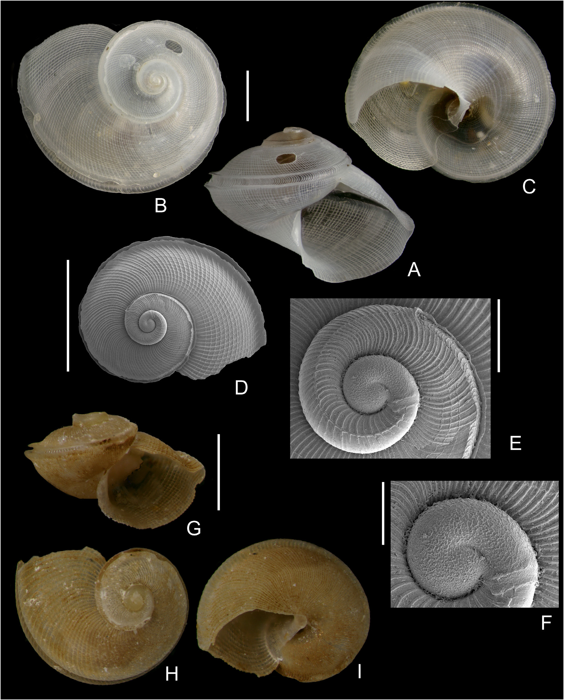
Fig. 7. Anatomidae 1. A–C. Anatoma richardi (Dautzenberg & H. Fischer, 1896), SEAMOUNT 1, DW116, 985–1000 m, diameter 4.2 mm. D–F. A. richardi, shell and detail of the protoconch and early teleoconch whorls, SEM micrograph, same locality, diameter 1.7 mm. G–I. A. cf. richardi, BANGAL 0711, GOC6, 903 m, diameter 2.3 mm. Scale bars: A–D, G–I = 1 mm; E = 200 µm; F = 100 µm.
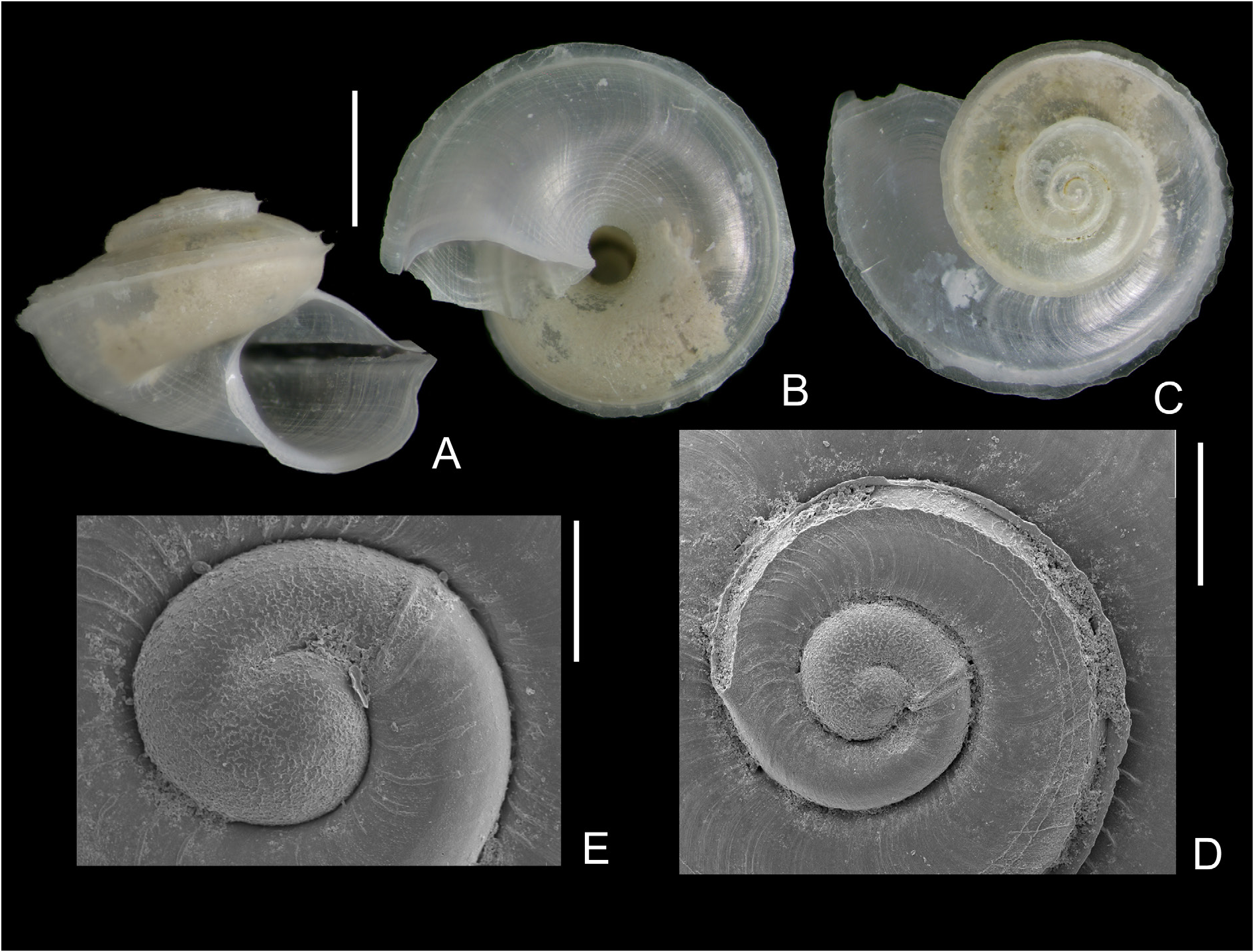
Fig. 9. Anatomidae 3. A–C. Anatoma umbilicata (Jeffreys, 1883), BANGAL 0711, V10, 1720 m, diameter 3.1 mm. D–E. Details of protoconch and early teleoconch whorls of another shell, same locality. Scale bars: A–C = 1 mm; D = 200 µm; E = 100 µm.
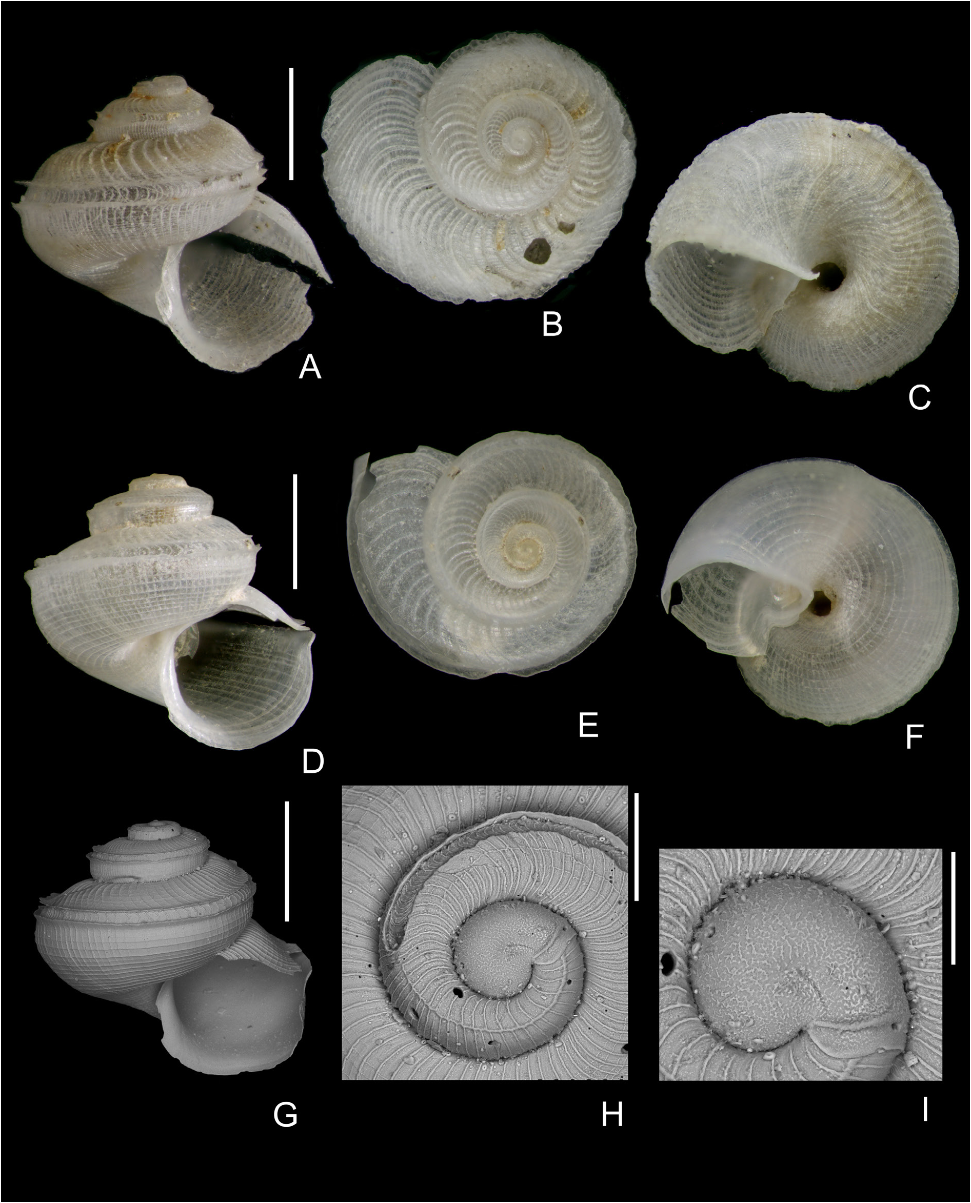
Fig. 8. Anatomidae 2. A–C. Anatoma aspera (Philippi, 1844), SEAMOUNT 1, DW116, 985–1000 m, diameter 2.8 mm. D–F. Anatoma eximia (Seguenza, 1880), SEAMOUNT 1, DW116, 985–1000 m, diameter 2.5 mm. G. A. eximia, SEM micrograph, BANGAL 0711, V4, 744 m, diameter 2.3 mm. H–I. A. eximia, SEM micrograph, details of protoconch and early teleoconch whorls of another shell, same locality. Scale bars: A–G = 1 mm; H = 200 µm; I = 100 µm.
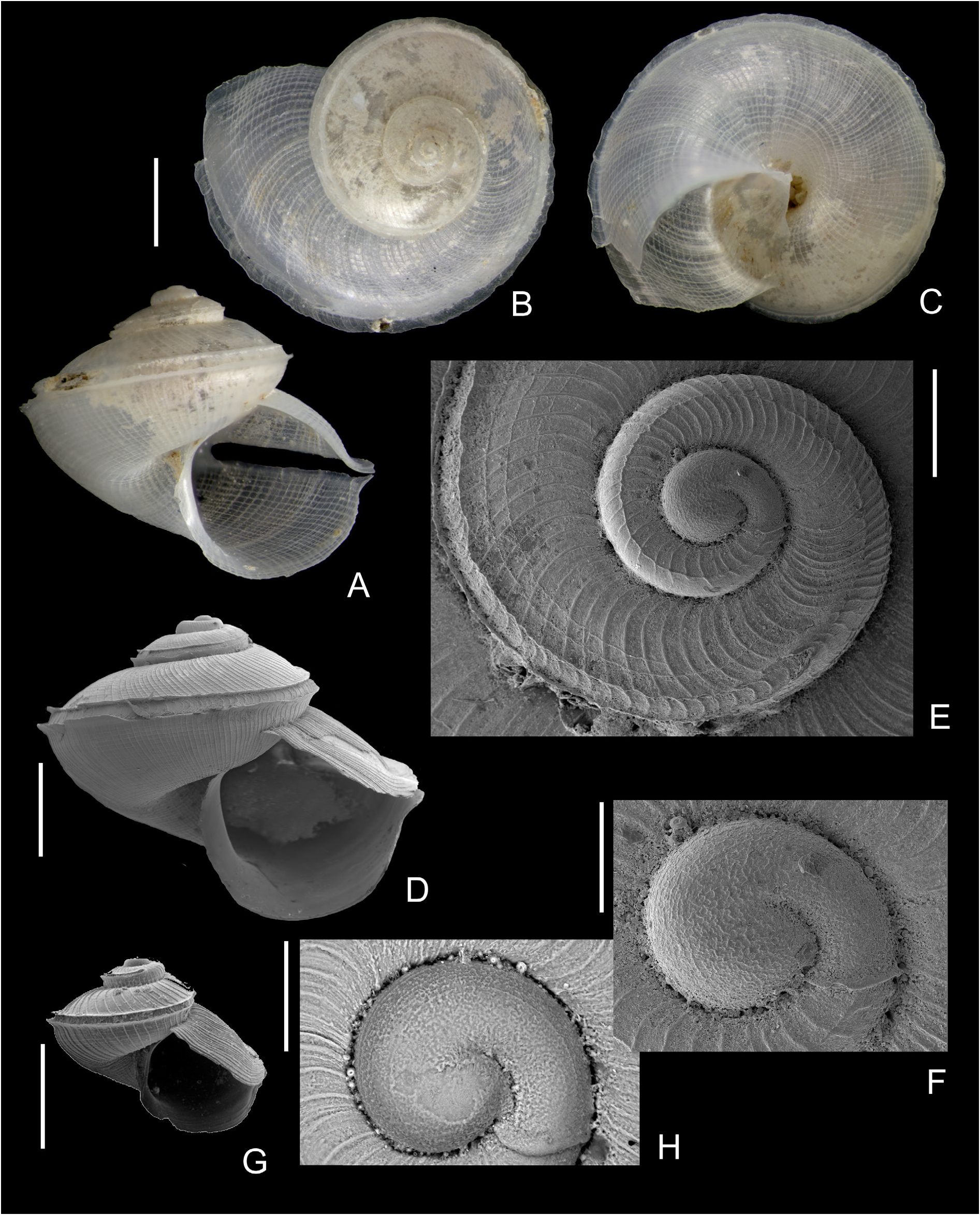
Fig. 10. Anatomidae 4. Anatoma corralae Gofas & Luque sp. nov. A–C. Paratype (MNCN 15.05/200138P), BANGAL 0711, V10, 1720 m, 4.0 mm diameter. D–F. Holotype (MNCN 15.05/200138H), SEM micrograph, BANGAL 0711, V10, 4.1 mm diameter. E–F. Details of early teleoconch whorls and protoconch of the holotype. G–H. Shell and detail of the protoconch, SEM micrographs, from BANGAL 0711, V5, 1631 m, 2.1 mm diameter. Scale bars: A–D, G = 1 mm; E = 200 µm; F, H = 100 µm.
No known copyright restrictions apply. See Agosti, D., Egloff, W., 2009. Taxonomic information exchange and copyright: the Plazi approach. BMC Research Notes 2009, 2:53 for further explanation.
|
Kingdom |
|
|
Phylum |
|
|
Class |
|
|
SubClass |
Vetigastropoda |
|
Order |
|
|
Family |
1 (by felipe, 2021-12-22 14:09:58)
2 (by ExternalLinkService, 2021-12-22 14:19:45)
3 (by valdenar, 2021-12-22 20:01:24)
4 (by ExternalLinkService, 2022-01-11 12:35:50)
5 (by ExternalLinkService, 2022-01-11 12:48:13)
6 (by ExternalLinkService, 2022-01-11 13:14:03)
7 (by plazi, 2023-11-08 11:47:52)
8 (by ExternalLinkService, 2023-11-08 19:50:43)
9 (by jonas, 2024-10-22 18:21:59)