
Notanisus yemenensis, Gibson, Gary A. P., 2015
|
publication ID |
https://doi.org/ 10.11646/zootaxa.3948.3.4 |
|
publication LSID |
lsid:zoobank.org:pub:E349818A-165B-4CA8-BA29-0E345AFDF6C6 |
|
DOI |
https://doi.org/10.5281/zenodo.5275707 |
|
persistent identifier |
https://treatment.plazi.org/id/D4478723-FF92-D177-299D-A9DFFDDFF8BB |
|
treatment provided by |
Plazi |
|
scientific name |
Notanisus yemenensis |
| status |
sp. nov. |
Notanisus yemenensis n. sp.
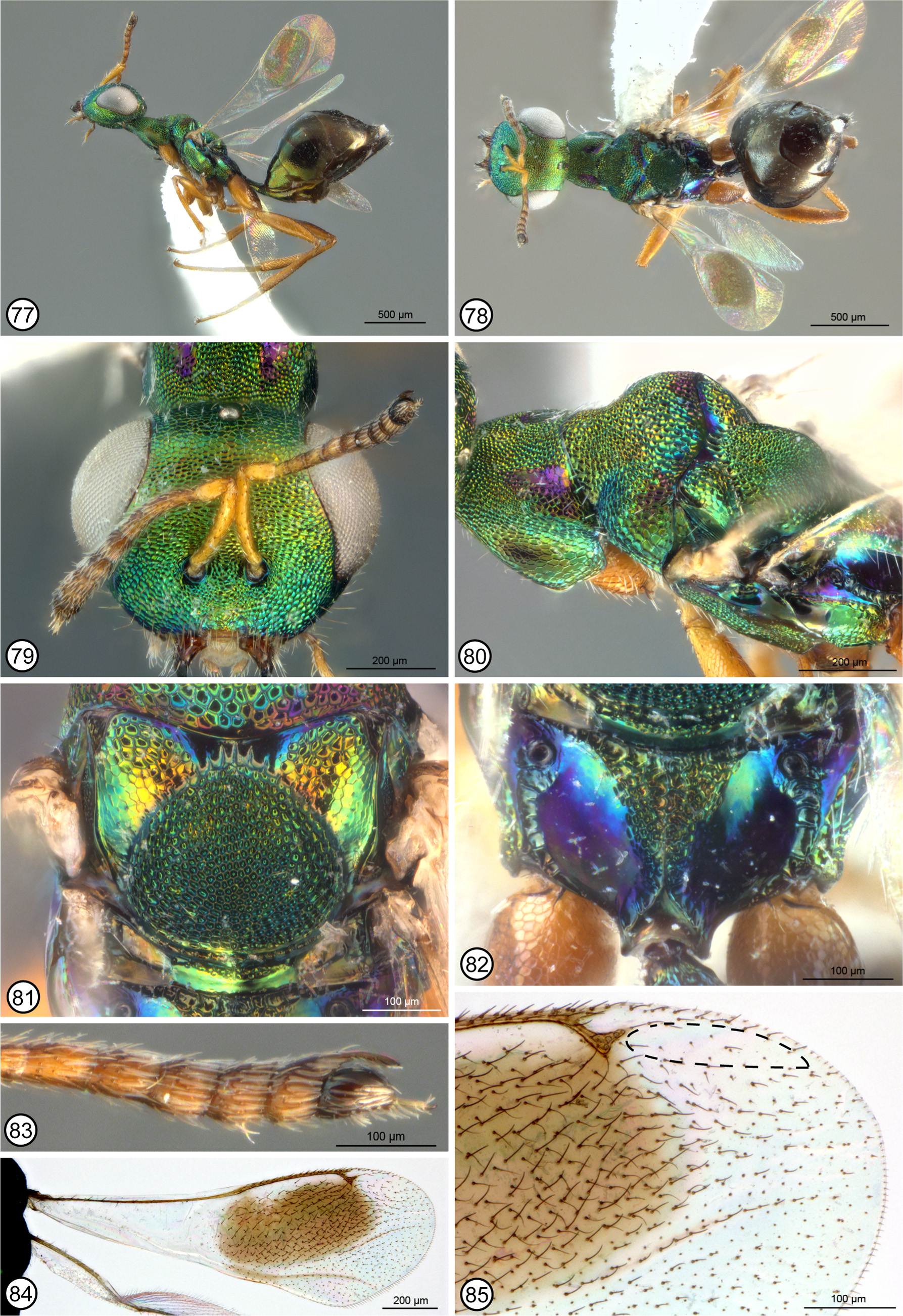
Figs 77–85 View FIGURES 77 – 85
Type material. Holotype ♀ ( CNC). YEMEN Lahj, IX.2000, A. van Harten, A. Sallam, MT (point-mounted; entire).
Paratype (1♂ CNC). Same data as holotype.
Etymology. Named after the country from which the species was collected.
Description. FEMALE ( Fig. 77 View FIGURES 77 – 85 ). Length about 2.7 mm. Head with face and frontovertex green from direct or oblique viewing angle ( Figs 78, 79 View FIGURES 77 – 85 ), though with slight coppery luster above level of toruli, and in dorsal view with small reddish-violaceous region behind anterior ocellus within ocellar triangle; frontovertex distinctly differentiated by difference in sculpture at level about midway between toruli and anterior ocellus, with larger, and more isodiametric meshlike reticulations dorsad level and smaller, more transverse reticulations ventrad level, the sculpture between frontovertex and torulus in particular more transverse reticulate-rugose; in lateral view lower face and gena posterior to malar sulcus similarly strongly sculptured and colored; in dorsal view OOL only about 1.1× maximum diameter of posterior ocellus. Antenna with scape and pedicel yellow, funicle somewhat darker yellowish-brown and clava dark brown; fl1 slightly but distinctly longer than wide, fl4 slightly longer than combined length of fl2 and fl3, and funiculars increasing in width and beyond fl4 decreasing in length such that apical funicular slightly transverse in dorsal view; apical funicular ventrally extending under clava as apically tapered, ventrally bare and shiny, finger-like projection to claval apex; clava with slender, terminal, setose, spiniform process ( Fig. 83 View FIGURES 77 – 85 ). Mandible with two similar teeth ventrally and more obscure, obliquely angled margin dorsally.
Pronotal collar ( Figs 78, 80 View FIGURES 77 – 85 ) in lateral view shallowly concave and in dorsal view not distinctly “shoulder-like” posterolaterally; almost uniformly punctulate-reticulate mediolongitudinally and mostly green with slight coppery luster except for elongate-triangular and though comparatively slender, more shallowly reticulate to partly smooth, violaceous regions posterolaterally, these with only a couple of distinct setae but with longitudinal inner margins differentiating quadrangular median sculptured region. Mesoscutum ( Fig. 80 View FIGURES 77 – 85 ) anteriorly between incomplete notauli similarly colored and punctate-reticulate as for pronotum dorsomedially, but with much larger reticulations posteriorly and partly bluish to reddish-violaceous along transscutal articulation and posteriorly on lateral lobes; scutellar-axillar complex ( Fig. 81 View FIGURES 77 – 85 ) mostly green with variably distinct coppery luster on axillae under different angles of light except crenulate region between axillae bright purple to reddish-violaceous laterally, axilla with reticulate dorsal surface longer than medial crenulate region between axillae, and with obliquely angled posterior surface increasingly finely sculptured ventrally, and scutellum comparatively flat dorsally ( Fig. 77 View FIGURES 77 – 85 ) and similarly uniformly punctate except for slightly larger punctures, more reticulate-punctate, laterally ( Fig. 81 View FIGURES 77 – 85 ). Tegula yellow. Macropterous; fore wing ( Fig. 84 View FIGURES 77 – 85 ) marginal vein about 7.5× length of stigmal vein; postmarginal vein shorter than stigmal vein; uncus ( Fig. 85 View FIGURES 77 – 85 ) diverging from stigmal vein apically so distinct stigma not differentiated and apex separated from wing margin by about width of stigma plus uncus, but by more than twice width of uncus; costal cell ventrally with 6 setae within about basal third; disc ( Fig. 84 View FIGURES 77 – 85 ) with oval brown region behind venation to medial fold and cubital fold also infuscate, but narrowly hyaline along marginal vein, the hyaline region slightly expanded posteriorly behind marginal vein subbasally but not distinctly subdividing infuscation, and with distinct brownish setae except for mostly much shorter, more spiculate setae apically and toward posterior margin, and with narrow bare band along parastigma and marginal vein, and elongate bare region dorsally beyond stigmal vein ( Fig. 85 View FIGURES 77 – 85 ); marginal fringe distinct only along posteroapical margin. Prepectus extensively setose dorsoapically, but with relatively inconspicuous, short white setae. Mesepimeron bare along posterior margin ( Fig. 80 View FIGURES 77 – 85 ). Metapleuron bare, with about basal half reticulate and dorsal half smooth and shiny ( Fig. 80 View FIGURES 77 – 85 ). Metasternum comparatively long, with base of mesocoxa distinctly anterior to, and apex of mesocoxa about level with base of metacoxa. Legs ( Fig. 77 View FIGURES 77 – 85 ), including coxae, mostly orange with dorsal surface of metacoxa and metafemora more brownish-orange under some angles of light, but both meso- and metatarsi paler, the basal three tarsomeres yellowish-white and apical two tarsomeres more distinctly yellow; metacoxa dorsobasally bare or (right coxa) with only two white setae ( Fig. 82 View FIGURES 77 – 85 ). Propodeum ( Fig. 82 View FIGURES 77 – 85 ) with crenulate band along anterior margin recurved posteromedially into anteriorly broad, posteriorly tapered, V-shaped sculptured region on either side of complete median carina, the region anteriorly crenulate to rugose but minutely rugulose to punctulate over about posterior half; panels otherwise smooth and shiny, and callus smooth and shiny; callus anteriorly and propodeal plical region posteriorly and laterally between foramen and postspiracular furrow reddish-violaceous, with medial sculptured region green with coppery luster anteriorly and callus posterior to level of spiracle and smooth part of panel anteriorly adjacent to medial sculptured region variably blue to greenish.
Petiole ( Figs 77, 80 View FIGURES 77 – 85 ) dorsally blue to bluish-green and quite extensively and distinctly reticulate, with sides slightly divergent posteriorly, but about 1.66× as long as medial width, uniformly dark brown; presyntergal tergites isodiametric meshlike coriaceous except smooth along posterior margins, with Gt2 subequal in length to Gt3.
MALE (Figs). See description of N. vanharteni .
Biology. Unknown.
Remarks. See under N. vanharteni .
| CNC |
Canadian National Collection of Insects, Arachnids, and Nematodes |
No known copyright restrictions apply. See Agosti, D., Egloff, W., 2009. Taxonomic information exchange and copyright: the Plazi approach. BMC Research Notes 2009, 2:53 for further explanation.
|
Kingdom |
|
|
Phylum |
|
|
Class |
|
|
Order |
|
|
Family |
|
|
Genus |