
Psechrus Thorell, 1878
|
publication ID |
https://doi.org/ 10.11646/zootaxa.3379.1.1 |
|
persistent identifier |
https://treatment.plazi.org/id/D0272654-FF87-584E-FF20-2F7CFEB043F6 |
|
treatment provided by |
Felipe |
|
scientific name |
Psechrus Thorell, 1878 |
| status |
|
Psechrus Thorell, 1878 View in CoL View at ENA
Psechrus Thorell 1878: 170 View in CoL . [Type species: Tegenaria argentata Doleschall, 1857 , transferred to Psechrus View in CoL by Thorell (1878)]. Simon 1890: 80 (Transfer to Psechridae View in CoL ). Simon 1892: 226. Dalmas 1917: 324. Homann 1950: 66. Lehtinen 1967: 260, 383. Levi 1982: 118. Coddington 1990: 7. Griswold 1993: 539. Murphy and Murphy 2000: 264. Griswold et al. 2005: 37.
Lancaria Karsch 1879: 557 . [Type species: Tegenaria torva O. Pickard-Cambridge, 1869 , transferred to Lancaria by Karsch (1879)]. Simon 1887: 194 (Syn., formal transfer of Lancaria torva to Psechrus View in CoL ).
Species transferred to other genera:
Psechrus nicobarensis Tikader, 1977 to Fecenia ( Levi 1982: 138) View in CoL .
Note: Fecenia nicobarensis ( Tikader, 1977) is junior synonym of Fecenia protensa Thorell, 1891 ( Bayer 2011) .
Diagnosis. Psechrus is distinguished from Fecenia by the following characters: AME at most equal to other eyes, mostly smaller; white longitudinal line ventrally on opisthosoma; clypeus (relatively) high, 2–3.5 times diameter of AME, consequently cephalic region of carapace higher than in Fecenia ; length of leg IV more or less equal to leg II; males (mostly) without RTA or apophyses on other palp limbs (except femoral extension, see below) and always without median apophysis on tegulum; females with rather simple median septum of epigyne; vulva mostly with spherical spermathecal heads.
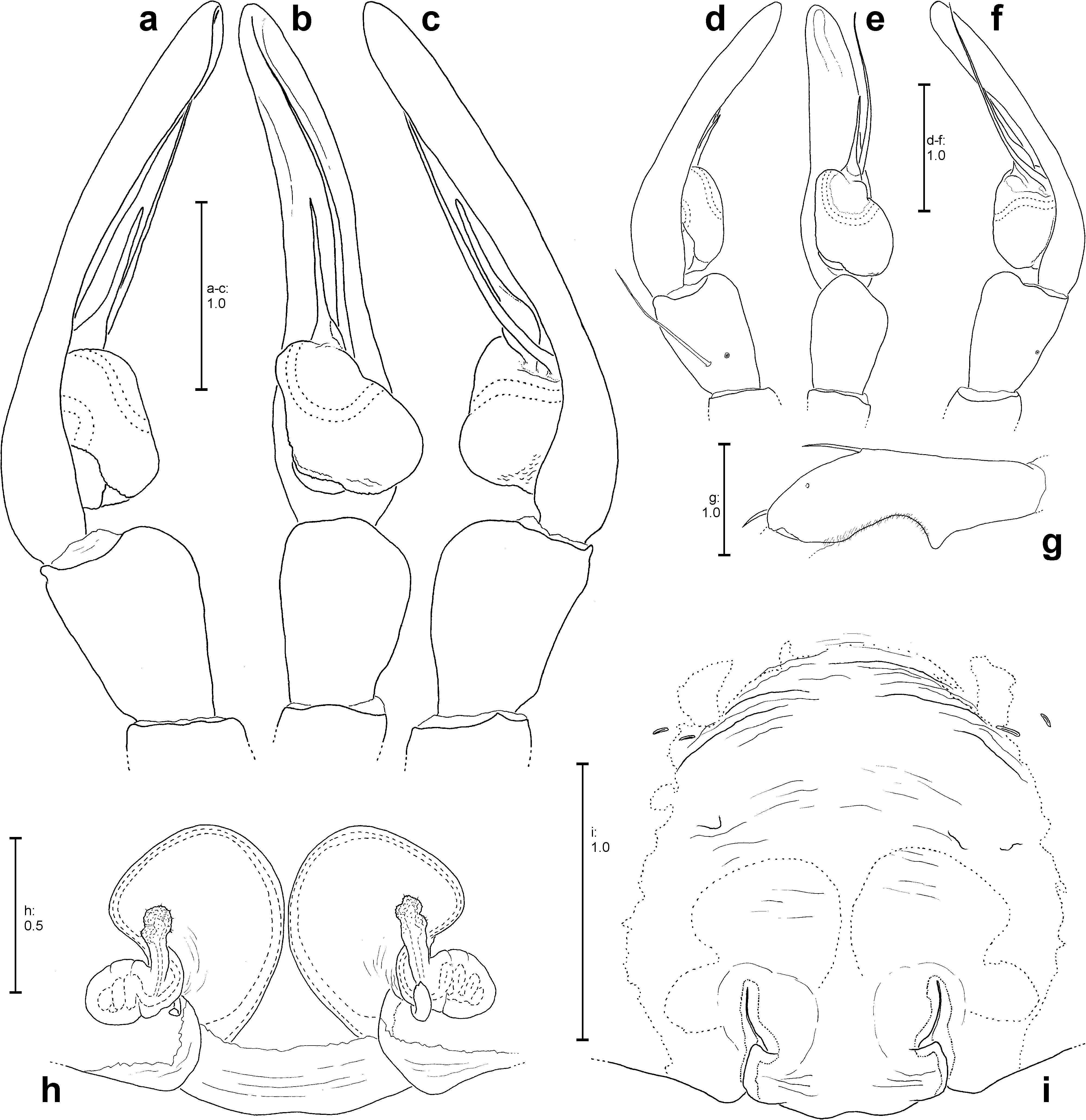
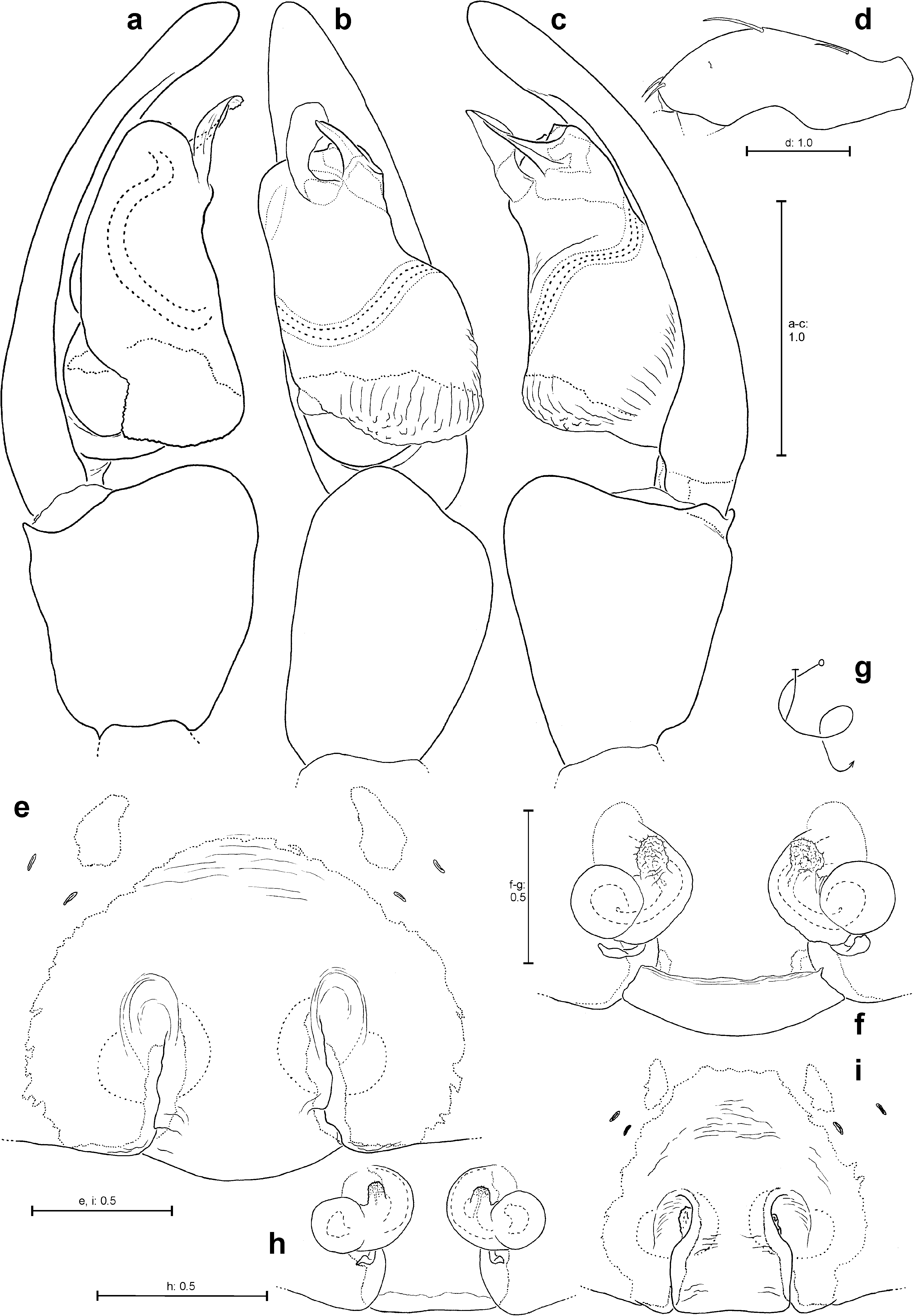
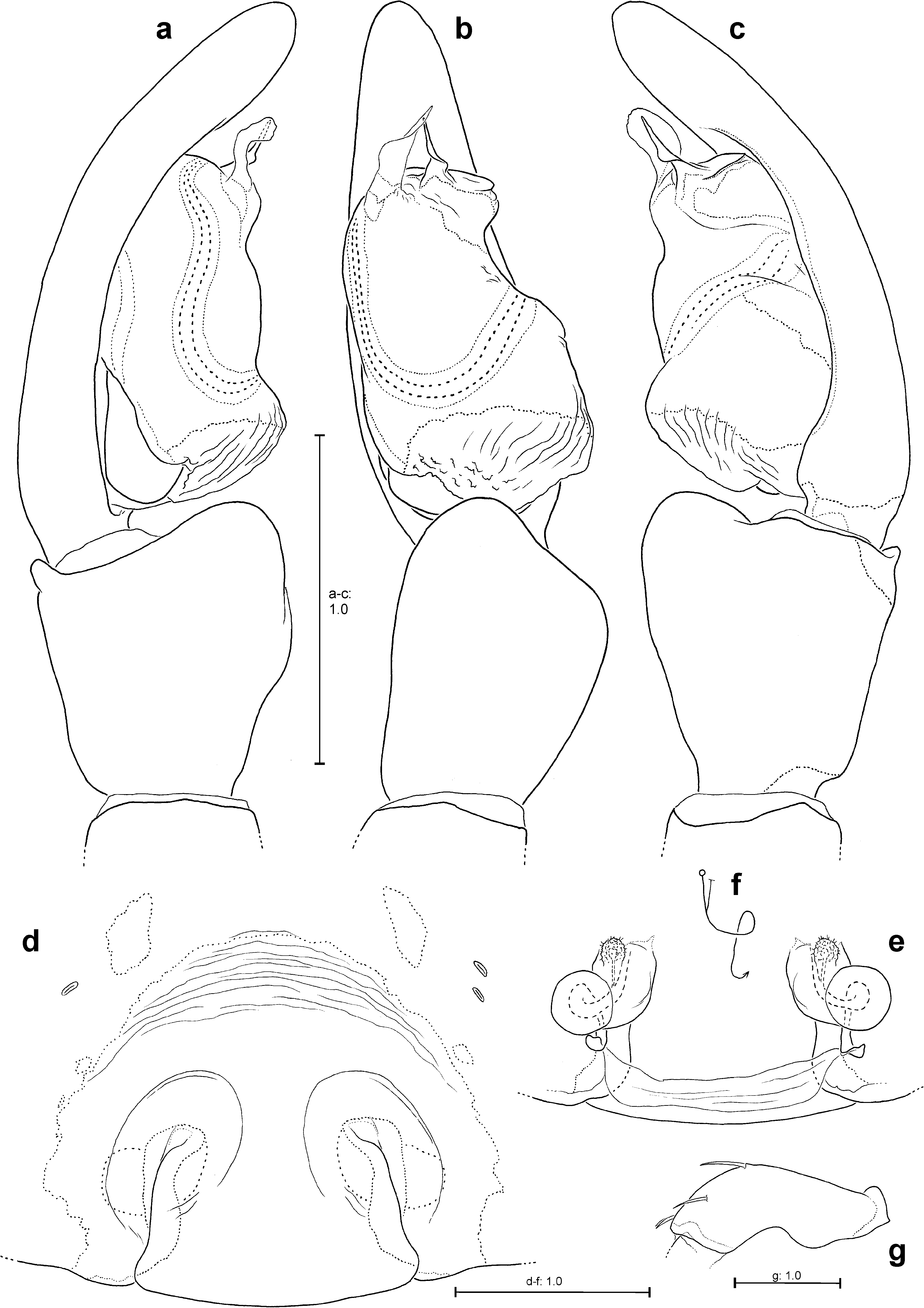
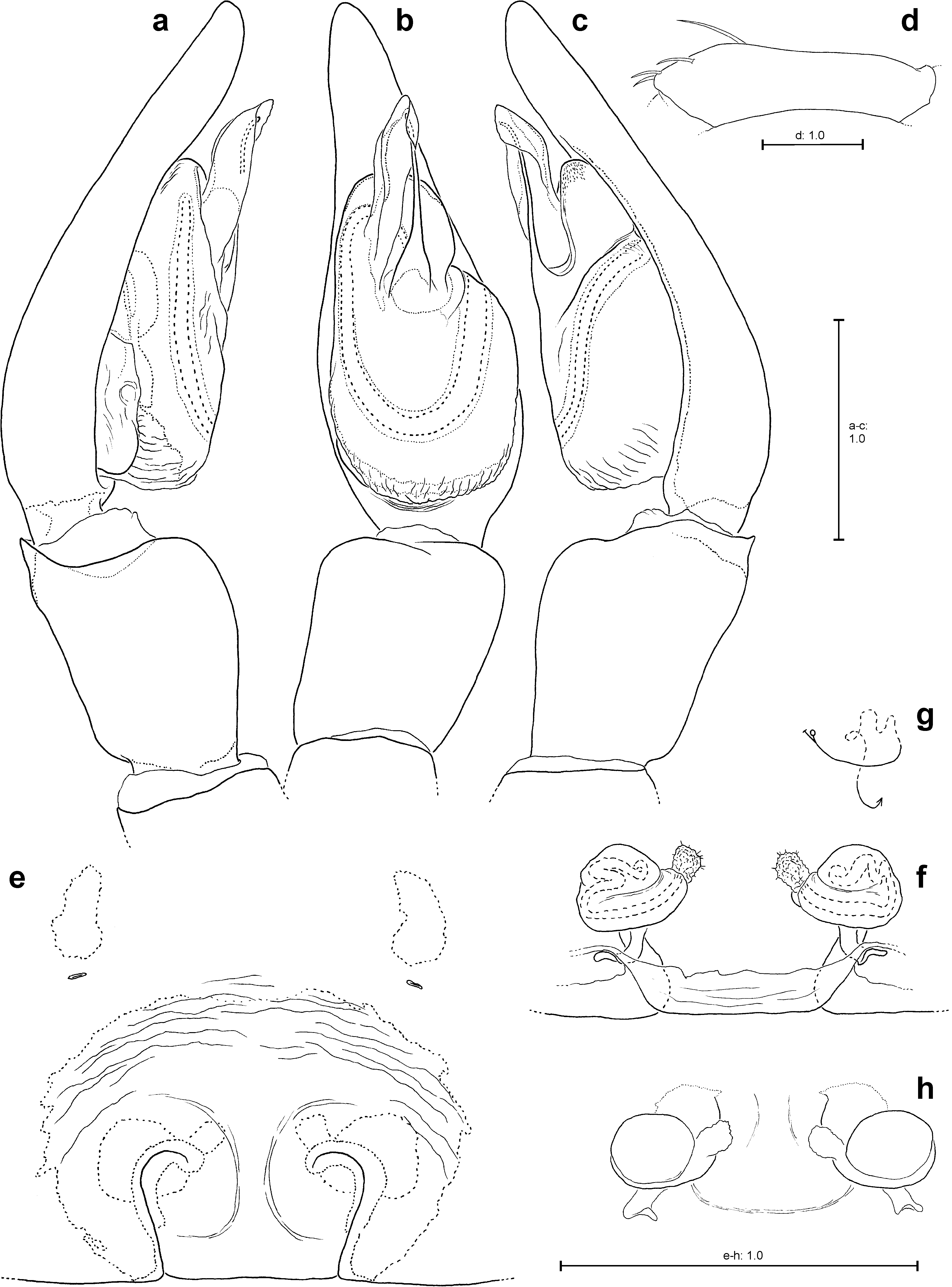
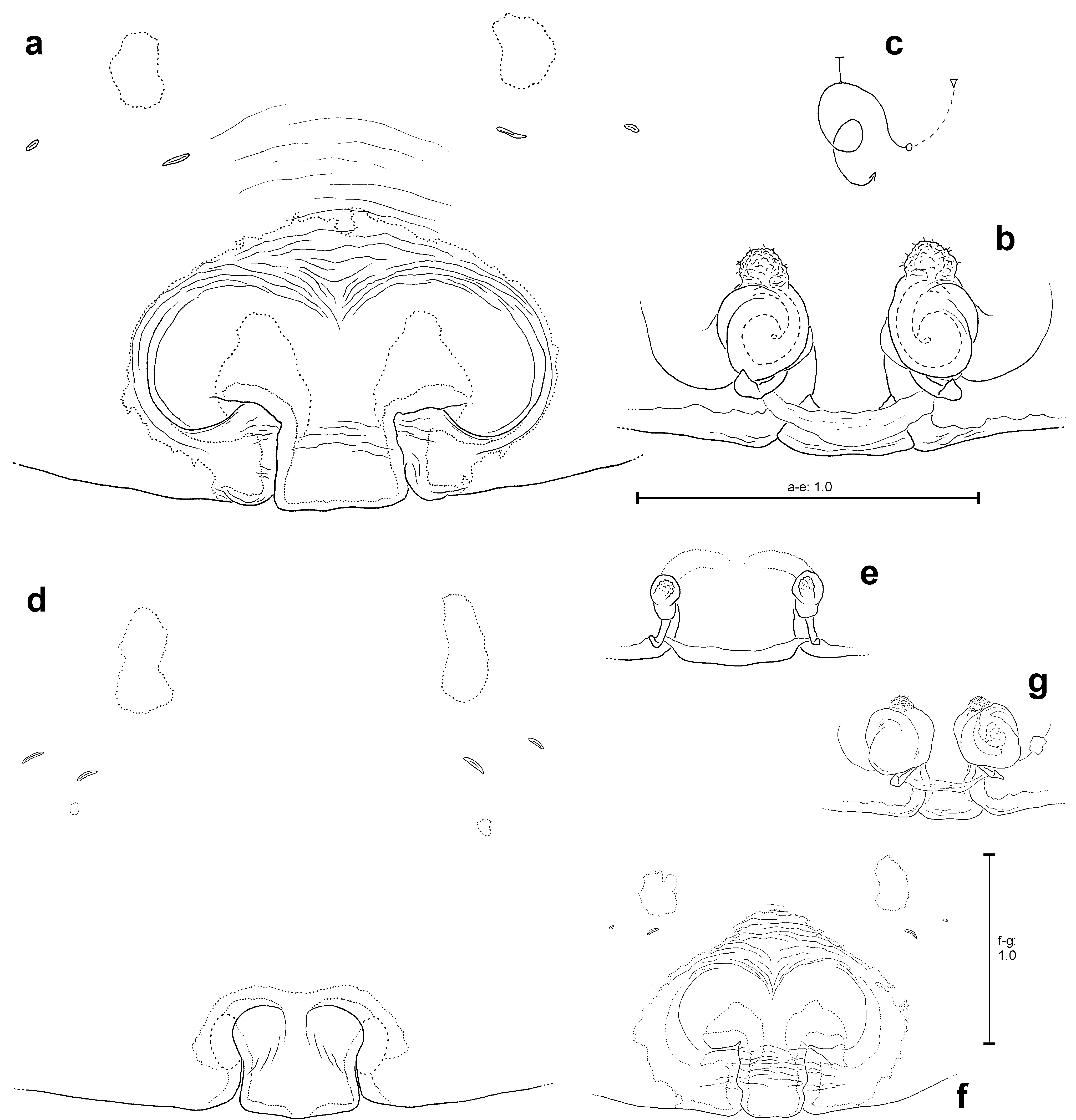
Description. Large Psechridae , body length in males: 8.5–23.8 mm; females: 11.0– 31.1 mm. Width of anterior part of carapace smaller than broadest section of carapace. Both anterior and posterior eye row recurved. Chelicerae long and strong, basal limb at least 2.5 times longer than broad. Cheliceral furrow with three teeth anteriorly, four to five posteriorly and a longitudinal row of 5–10 small denticles in between both rows of teeth ( Fig. 66e View FIGURES 66 ). Their number varies intraspecifically. Basal limb of chelicerae ventrally with long field of short, transversal striae. Labium longer than broad ( Fig. 2e View FIGURES 2 ). Gnathocoxae ca. 2.5 times longer than broad ( Fig. 2e View FIGURES 2 ). Sternum longer than broad, with pointed posterior ending and broad-angled (160°) anterior ending ( Fig. 2f View FIGURES 2 ). Pedipalp in females with single, toothed claw ( Fig. 21g View FIGURES 21 ), which is similarly shaped as the pair of distal tarsal claws of the legs. The tarsi of the legs long, gracile, elastic, and apically additionally with a third, small, toothed and short tarsal claw (median hook). Legs very long in males (metatarsus I ca. 2.5–3.5x carapace length, Fig. 81c View FIGURES 81 ), long in females (metatarsus I ca. 2x carapace length, Fig. 81d View FIGURES 81 ). Leg formula 1243 or 1423. Coxae of legs I and II slightly broader than of IV and especially III, which is smallest. Calamistrum dorso-retrolaterally on metatarsus IV consisting of 4–5 rows of setae, inner rows generally with irregular arrangement. In adult males calamistrum rudimentary, if not completely reduced.
Spination of palp and legs: Variable within each species. Mostly no species-specific spination pattern recognisable. The spination pattern is in parts useful for the characterisation of the different species groups (see below). At the following positions spines are always absent: All patellae, tarsi, dorsal surface of all metatarsi, ventral surface of all femora. Ventral spines on tibiae and metatarsi generally paired, except for most distal one (in the case of odd numbers).
Males of many species possess macrosetae ventrally on coxae of legs I (MC-I) and/or II (MC-II) as well as an apical row of macrosetae ventrally on trochanter I (MT-I).
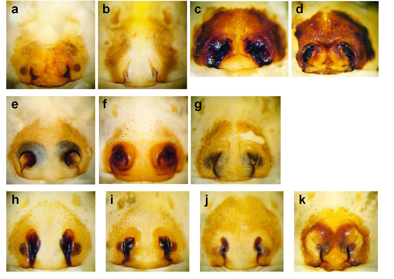
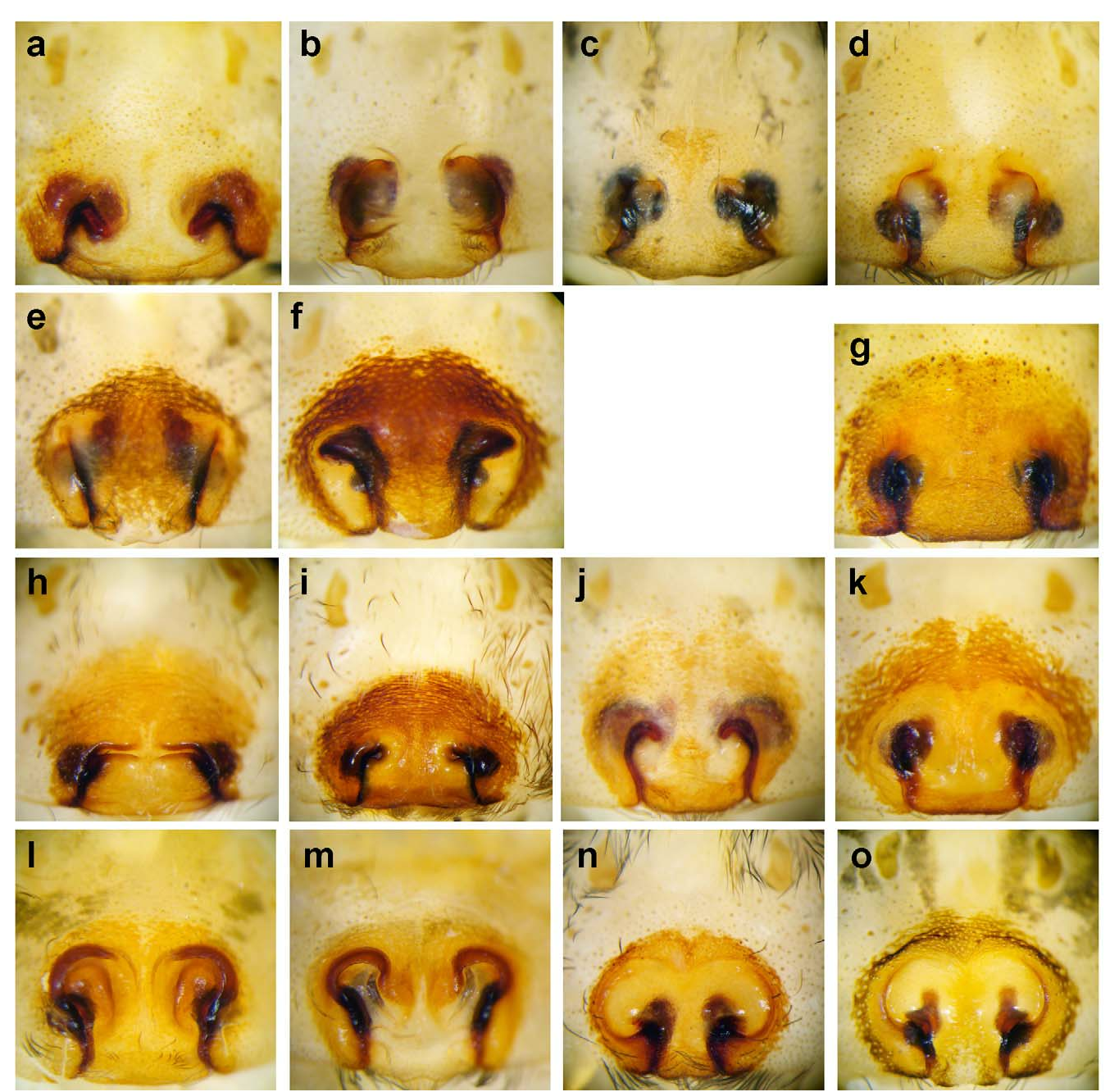
Colouration: Chelicerae brown to dark red-brown. Sternum mostly yellowish brown at lateral margins and with (dark) brown, tapered patch centrally ( Fig. 82h View FIGURES 82 ). Rarely unicoloured light brown ( Fig. 82j View FIGURES 82 ) and even more rarely brown with light longitudinal line ( Fig. 82k View FIGURES 82 ). Carapace yellowish brown, always with two dark brown median bands ( Figs 82a–g View FIGURES 82 ), which may be serrated laterally. Most distal lateral margins often also with dark bands ( Figs 82a,f–g View FIGURES 82 ), however these are (much) narrower than median ones. Palpal femur ventrally with a longitudinal row of 6–10 long bristles, which may be reduced in adult males. Legs from yellowish brown or light brown to brown, often dark brown annulated ( Figs 81a–d View FIGURES 81 ). Opisthosoma dorsally yellowish-brown with (dark) brown patches ( Figs 81a–b View FIGURES 81 ), ventrally brown with a straight, light, longitudinal line ( Figs 81e–i View FIGURES 81 ). On each side next to that line with a longitudinal row of small patches of muscle sigillae ( Figs 81h–i View FIGURES 81 ). Opisthosoma ventrally very rarely unicoloured brown ( Fig. 81j View FIGURES 81 ).
Anterior lateral spinnerets are short (broader than long) and more or less conical, posterior median spinnerets distinctly smaller, slender and cylindrical. Posterior lateral spinnerets also cylindrical and bipartite, similar size to anterior lateral spinnerets. Cribellum divided into two very narrow parts ( Fig. 81h View FIGURES 81 ).
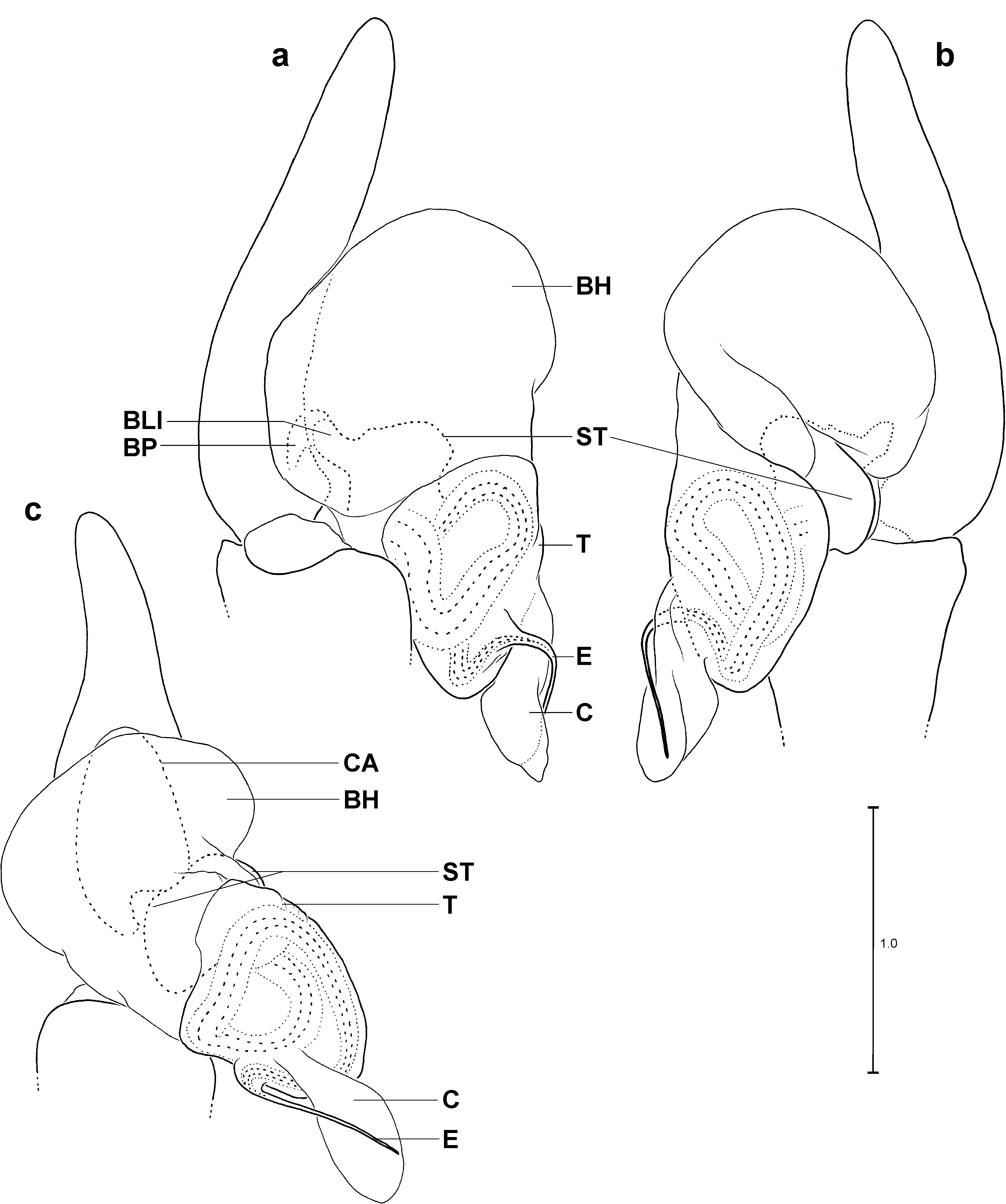
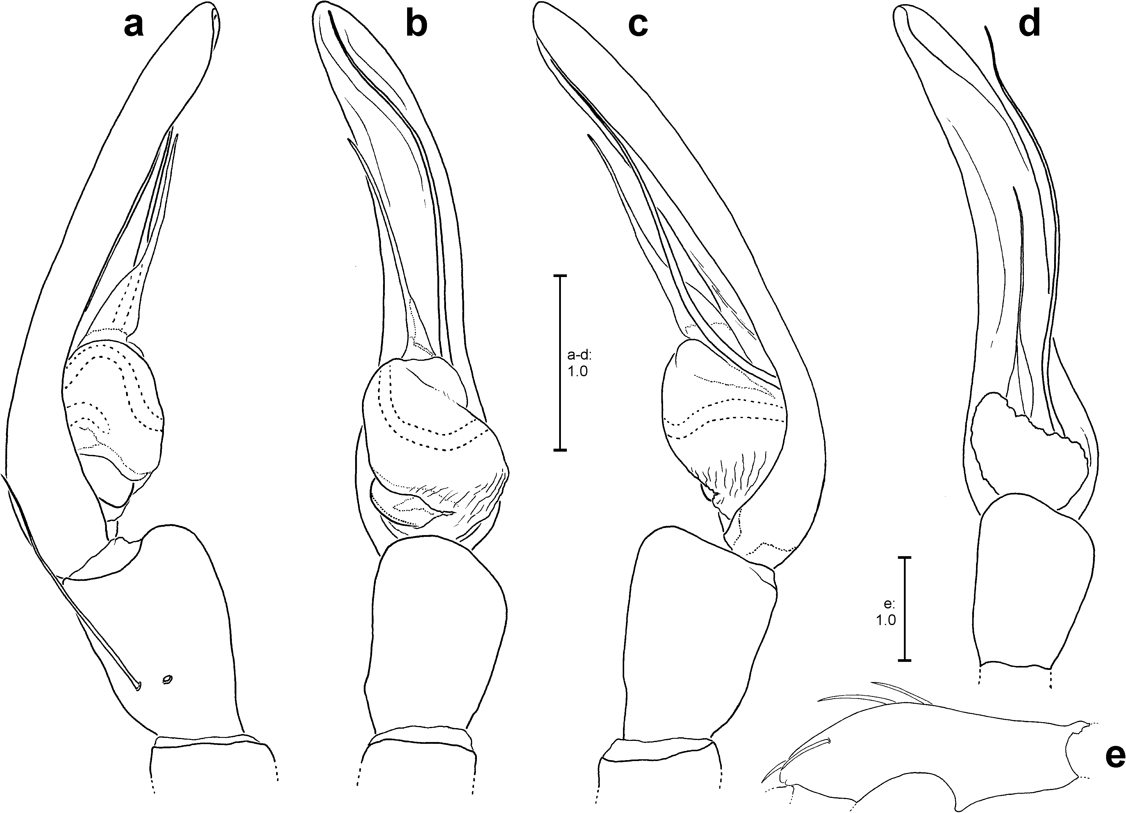
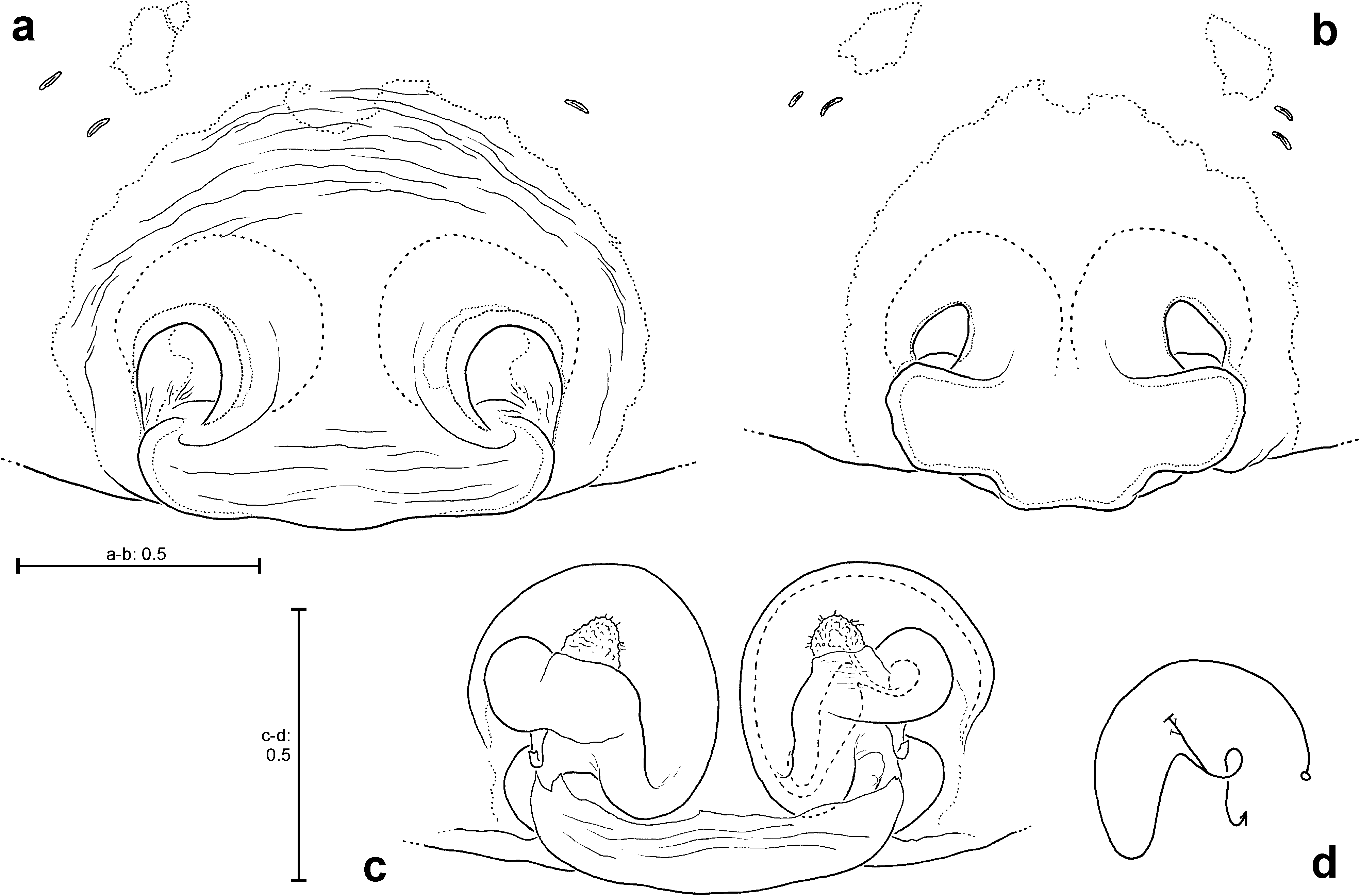
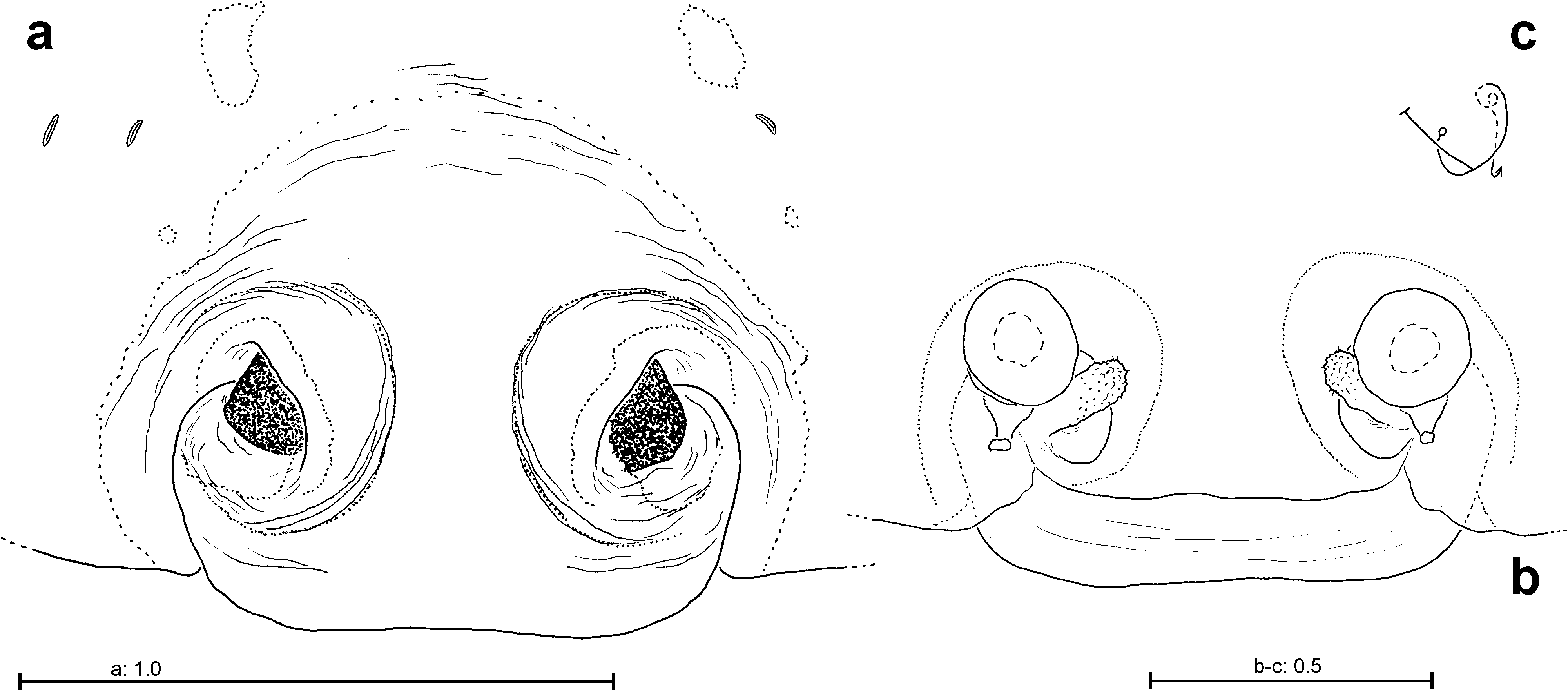
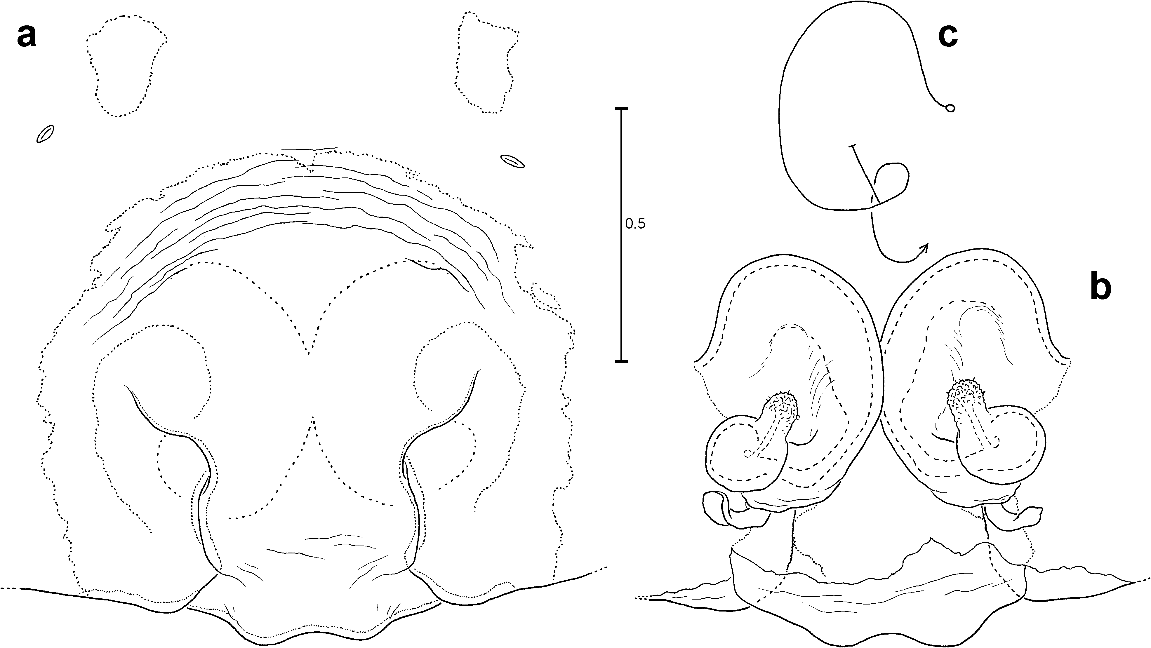
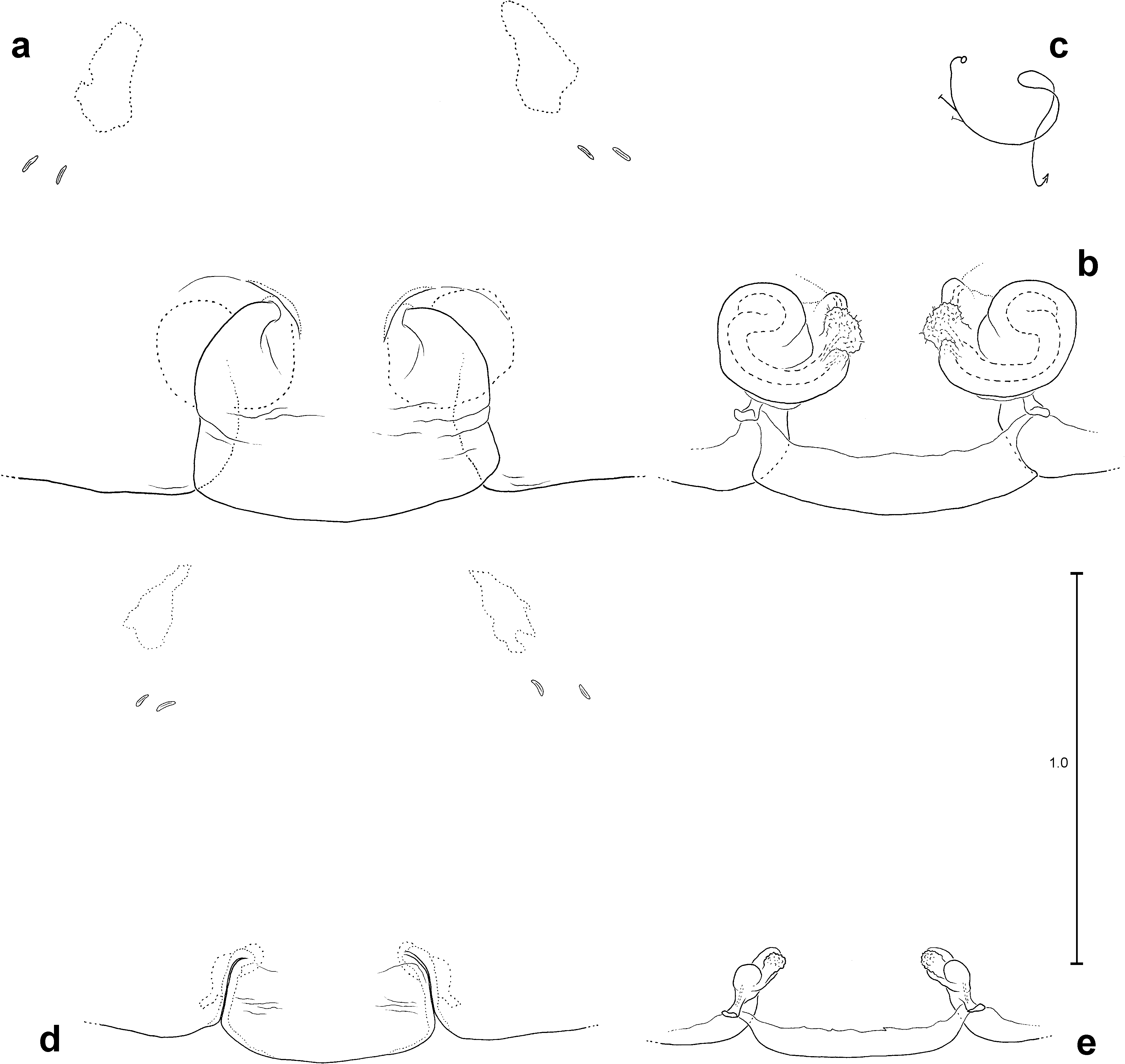
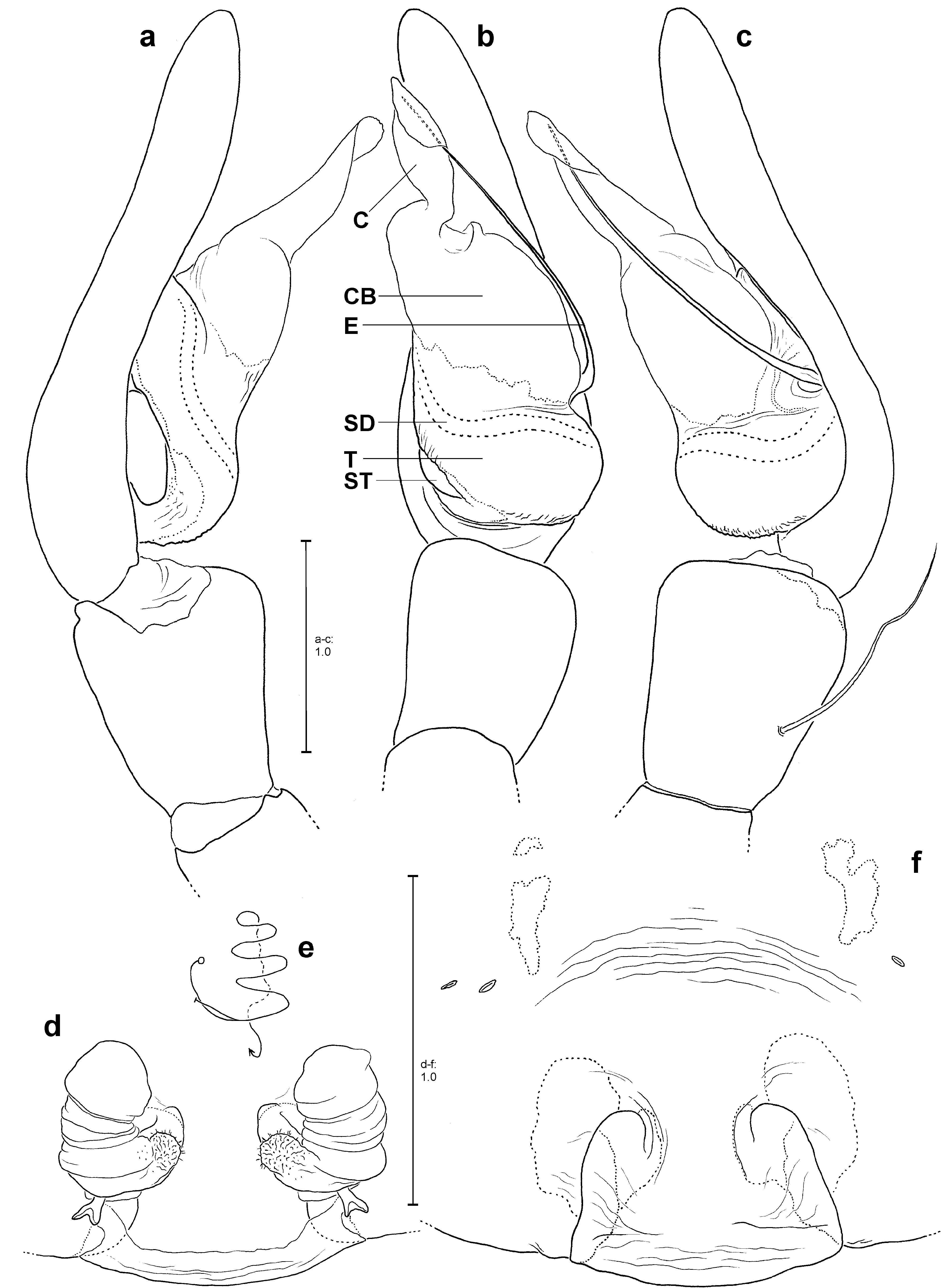
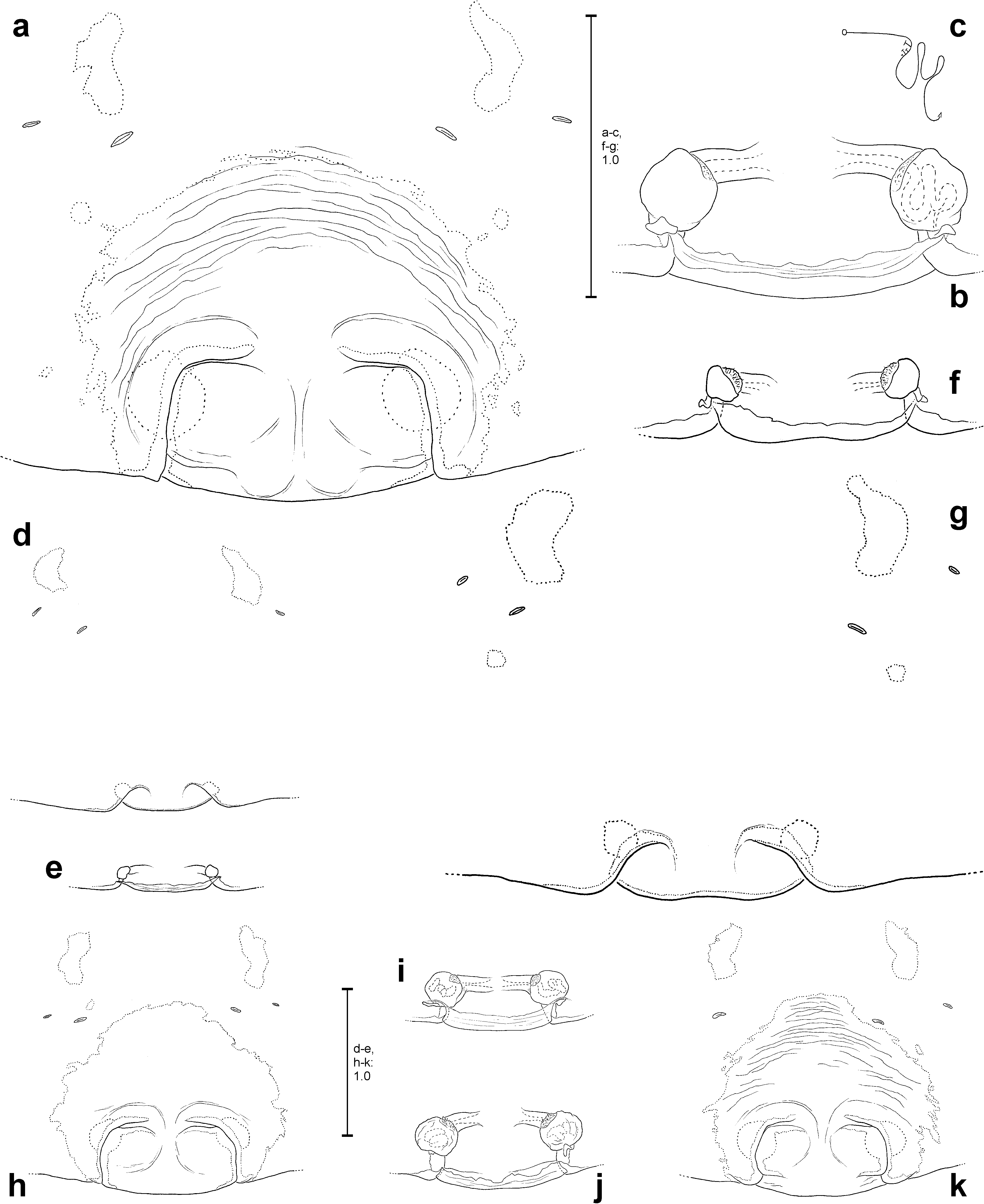
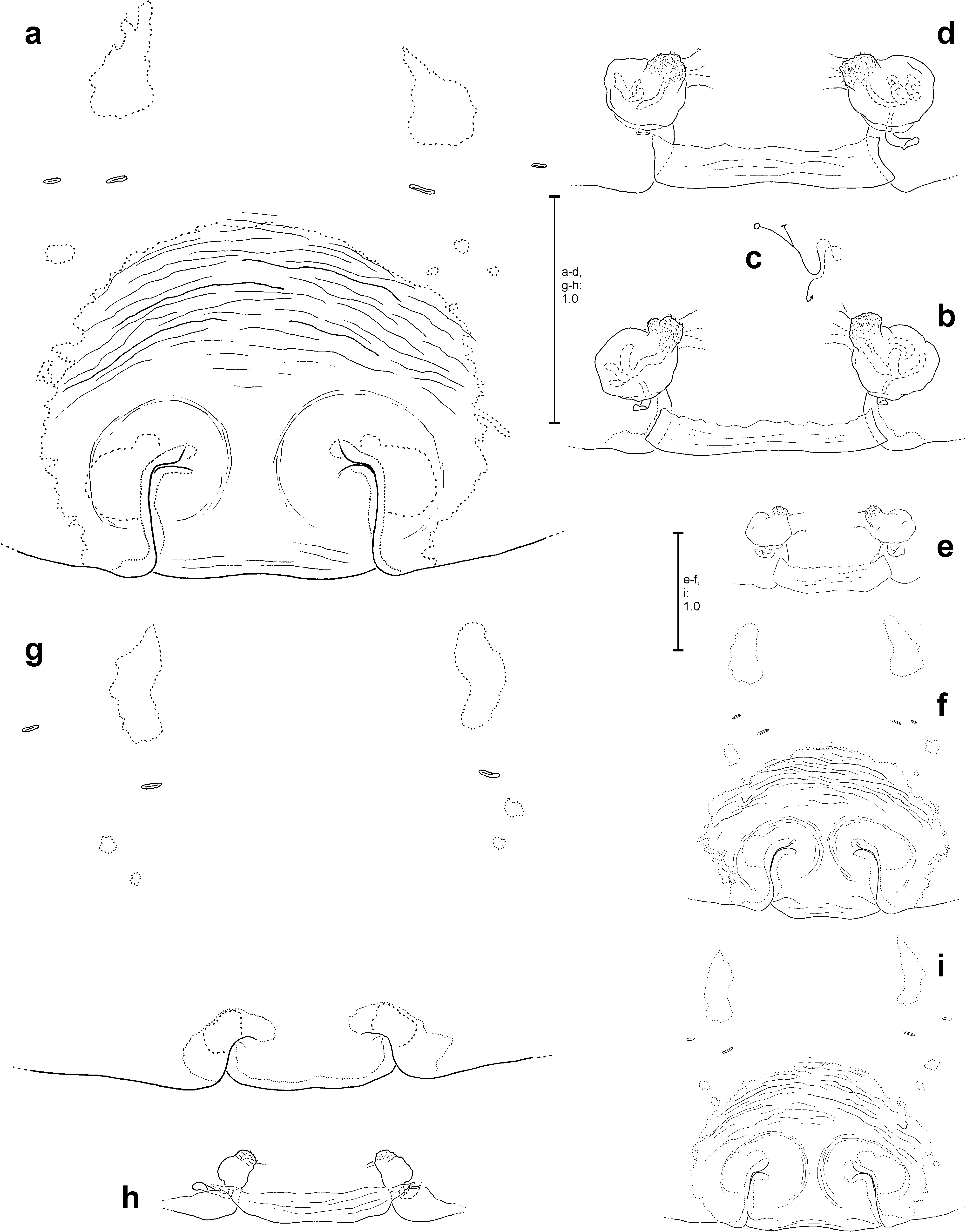
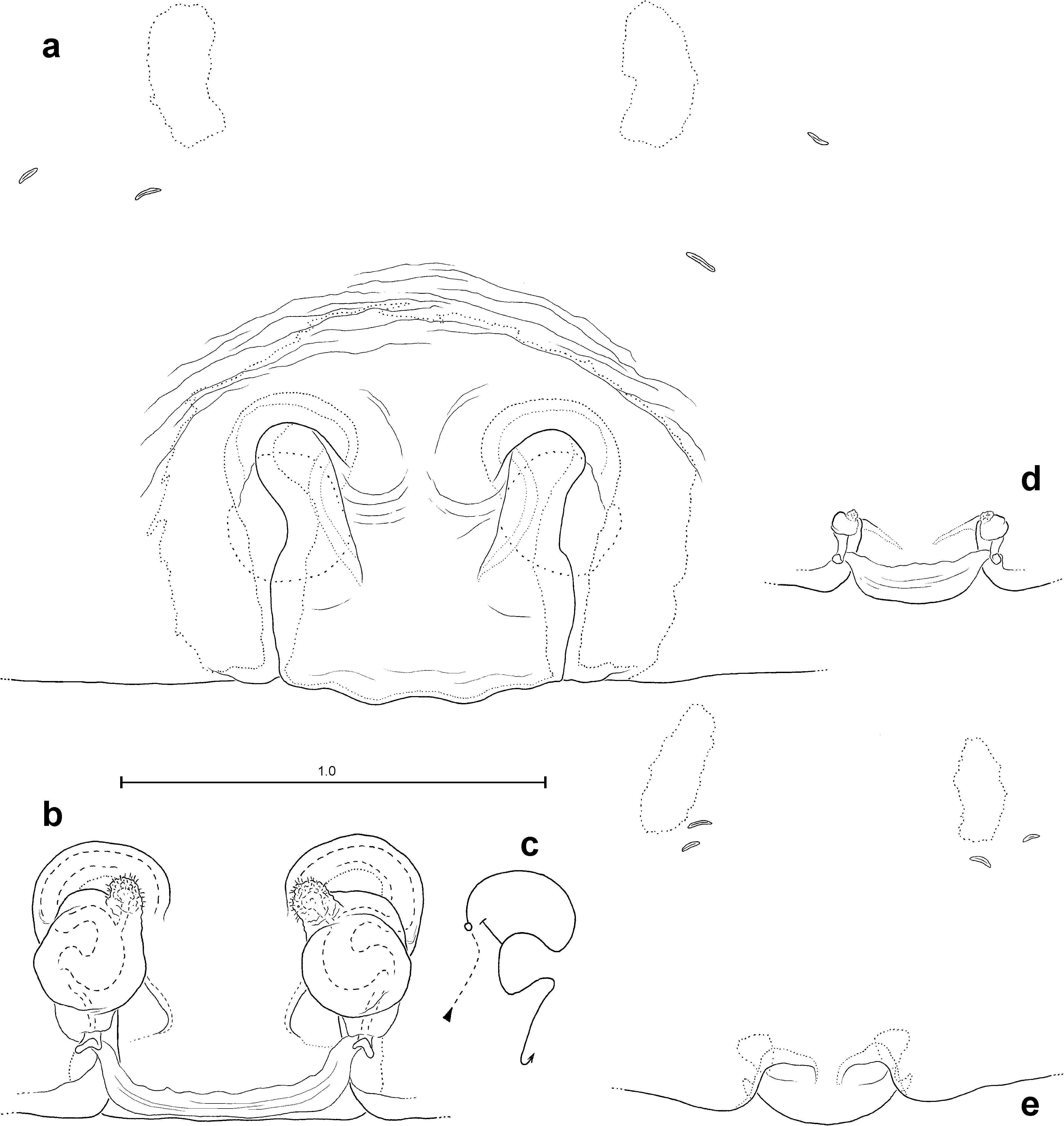
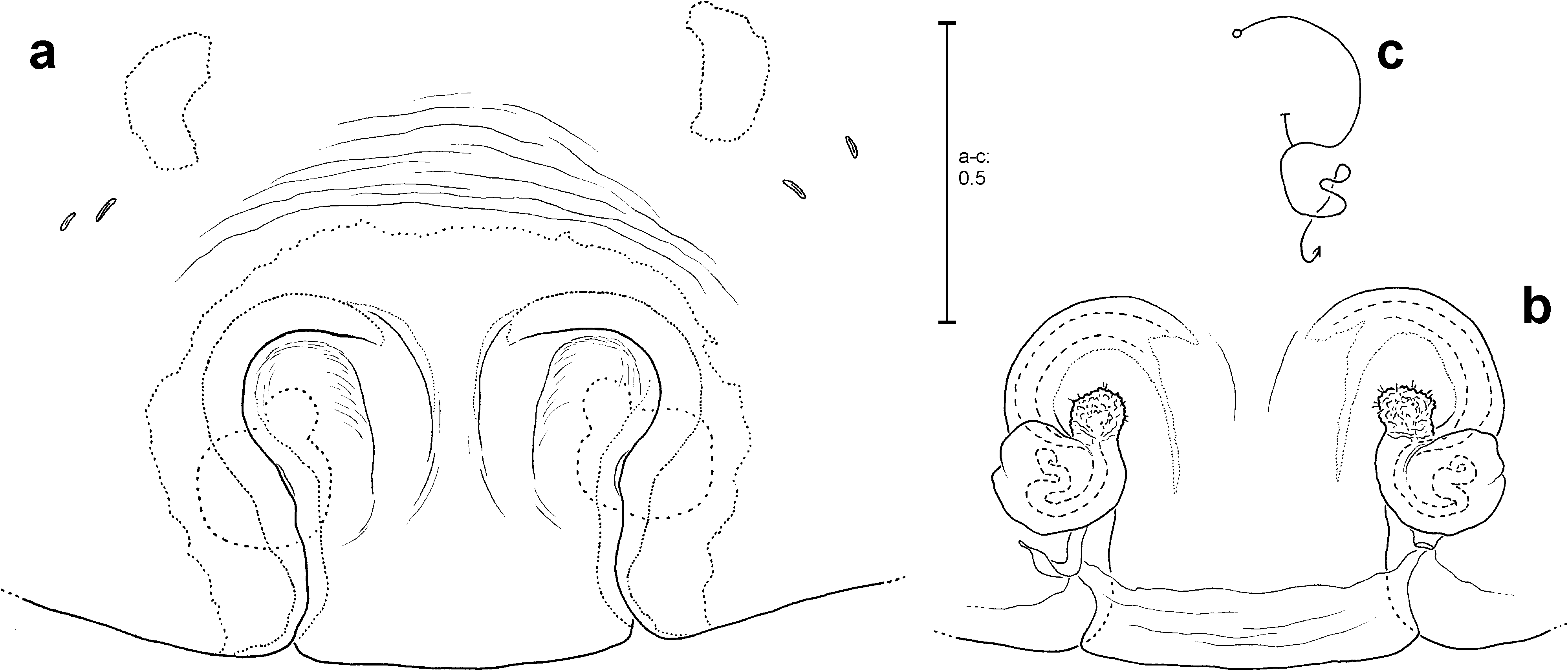
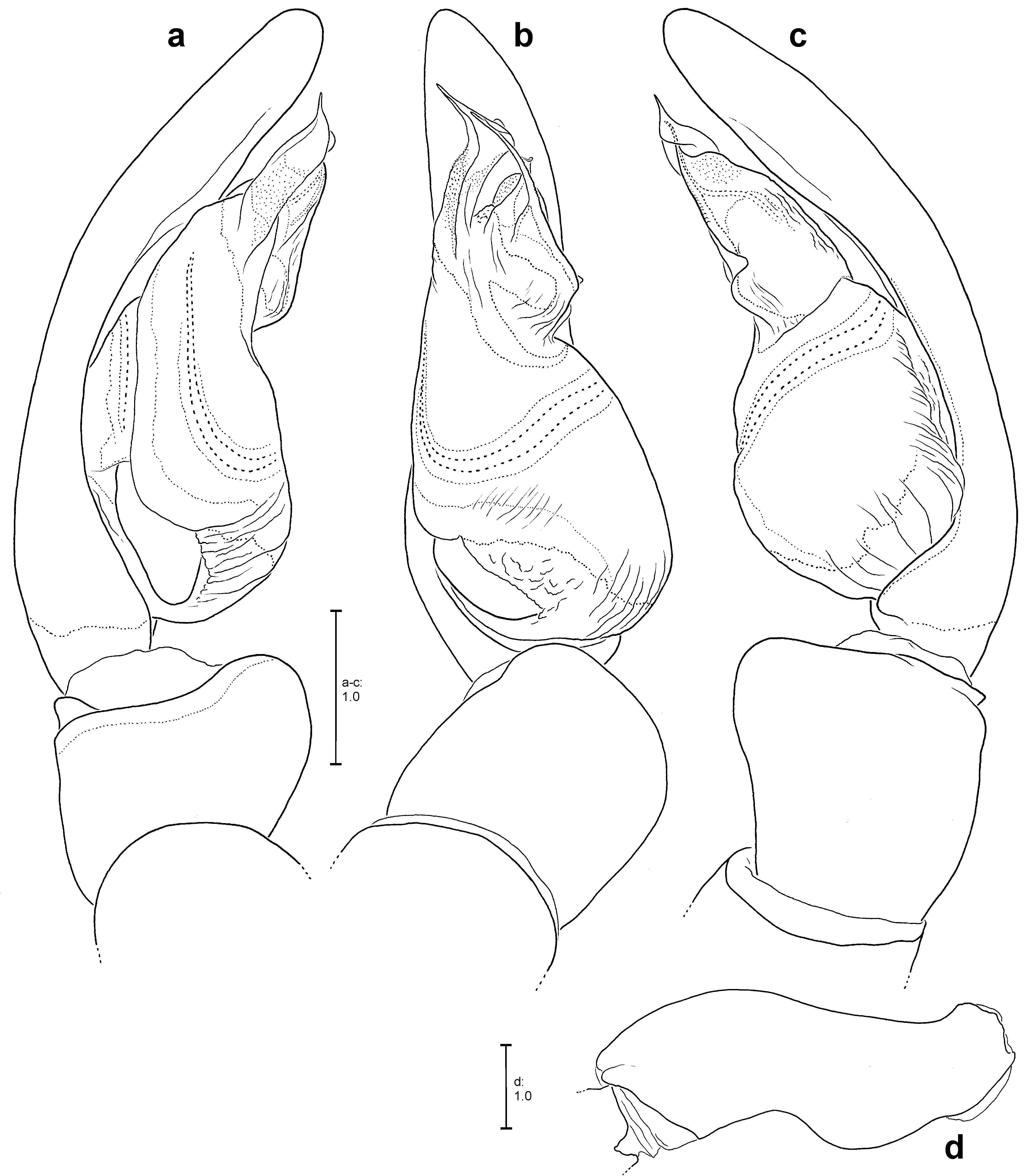
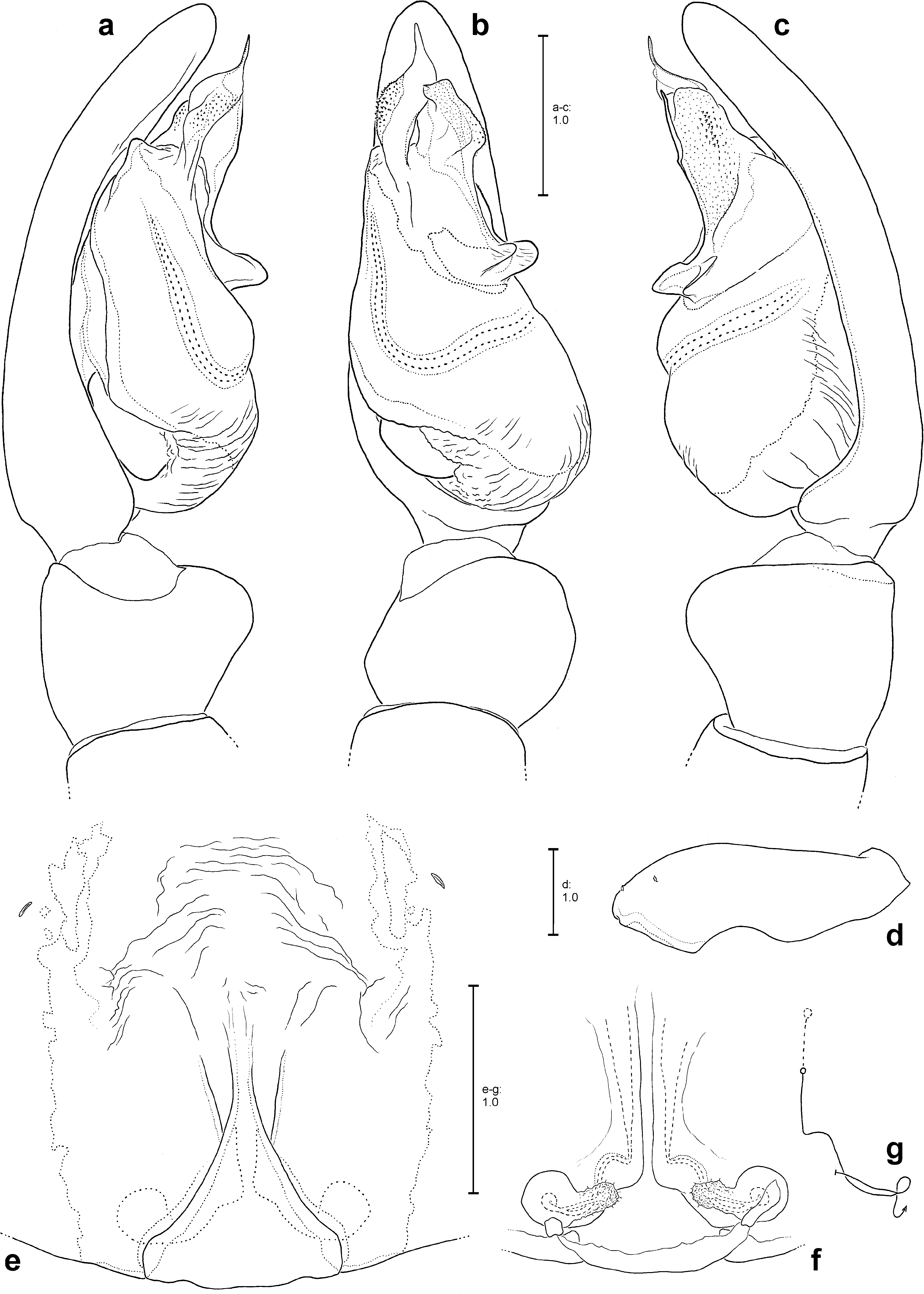
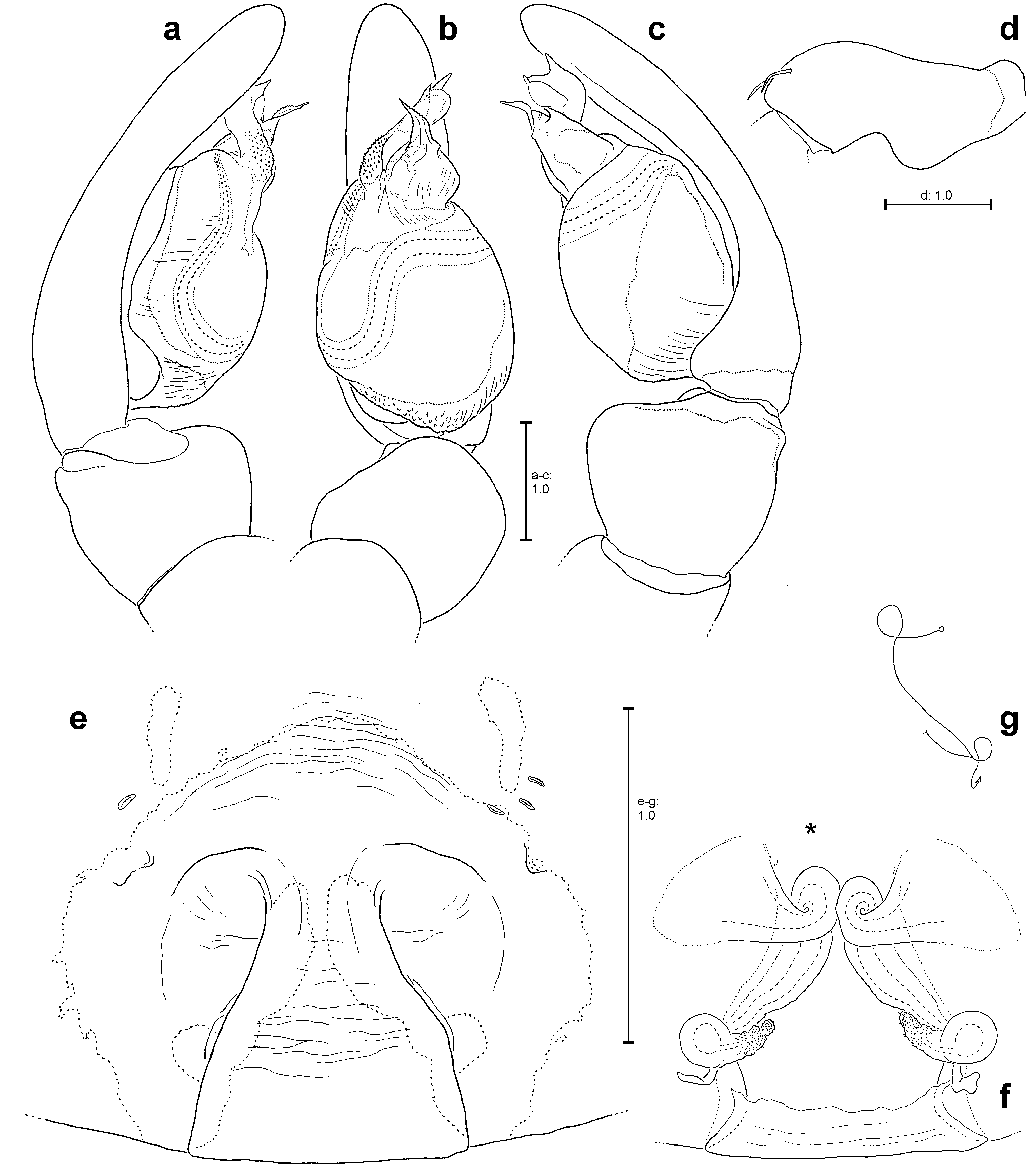
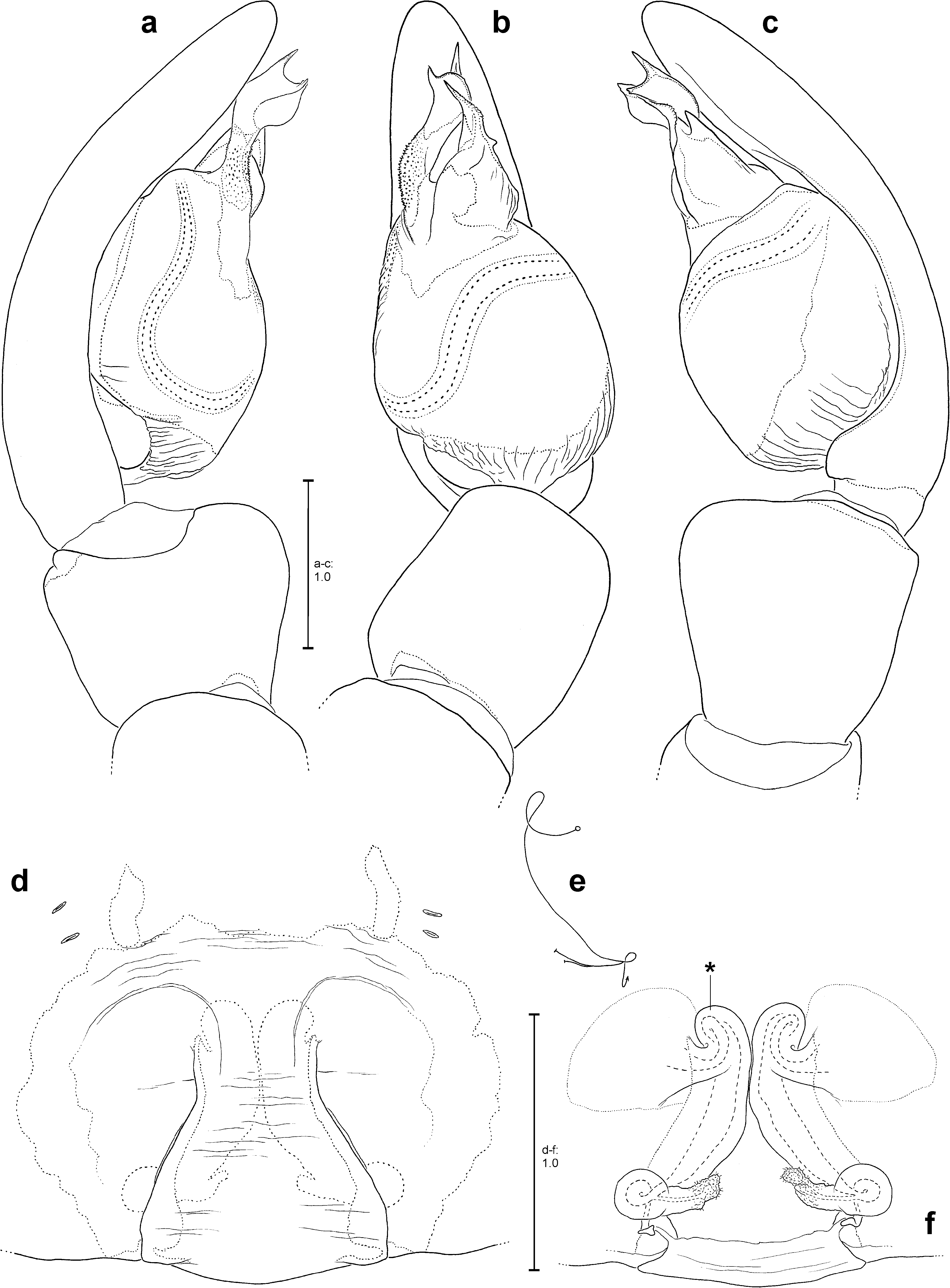
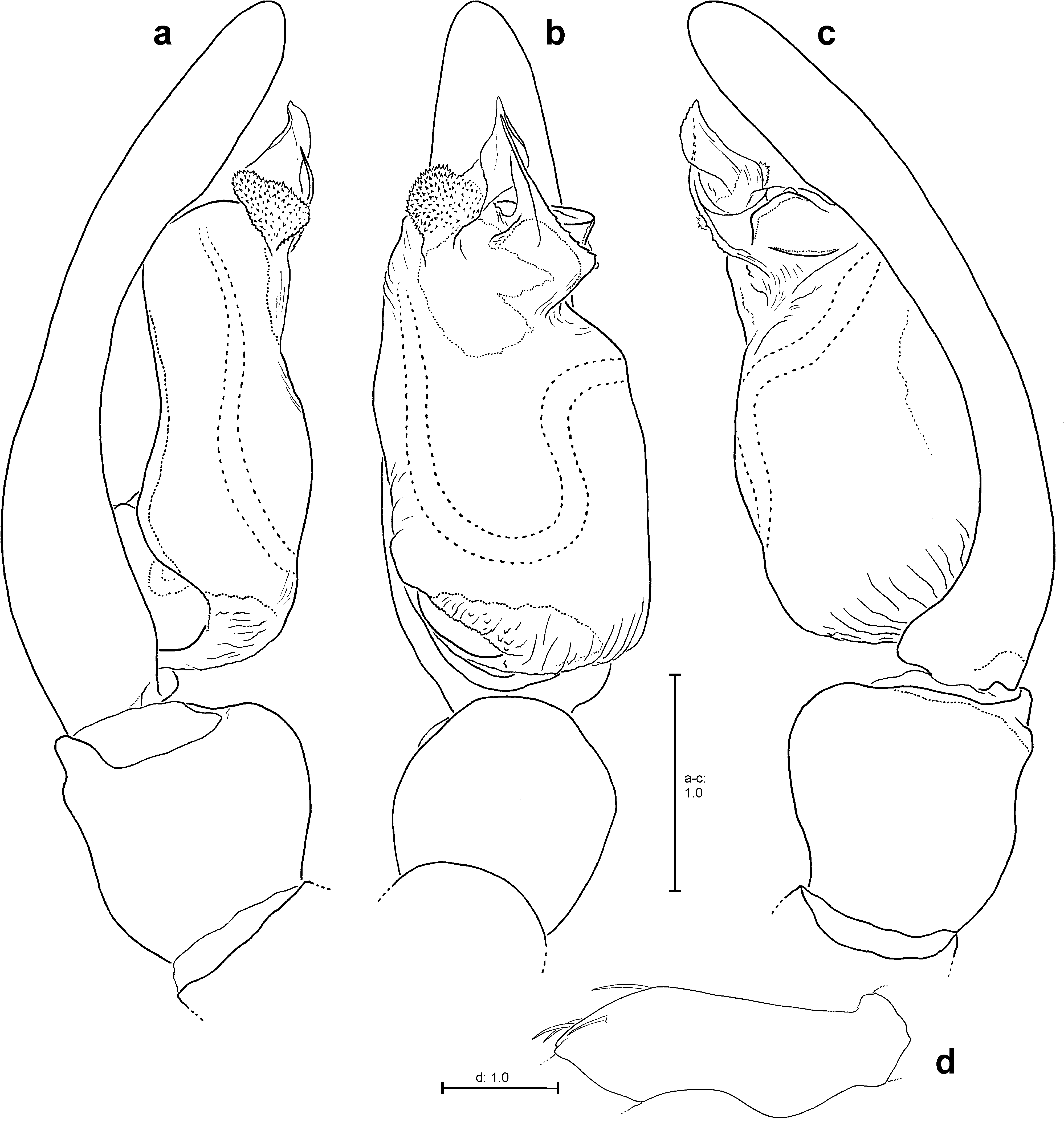
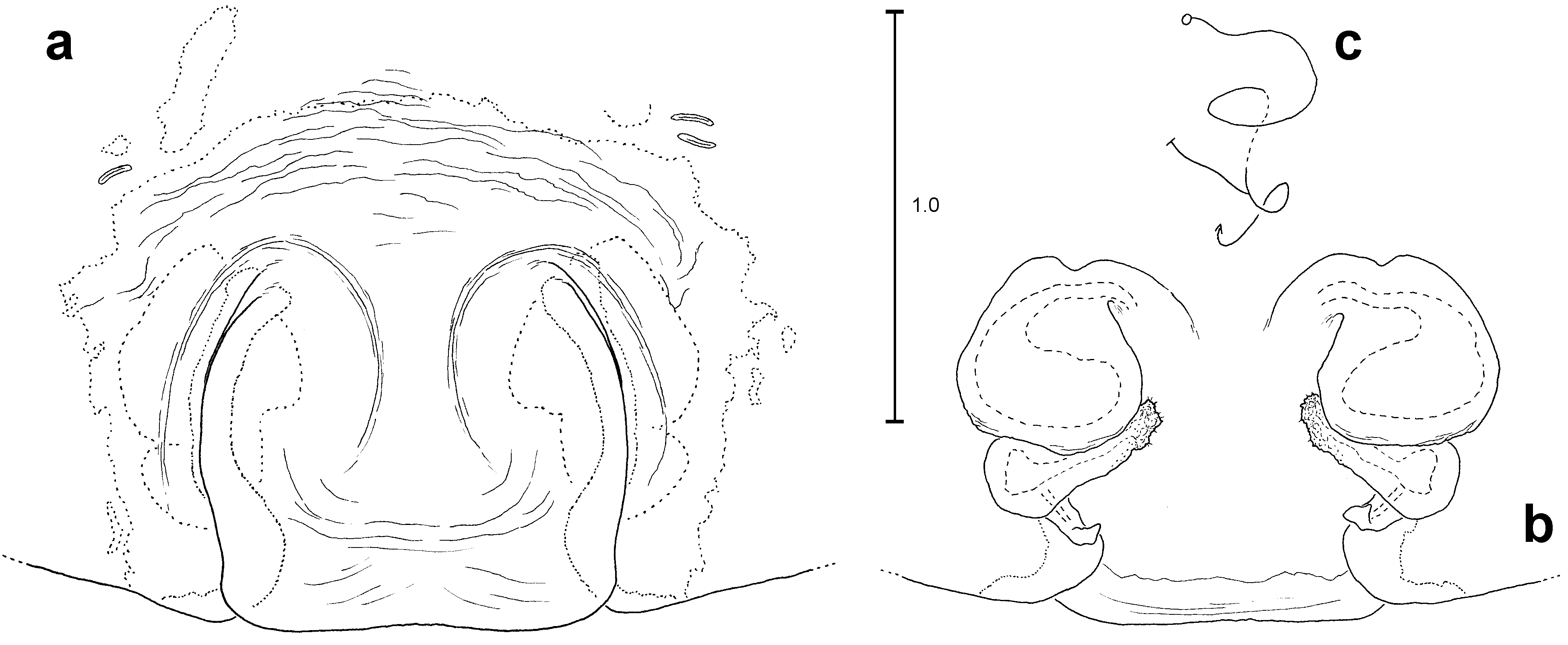
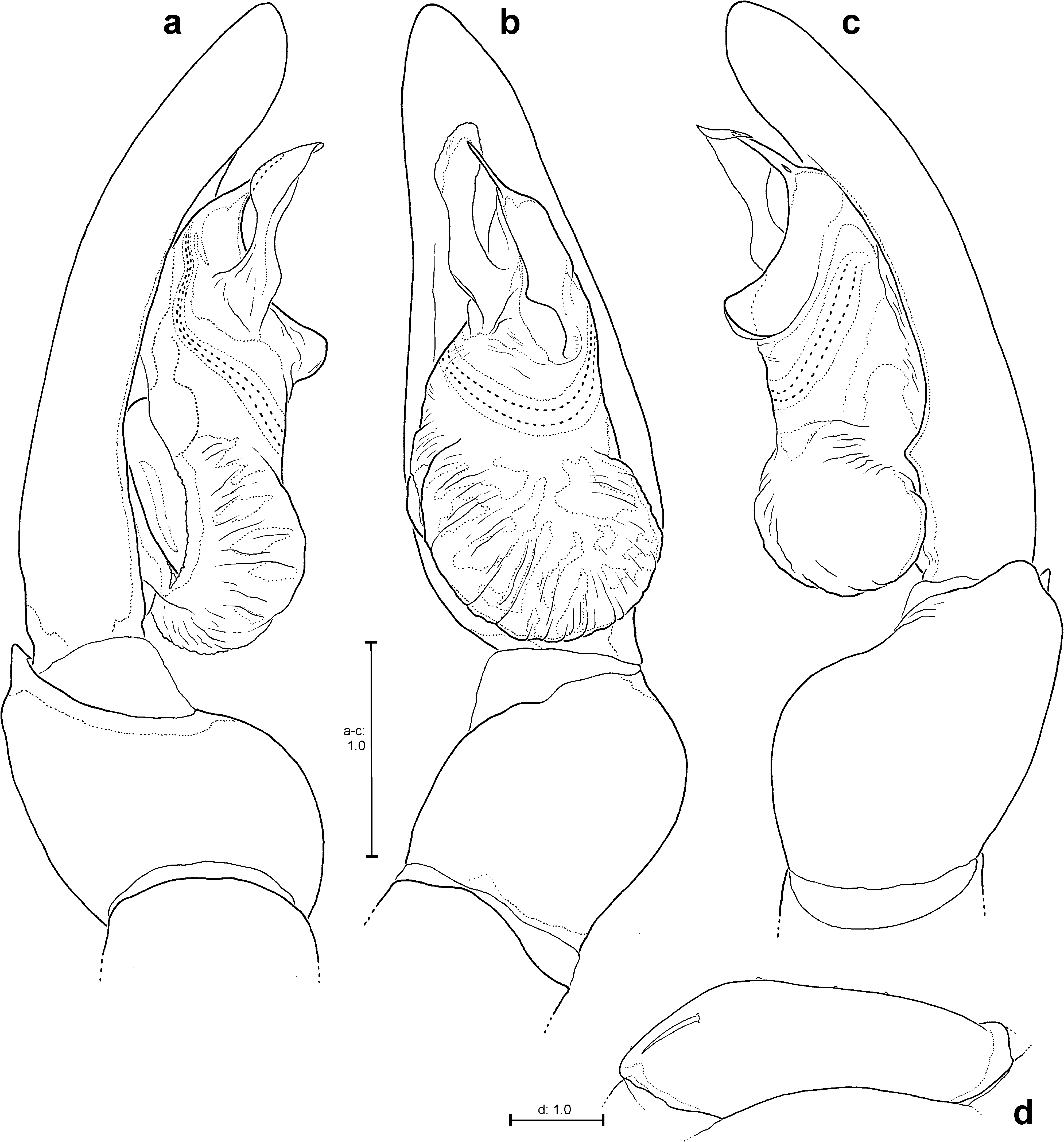
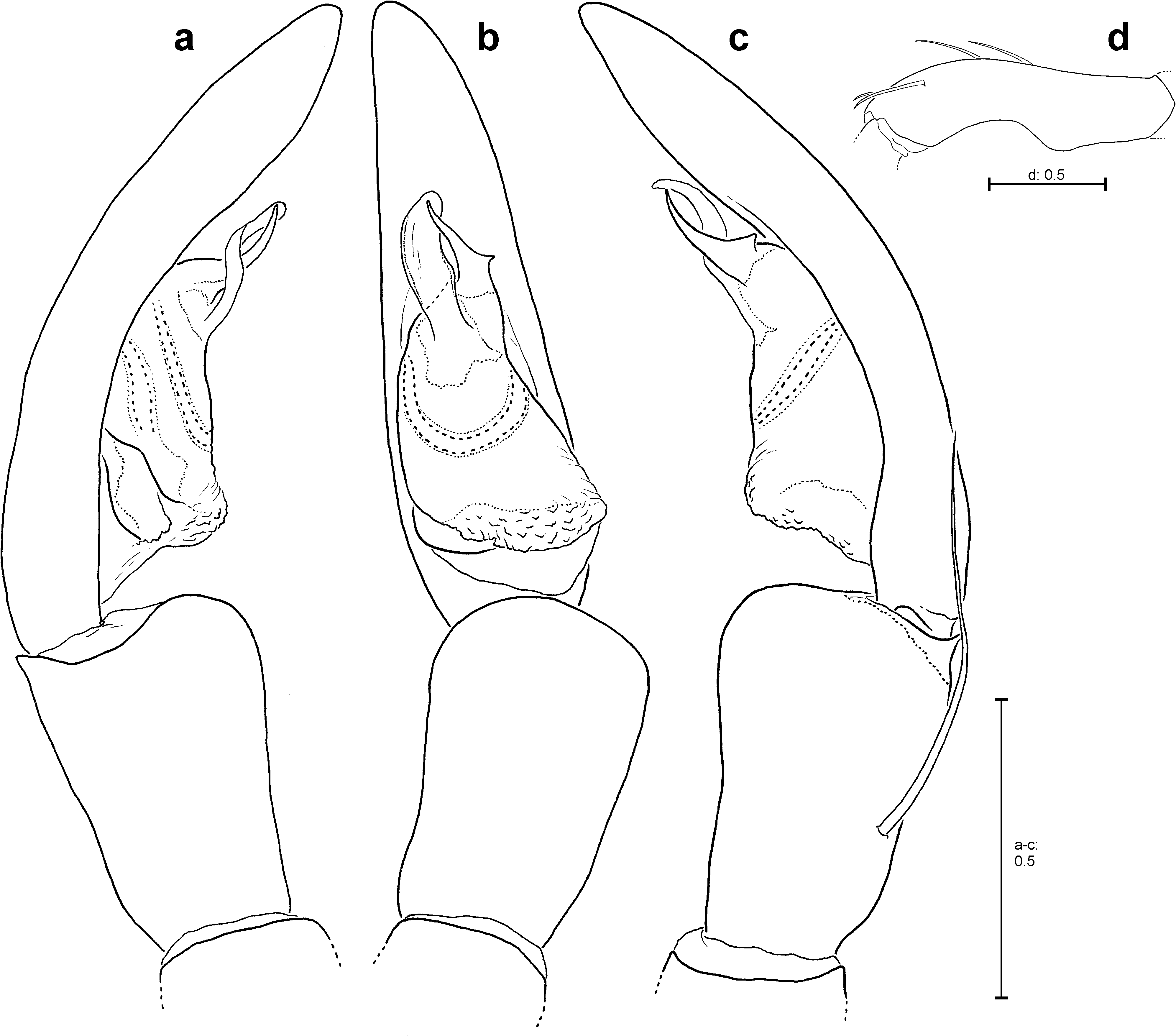
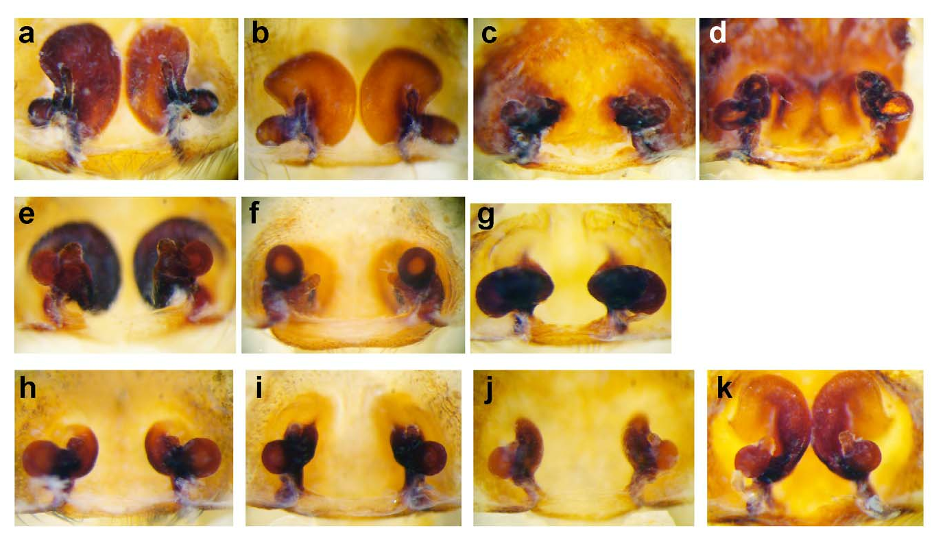
Copulatory organs: Male palp with more or less oval tegulum (T). Embolus (E) filiform ( Figs 2b–c View FIGURES 2 , 25b–c View FIGURES 25 ), broad and strong ( Figs 10a–c View FIGURES 10 , 54b View FIGURES 54 ), or of intermediate shape. E arising in retrolateral half of T or centrally. Conductor (C) mostly membranous, rarely sclerotised, rudimentary or completely reduced. C arising medially in upper half of T ( Figs 2b View FIGURES 2 , 70b View FIGURES 70 ). Expanded bulb clearly showing the large basal haematodocha (BH), surrounding subtegulum partly ( Figs 1a–c View FIGURES 1 ). A median haematodocha, like present e.g. in Araneidae ( Grasshoff 1968) , Oecobius Lucas , Uroctea Dufour ( Baum 1972) , Liphistius Schiödte ( Kraus 1978) or in Pisauridae ( Sierwald 1987) , is absent in Psechrus . Bulbal ligament (BL) and bulbal petiolus (BP) are visible through the BH ( Figs 1a–b View FIGURES 1 ). Cymbium (slightly) broader than palpal tibia and patella or more or less equal in width. RTA usually absent, but a few species belonging to the mulu -group (see below) with a tibial process (TP) with a bunch of long setae ( Figs 10b–c View FIGURES 10 ). It is not clarified if this process represents a RTA, if it is homologous to a RTA or if it is a completely different structure. A “regular” RTA usually lacks setae and is generally strongly sclerotised. Palpal femur may be modified (e.g. Figs 2d View FIGURES 2 , 10d View FIGURES 10 , 15d View FIGURES 15 or 55d). The respective modifications may be species-specific. Cymbium dorsally mostly with scopula (CS) ( Figs 83a–b, d–g View FIGURES 83 ). There are differences in density among the different species: very dense ( Figs 83d–g View FIGURES 83 ) or moderate dense ( Figs 83a–b View FIGURES 83 ). CS can be of different length (covering cymbium from 1/4 up to 6/7, Figs 83d–g View FIGURES 83 ). In a few species CS is absent ( Fig. 83c View FIGURES 83 ).
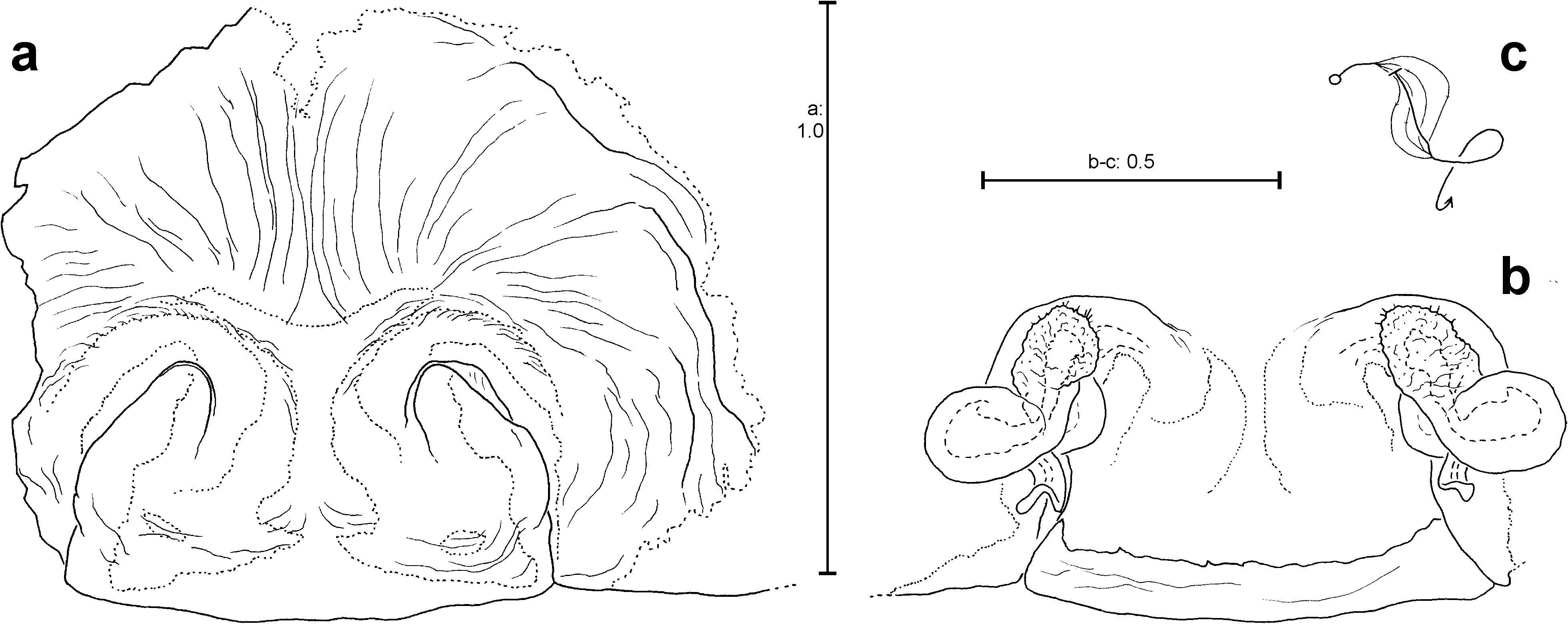
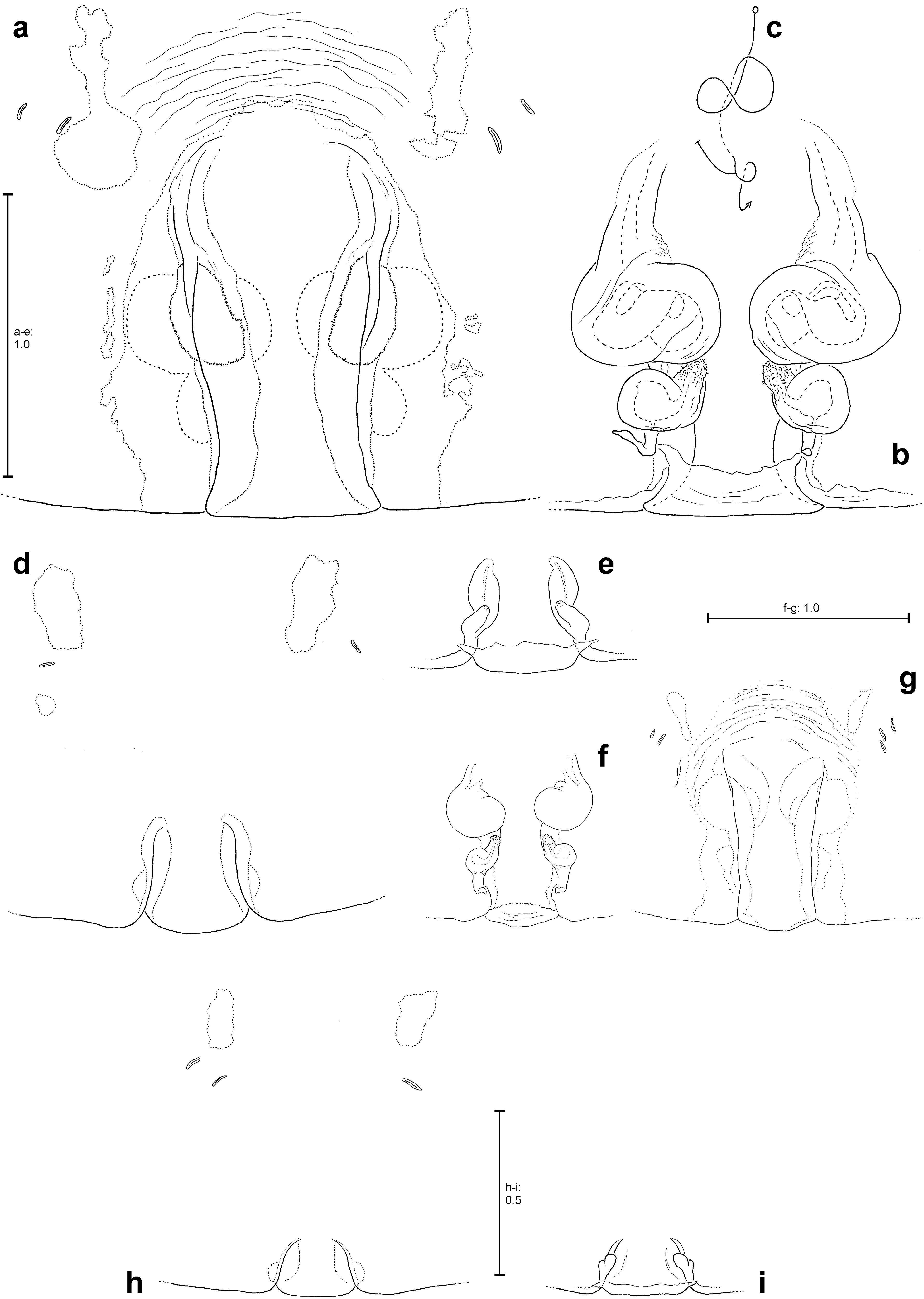
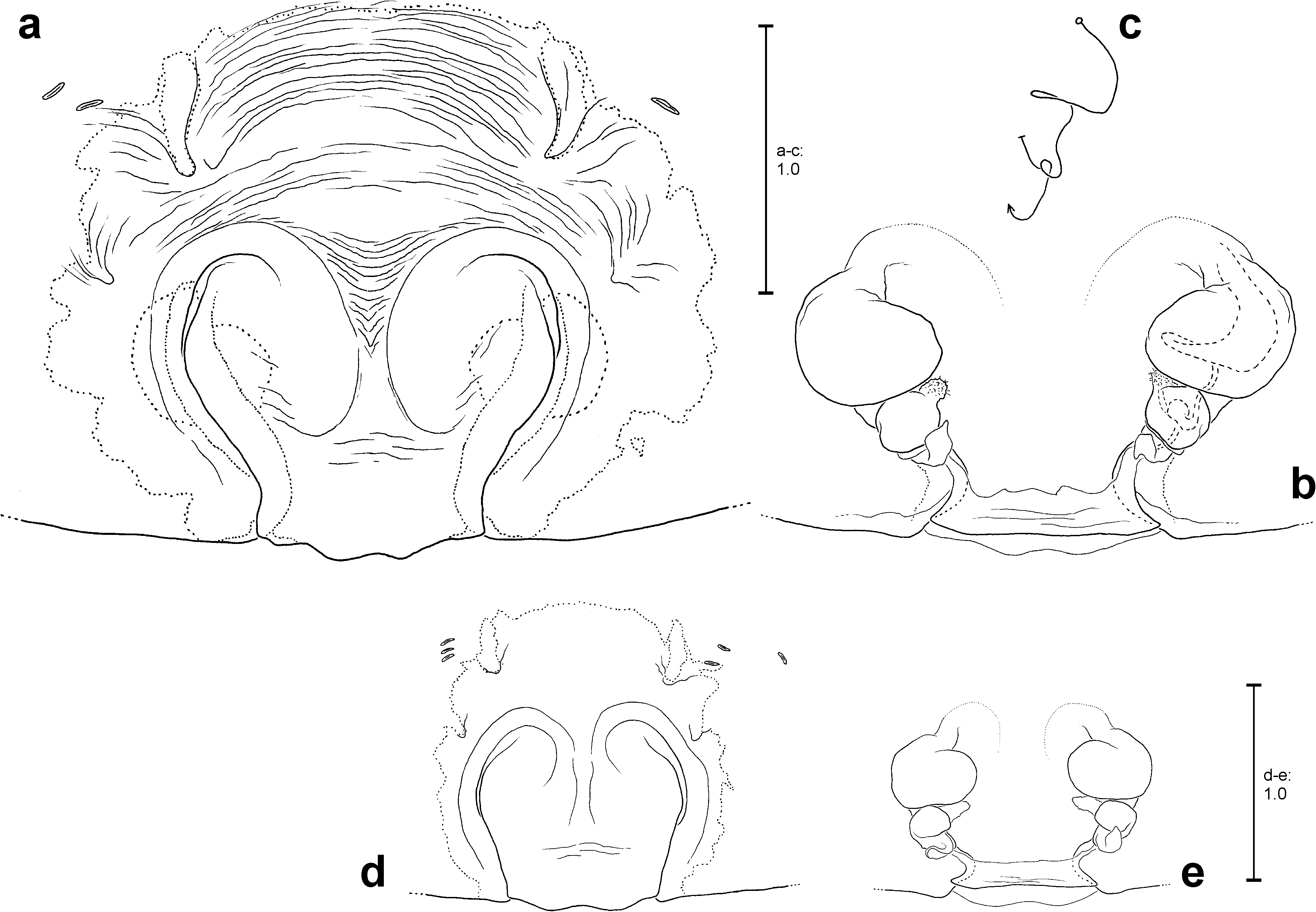
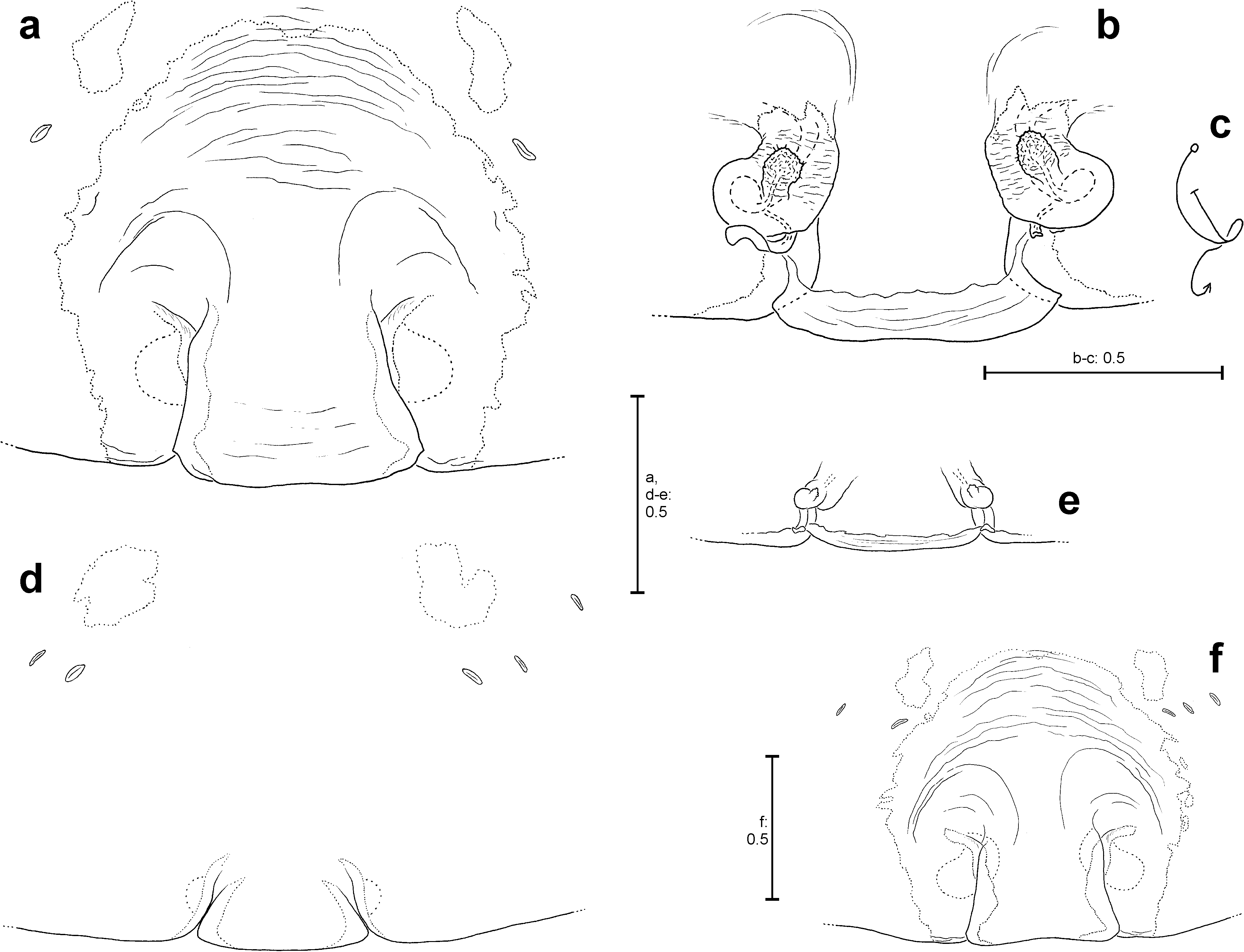
Female epigyne mostly with simple septum (e.g. Fig. 29e View FIGURES 29 ). Anterior to septum mostly lots of curved wrinkles ( Figs 29e View FIGURES 29 , 32a View FIGURES 32 ). Vulva specifically shaped (see each species description) with internal duct system generally divided into three sections: copulatory duct, receptaculum with spermathecal head and fertilisation duct ( Figs 2h View FIGURES 2 , 29d View FIGURES 29 ).
Biology. The lace-sheet-weavers generally live in shady habitats near the ground. They can be found in forests (e.g. between tree roots, in holes of tree trunks or underneath dead wood), between rocks, boulder, at rock walls or at clay escarpments. Sometimes they appear in untended barracks or huts, too. Several species, especially those that are often found at rock walls or between boulders, can be found in the entrance areas of caves, too. According to colleagues’ and my own observations in the field in Laos, there are several species, Psechrus laos sp. nov., P. ancoralis Bayer & Jäger, 2010 , P. antraeus Bayer & Jäger, 2010 , P. khammouan Jäger, 2007 and P. steineri Bayer & Jäger, 2010 , which prefer these habitats (rock walls etc., see above). Others, P. luangprabang Jäger, 2007 , P. ghecuanus Thorell, 1897 and P. jaegeri sp. nov., are found mainly in forests, between roots or in tree holes or at escarpments. There are a few Psechrus species reported only from caves, e.g. P. mulu Levi, 1982 , P. cebu Murphy, 1986 or P. steineri . However, they had mostly been reported from the entrance areas of caves (rarely from the aphotic zone). Presumably they can be found on rock walls or boulders outside of the caves, too. I would not assume that there is any Psechrus species that is restricted to caves. The lace-sheet weavers build a horizontal sheet web, which reaches up to 1.2 m length (in rare cases even 2 m [ Robinson and Lubin 1979]). At one side the web turns into a long tube, which leads into a narrow crevice or hole, where the spider is safe from invaders. This tuberetreat is generally located in a rigid environment, e.g. rocks, stones, rigid soil or wood. It never appears between leaves and only very rarely between grass. Psechrus moves upside down underneath the sheet web like representatives of Linyphiidae do. Psechrus behaves extremely shyly and careful in its sheet web. At the slightest disturbance it runs back to its retreat with extreme speed. This explains why Psechrus , though it is abundant in many regions, is not easy to catch. Females carry their egg sacs, which can be up to 25 mm in diameter, in their chelicerae ( Fig. 93b View FIGURES 93 ). According to my own observations in P. jaegeri sp. nov., P. argentatus ( Doleschall, 1857) , P. mulu Levi, 1982 , P. ghecuanus Thorell, 1897 and according to Jäger (2007) for P. khammouan Jäger, 2007 , egg sacs contain 70– 96 eggs. Yoshida (2009) counted 174 spiderlings in an egg sac of P. clavis sp. nov. (sub P. taiwanensis ).
Robinson and Lubin (1979) observed and described the predatory behaviour of Psechrus argentatus ( Doleschall, 1857) . Most of the behavioural units described therein, were also observed in my own trials using several P. laos sp. nov., P. luangprabang Jäger, 2007 and P. torvus (O. Pickard-Cambridge, 1869) specimens. Each spider was transferred into a glass terrarium with a piece of wood in one corner. A sheet web was built by the second day at the latest, with the retreat situated between the piece of wood and the corner of the terrarium. Crickets or large flies were used as prey, which were placed in the centre of the web. After a fly was put into the web, it took at most one second until the spider moved slightly forward in its retreat. A few seconds later it moved to the mouth of the retreat and pulled slowly at the web with the forelegs. Finally it ran out very fast to the respective site of the sheet web, grasped the fly with its forelegs and bit it. Immediately it ripped the fly out of the web with the chelicerae and ran back with it forward into the retreat. Later, it turned its direction within the retreat to be ready for the next prey attack (in some cases not before finishing up eating the fly). When larger prey items, e.g. large crickets, were offered, the behaviour was the same up to the point before grasping the prey. In this case the spider moved more slowly before directly encountering the prey. After a few attempts of stretching out and drawing back the forelegs over the cricket, it was bitten. The bite was mostly located between head and thorax or at the base of antennae and lasted for ca. 5 seconds. Then shorter bites at other sites of the cricket’s body (e.g. legs, antennae) followed. After ca. 1 minute the prey seemed to be paralysed and Psechrus began to bind its prey with threads produced from the spinnerets but also with threads taken from the web. Subsequently the prey was cut out of the web and carried within the chelicerae back to the retreat. The binding behaviour was not executed in every trial, but in ca. 50 % of prey attacks upon large prey items. The approaching behaviour in the terrarium is rapid and does not pass off stepwise as has been observed in the natural environment by Robinson and Lubin (1979) and myself. In the wild the webs are far larger in size and consequently prey are much more difficult to localise. The three Psechrus species examined showed no significant differences in web structure and predatory behaviour.
Mating behaviour was observed only once. In the respective trial a male of Psechrus luangprabang was released into the terrarium of a conspecific female (ca. 11:00 o’ clock a.m). It moved to an upper corner of the terrarium and stayed there for 4 ½ hours. At ca. 3: 30 p. m. the male slowly approached from a peripheral section of the web. The approach was interrupted by stops, from about 5–15 minutes duration. At each resting position the web was gently pulled rhythmically with the two pairs of forelegs. Meanwhile the female appeared at the mouth of the tube retreat. Finally the male reached the females legs and stroked them with his tarsi for about 5–10 minutes. Then he crawled underneath the female, both specimens facing the same direction. He surrounded her body with his long legs. They both swung violently up and down. Suddenly the male turned underneath the female and once again surrounded the latter with his legs. The spines on his legs, especially the femora, were erected to ca. 45°. The male turned again and pulled down the two first legs of the female with the metatarsus of his right leg I. Once again he turned and both spiders showed trembling movements. Then the male approached very closely to the female and a few seconds after that he departed again. This sequence was repeated about three times. Finally he moved his body perpendicular to the female and cleaned his two first legs and the palps. Both specimens trembled, even more intensely than before and the male’s spines erected to almost 90°. The male changed his position to a 45° angle towards the female and in this position he pulled her towards him and copulation took place. It was very rapid, lasting about 20 seconds. During copulation the first two pairs of the female’s legs and the second pair of the male’s legs were stretched straight forward. The expansion of the bulb and the exact position of insertion could not be observed with the naked eye. After copulation the male was chased away by the female and moved to an upper corner of the terrarium ( Fig. 93a View FIGURES 93 shows a Psechrus couple in the field observed by a colleague).
Species groups. The species groups are defined mainly by the basic structures of the copulatory organs of their representatives (see diagnoses of each species group below). A few somatic characters are in parts useful for such a “classification”, but often only by trend as there are exceptions in character patterns. These useful characters are: 1) the shape of median and lateral bands on carapace, 2) the shape of the light, longitudinal line ventrally on opisthosoma, 3) the relative leg length (measured as ratio between femur I + metatarsus I / carapace length), 4) dorsal spines on tibia III and IV. The length of legs is variable among different specimens of the same sex, which is the case for every species. It will be noted as an approximate ratio for males and females in the description of each species-group, with the following convention: Ratio between femur + metatarsus of leg I / carapace length (FEM-I + MTT-I / CL). Presently, eight groups, the argentatus-, mulu-, annulatus-, singaporensis-, ancoralis-, himalayanus-, sinensis-, and the torvus- group are differentiated.
Key to species of Psechrus :
1 Male [unknown in: borneo View in CoL , annulatus View in CoL , aluco View in CoL sp. nov., norops View in CoL sp. nov., arcuatus View in CoL sp. nov., jinggangensis View in CoL , fuscai View in CoL sp. nov., kenting View in CoL , taiwanensis View in CoL , tauricornis View in CoL sp. nov.; identification not absolutely certain in: demiror View in CoL sp. nov., zygon View in CoL sp. nov.; those of demiror View in CoL sp. nov. and zygon View in CoL sp. nov. are included in the present key, but with question mark].......................... 2
- Female [unknown in: ulcus View in CoL sp. nov., kinabalu View in CoL , schwendingeri View in CoL sp. nov.]......................................... 37
2 Harpago ( Figs 70b–c View FIGURES 70 , 72b–c View FIGURES 72 , 74b–c View FIGURES 74 ) present................................................................ 3
- Harpago absent....................................................................................... 5
3 C longer than width of T........................................................................... torvus View in CoL
- C shorter than width of T............................................................................... 4
4 C centrally as broad as in distal fourth and located medially in upper half of T....................... hartmanni View in CoL sp. nov.
- C broadest in distal fourth and located in retrolateral half of T..................................... zygon View in CoL sp. nov. (?)
5 C absent or strongly reduced............................................................................ 6
- C well developed...................................................................................... 9
6 C absent............................................................................................ 7
- C rudimentary (a very short and stout structure still recognisable), E quite broad and strongly sclerotised, resting in CA ( Figs 78a–c View FIGURES 78 )............................................................................ schwendingeri View in CoL sp. nov.
7 With three apophyses close to E ( Figs 7a–b View FIGURES 7 )............................................................ mulu View in CoL
- With less than three apophyses close to E.................................................................. 8
8 E retrolaterad; E and EB constitute an extremely bulky structure ( Fig. 10b View FIGURES 10 ).............................. ulcus View in CoL sp. nov.
- E rather slim, prolatero-apicad..................................................................... kinabalu View in CoL
9 C with numerous small or very small, short spines or tubercles ( Figs 52a–b View FIGURES 52 , 54a–b View FIGURES 54 , 57a–b View FIGURES 57 , 66a–b View FIGURES 66 ).................... 31
- C without such structures.............................................................................. 10
10 Palpal femur modified with ventral bulge (the latter may be flat) ( Figs 14d View FIGURES 14 , 15d View FIGURES 15 ) or with pointed, ventral extension ( Fig. 2d View FIGURES 2 ). ................................................................................................... 11
- Palpal femur without modification....................................................................... 16
11 E dorsally with one distinct, pointed apophysis ( Fig. 79b View FIGURES 79 )................................................... cebu View in CoL
- E different.......................................................................................... 12
12 C strongly sclerotised and narrow, its distal half just as broad as E ( Figs 2b View FIGURES 2 , 6b View FIGURES 6 )................................... 13
- C membranous and/or fleshy, its distal half (distinctly) broader than E........................................... 14
13 C more than half as long as E........................................................................ libelti View in CoL
- C less than half as long as E..................................................................... argentatus View in CoL
14 C ca. as long as T....................................................................... decollatus View in CoL sp. nov.
- C far shorter than T................................................................................... 15
15 EB in ventral view in alignment with upper retrolateral margin of T ( Fig. 15b View FIGURES 15 )........................... singaporensis View in CoL
- EB protruding beyond upper retrolateral margin of T ( Fig. 17b View FIGURES 17 )..................................... elachys View in CoL sp. nov.
16 Bulb with elongated EB possesing a distinct ventral protrusion basally ( Figs 76a–c View FIGURES 76 ).................... crepido View in CoL sp. nov.
- Bulb different....................................................................................... 17
17 E arising medially on upper half of T, coxa of leg I ( Figs 82l,r View FIGURES 82 ) or proximal part of palpal femur ( Fig. 35d View FIGURES 35 ) ventrally with distinct field of macrosetae............................................................................... 23
- E arising retrolaterally on T, neither coxa of leg I nor proximal part of palpal femur ventrally with distinct field of macrosetae (may be with few unconspicuous macrosetae subdistally in addition to an apical row of macrosetae)................... 18
18 C with distinct, broadened base ( Figs 23b View FIGURES 23 , 25b View FIGURES 25 )............................................................ 19
- C without distinct, broadened base....................................................................... 20
19 E longer than T.............................................................................. laos View in CoL sp. nov.
- E shorter than T..................................................................................... rani View in CoL
20 C broader than 1/3 the width of T................................................................. ancoralis View in CoL
- C narrower than 1/3 the width of T....................................................................... 21
21 E longer than width of T......................................................................... antraeus View in CoL
- E shorter than width of T............................................................................... 22
22 E longer than half the width of palpal tibia........................................................ khammouan View in CoL
- E shorter than half the width of palpal tibia............................................................ steineri View in CoL
23 E (almost) straight.................................................................................... 26
- E curved........................................................................................... 24
24 EB with particular flat, elongated and proximally curved extension ( Fig. 43b View FIGURES 43 )............................ luangprabang View in CoL
- EB different......................................................................................... 25
25 Distal section of E curved and distinctly narrower than central section ( Fig. 50f View FIGURES 50 )....................... jaegeri View in CoL sp. nov.
- Distal section of E hardly curved and not significantly narrower than central section ( Fig. 50e View FIGURES 50 ).............. vivax View in CoL sp. nov.
26 C as long as T......................................................................... demiror View in CoL sp. nov. (?)
- C shorter than T...................................................................................... 27
27 T apically with quite strongly sclerotised, semicircular extension ( Figs 37b View FIGURES 37 , 39g View FIGURES 39 , 41b View FIGURES 41 ).............................. 28
- T apically without such an extension..................................................................... 29
28 E arising distinctly further distally than C.......................................................... ghecuanus View in CoL
- E arising at most at the same level as C, but mostly further proximally.............................. pakawini View in CoL sp. nov.
29 Sperm duct simply U-shaped................................................................... himalayanus View in CoL
- Sperm duct with at least two loops....................................................................... 30
30 C with broad and large proximal section and small and narrow distal section ( Fig. 35b View FIGURES 35 ).................. inflatus View in CoL sp. nov.
- C broadest distally............................................................................ marsyandi View in CoL
31 C apically (at least slightly) bifid ( Figs 56a–c View FIGURES 56 , 57a–c View FIGURES 57 )........................................................ 32
- C apically not bifid................................................................................... 33
32 EB with apophyses only in its distal half ( Figs 55b–c View FIGURES 55 , 56b–c View FIGURES 56 )......................................... tingpingensis View in CoL
- EB with apophyses only in its proximal half ( Figs 57b–c View FIGURES 57 )........................................ obtectus View in CoL sp. nov.
33 C in ventral view proximally with striking, hemispherical bulge ( Fig. 62b View FIGURES 62 )............................... senoculatus View in CoL
- C without such a bulge................................................................................ 34
34 Sperm duct with distinctly U-shaped section in prolateral half of T ( Figs 66b View FIGURES 66 )........................... clavis View in CoL sp. nov.
- Sperm duct different (e.g. as in Figs 52b View FIGURES 52 , 54b View FIGURES 54 , 86m,o View FIGURES 86 )....................................................... 35
35 EB with long, apically bifurcated apophysis ( Fig. 86n View FIGURES 86 ).............................................. kunmingensis View in CoL
- EB without long, apically bifurcated apophysis............................................................. 36
36 E in ventral view short, broad and blunt ( Fig. 54b View FIGURES 54 )................................................... triangulus View in CoL
- E in ventral view with filiform distal section ( Fig. 52b View FIGURES 52 )................................................... sinensis View in CoL
37 Epigyne complex, tegimentum (TM) present ( Figs 69a,d View FIGURES 69 , 71a,d View FIGURES 71 , 73a,e View FIGURES 73 , 75a,e View FIGURES 75 , 89m –p View FIGURES 89 ).............................. 38
- Epigyne without TM................................................................................. 41
38 Branches of TM distally rounded ( Figs 69a View FIGURES 69 , 73a View FIGURES 73 )............................................................ 39
- Branches of TM distally pointed, resembling the horns of a bull ( Figs 75a,e View FIGURES 75 )........................ tauricornis View in CoL sp. nov.
39 Branches of TM directed anterio-mediad............................................................... torvus View in CoL
- Branches of TM directed (anterio-) laterad................................................................ 40
40 Distal section of CD located medial to SH....................................................... zygon View in CoL sp. nov.
- Distal section of CD located lateral to SH.................................................... hartmanni View in CoL sp. nov.
41 Epigyne with flat, large-area bulge in front of MS ( Figs 77a,e View FIGURES 77 , 89k View FIGURES 89 )................................. crepido View in CoL sp. nov.
- Epigyne without such a bulge in front of MS............................................................... 42
42 MS longer than broad................................................................................. 43
- MS broader than long................................................................................. 51
43 Anterior half of MS broader than posterior half ( Figs 61a View FIGURES 61 , 63a View FIGURES 63 , 65a View FIGURES 65 , 67a View FIGURES 67 )......................................... 44
- Posterior half of MS broader than anterior half ( Figs 53a View FIGURES 53 , 56d View FIGURES 56 , 60a,e View FIGURES 60 )........................................... 48
44 CD with twisted section ( Fig. 61b View FIGURES 61 ), the latter narrower than spermatheca............................... jinggangensis View in CoL
- CD with bulbous section, the latter broader than spermatheca................................................. 45
45 MS distinctly longer than broad (more than 1.6 times, Fig. 63a View FIGURES 63 ), initial section of CD straight ( Fig. 63b View FIGURES 63 )........ senoculatus View in CoL
- MS less than 1.6 times longer than broad, initial section of CD either integrated within bulbous section ( Fig. 67b View FIGURES 67 ) or anteriorly curved mediad ( Figs 64b View FIGURES 64 , 65b View FIGURES 65 ).......................................................................... 46
46 Initial section of CD integrated in kidney-shaped, bulbous section.................................... clavis View in CoL sp. nov.
- Initial section of CD anteriorly curved mediad.............................................................. 47
47 Posterior half of MS just slightly narrower than anterior half ( Fig. 65a View FIGURES 65 ), bulbous section of CD anteriorly with small bulge ( Fig. 65b View FIGURES 65 )....................................................................................... taiwanensis View in CoL
- Posterior half of MS clearly narrower than anterior half ( Figs 64a,d View FIGURES 64 ), bulbous section of CD anteriorly without small bulge .................................................................................................. kenting View in CoL
48 CD with twisted section ( Figs 55f View FIGURES 55 , 58b View FIGURES 58 , 59b View FIGURES 59 )............................................................... 51
- CD without twisted section ( Figs 53b View FIGURES 53 , 54f View FIGURES 54 , 60b,d View FIGURES 60 )........................................................... 49
49 MS and LL complicatedly folded interleaved ( Figs 60a,e View FIGURES 60 )............................................ kunmingensis View in CoL
- MS and LL different.................................................................................. 50
50 Posterior half of MS anteriorly rounded ( Figs 53a,d View FIGURES 53 )..................................................... sinensis View in CoL
- Posterior half of MS triangular ( Fig. 54e View FIGURES 54 )............................................................ triangulus View in CoL
51 Epigyne anteriorly with two rounded, flattened, glossy fields near CO ( Figs 59a View FIGURES 59 , 89e View FIGURES 89 ).................... fuscai View in CoL sp. nov.
- Epigyne without rounded, flattened, glossy fields near CO.................................................... 52
52 CD at least 4x longer, but less than 2x broader than diameter of receptaculum.................................... 53
- CD different........................................................................................ 54
53 Twisted sections of CD, located anteriorly beyond transversal, initial sections of CD, shorter than half the diameter of one receptaculum ( Figs 58b,g View FIGURES 58 ).................................................................. obtectus View in CoL sp. nov.
- Twisted sections of CD, located anteriorly beyond transversal, initial sections of CD, almost as long as diameter of one receptaculum ( Figs 55f View FIGURES 55 , 56f View FIGURES 56 )....................................................................... tingpingensis View in CoL
54 CD with voluminous, spherical/bulbous section, the latter distinctly larger than receptaculum ( Figs 64b,e View FIGURES 64 , 65b View FIGURES 65 ).......... 47
- CD may be large and broad ( Figs 2h View FIGURES 2 , 5c View FIGURES 5 ), but without voluminous, spherical/bulbous section......................... 55
55 Epigyne and EF strongly sclerotised, with dark red-brown colour ( Figs 87c,d View FIGURES 87 ).................................... 56
- At least EF different.................................................................................. 57
56 CD running transversally and straight ( Figs 7e View FIGURES 7 , 8f View FIGURES 8 )........................................................ mulu View in CoL
- CD curved and flowing into receptaculum from anterior ( Fig. 9b View FIGURES 9 ).......................................... borneo View in CoL
57 Epigyne protruding, CO large ( Figs 12a,b View FIGURES 12 , 13a View FIGURES 13 , 14f View FIGURES 14 , 87e–g View FIGURES 87 )................................................... 58
- Epigyne not or hardly protruding, CO rather small.......................................................... 60
58 Vulva with spherical SH............................................................................... 59
- Vulva without spherical SH ( Fig. 14h View FIGURES 14 )....................................................... decollatus View in CoL sp. nov.
59 CD distinctly larger than receptaculum ( Fig. 12c View FIGURES 12 )..................................................... annulatus View in CoL
- CD not larger than receptaculum ( Fig. 13b View FIGURES 13 , 90f View FIGURES 90 ).................................................. aluco View in CoL sp. nov.
60 CD very large, broad and flat (CD at least 5x larger than receptaculum, Figs 2h View FIGURES 2 , 5c View FIGURES 5 , 20b View FIGURES 20 )............................ 61
- CD not or not distinctly larger than receptaculum........................................................... 63
61 CO located anterior to SH, receptacula round................................................... arcuatus View in CoL sp. nov.
- CO located posterior to SH, receptacula cross-oval.......................................................... 62
62 MS at most half as long as broad and with notches laterally ( Figs 2g View FIGURES 2 , 3i View FIGURES 3 ).................................. argentatus View in CoL
- MS not distinctly broader than long, with continuous lateral margins ( Figs 5a–b View FIGURES 5 )............................... libelti View in CoL
63 Lateral, dark bands on carapace clearly broader than diameter of PME ( Fig. 82a, f–g View FIGURES 82 ), receptacula remarkably round ( Figs 15f View FIGURES 15 , 17e View FIGURES 17 ), CD curved medially ( Figs 15h,f View FIGURES 15 , 19b View FIGURES 19 )................................................................ 64
- At least one of the features listed in item 63 different........................................................ 68
64 CD ca. 4–5x larger than receptaculum........................................................ arcuatus View in CoL sp. nov.
- CD at most 2x larger than receptaculum................................................................... 65
65 Anterior margins of LL strongly diverging ( Fig. 80a View FIGURES 80 ; Attention: anterior margins of LL may be confound with the anterior margins of MS, but the latter are short and pointing more or less anteriad)......................................... cebu View in CoL
- Anterior margins of LL not or hardly diverging............................................................. 66
66 SH located upon receptacula................................................................. norops View in CoL sp. nov.
- SH (still) located upon CD............................................................................. 67
67 CO small, pointing anteriorly ( Fig. 17d View FIGURES 17 ), penetration of E happens frontally.......................... elachys View in CoL sp. nov.
- CO elongated ( Fig. 15e,i View FIGURES 15 ), penetration of E happens laterally......................................... singaporensis View in CoL
68 Lateral bands on carapace quite broad (1.5–2.5x diameter PME)............................................. cebu View in CoL
- Lateral bands on carapace narrow to medium-sized (at most 1.2x diameter PME).................................. 69
69 MS mushroom-like shaped ( Figs 49a View FIGURES 49 , 51a View FIGURES 51 )................................................................ 70
- MS differently shaped................................................................................. 71
70 Anterior half of MS more than 2.5x broader than posterior section................................... jaegeri View in CoL sp. nov.
- Anterior half of MS less than 2.5x broader than posterior section...................................... vivax View in CoL sp. nov.
71 Lateral margins of MS anteriorly diverging................................................................ 72
- Lateral margins of MS anteriorly converging............................................................... 73
72 Lateral margins of MS strongly diverging anteriorly ( Fig. 30e View FIGURES 30 ), CD at most 1.5x longer than receptaculum.......... steineri View in CoL
- Lateral margins of MS moderately diverging anteriorly ( Figs 29a,e View FIGURES 29 ), CD 2.5–3x longer than receptaculum...... khammouan View in CoL
73 MS ca. 2x broader than long, its anterior margins running transversally mediad, parallel with anterior margins of LL ( Figs 32a View FIGURES 32 , 34a View FIGURES 34 , 88h–i View FIGURES 88 ).......................................................................................... 74
- MS and/or anterior margins of LL different................................................................ 75
74 CD shorter than distance in between both CO....................................................... marsyandi View in CoL
- CD longer than distance in between both CO....................................................... himalayanus View in CoL
75 MS more than 2x broader than long................................................................ ancoralis View in CoL
- MS less than 2x broader than long....................................................................... 76
76 Epigyne without EF................................................................................... 77
- Epigyne with EF..................................................................................... 79
77 Initial parts of CD clearly located anterior to spermathecae ( Fig. 27f View FIGURES 27 )....................................... antraeus View in CoL
- Initial parts of CD located posterior to spermathecae or (at most) at the same level................................. 78
78 Helical section of spermatheca high, with more than three windings ( Figs 25d View FIGURES 25 , 26d View FIGURES 26 )........................ laos View in CoL sp. nov.
- Helical section of spermathecae not or just slightly higher than broad, with less than two windings ( Fig. 24b View FIGURES 24 ).......... rani View in CoL
79 Spermathecae high, with at least three helical windings ( Figs 25d View FIGURES 25 , 26d View FIGURES 26 )................................. laos View in CoL sp. nov.
- Spermathecae compact, with at most one helical winding..................................................... 80
80 Anterior margins of LL constitute strongly sclerotised clasps ( Figs 44a View FIGURES 44 , 47a View FIGURES 47 , 88l–m View FIGURES 88 ), CD longer than diamter of one spermatheca 81
- Anterior margins of LL rather inconspicuous, CD shorter than diameter of one spermatheca......................... 82
81 CD ca. two times longer than diameter of one spermatheca ( Fig. 47b View FIGURES 47 )................................ demiror View in CoL sp. nov.
- CD at most 1.5x longer than diameter of one spermatheca ( Fig. 44b View FIGURES 44 , 45b View FIGURES 45 )............................... luangprabang View in CoL
82 SH very flat, hardly protruding out of spermathecae.............................................. inflatus View in CoL sp. nov.
- SH regularly spherical, protruding out of spermathecae....................................................... 83
83 CD extending medially (clearer in frontal view) ( Figs 41f,h View FIGURES 41 )...................................... pakawini View in CoL sp. nov.
- CD constitutes one compact structure together with spermathecae ( Fig. 37e,g View FIGURES 37 )............................. ghecuanus View in CoL argentatus- group


No known copyright restrictions apply. See Agosti, D., Egloff, W., 2009. Taxonomic information exchange and copyright: the Plazi approach. BMC Research Notes 2009, 2:53 for further explanation.
|
Kingdom |
|
|
Phylum |
|
|
Class |
|
|
Order |
|
|
Family |
Psechrus Thorell, 1878
| Bayer, Steffen 2012 |
Lancaria
| Simon, E. 1887: 194 |
| Karsch, F. 1879: 557 |
Psechrus
| Griswold, C. E. & Ramirez, M. J. & Coddington, J. A. & Platnick, N. I. 2005: 37 |
| Murphy, F. & Murphy, J. 2000: 264 |
| Griswold, C. E. 1993: 539 |
| Coddington, J. A. 1990: 7 |
| Levi, H. W. 1982: 118 |
| Lehtinen, P. T. 1967: 260 |
| Homann, H. 1950: 66 |
| Dalmas, C. R. de 1917: 324 |
| Simon, E. 1892: 226 |
| Simon, E. 1890: 80 |
| Thorell, T. 1878: 170 |