
Cribrinopsis olegi, Sanamyan & Sanamyan, 2006
|
publication ID |
https://doi.org/10.1080/00222930600703532 |
|
persistent identifier |
https://treatment.plazi.org/id/CF17AF78-FFBF-387C-FE37-B3CEFCFFCF9F |
|
treatment provided by |
Felipe |
|
scientific name |
Cribrinopsis olegi |
| status |
sp. nov. |
Cribrinopsis olegi View in CoL sp. nov.
( Figures 3 View Figure 3 , 4 View Figure 4 , 13 View Figure 13 )
Material examined
Holotype: East Kamchatka: Avacha Bay, Starichkov Island , 13 m, 5 June 2003 ( KBPIG 246 /8) . Paratypes: East Kamchatka: Avacha Bay, Starichkov Island , 6 m, 31 August 2004, one specimen ( KBPIG 248 /10) ; 13 m, 5 June 2003, one specimen ( KBPIG 247 /9) ; 20 m, 9 September 2003, two specimens ( KBPIG 243 /5); Bezymyannaya Bay , 10 m, 1 October 2004, three specimens ( KBPIG 244 /6, 245/7, 252/14) ; 9–12 m, one specimen ( KBPIG 249 /11) ; 15 m, 21 September 2004, three specimens ( KBPIG 250 /12, 251/13). Kronotsky Bay, Morzhovaya Bay , 12 m, 18 May 2003, seven specimens ( KBPIG 239 /1, 240/2, 241/3, 242/4) .
Other specimens. East Kamchatka: Avacha Bay, Bezymyannaya Bay, 9–12 m, 1 October 2004, one specimen ( KBPIG 254 /16) ; 10 m, one specimen ( KBPIG 255 /17). Commander Islands : Medny Island, Drovenskoy Point , 15 m, 23 June 1995, one specimen ( KBPIG 59 / 21); Gladky Point , 32 m, 24 July 1992, one specimen ( KBPIG 66 /18). North Kurile Islands : Shumshu Island , 30 m, 8 July 1985, one specimen ( KBPIG 127 /19) .
Description
External structure. This large species reaches 10 cm in diameter and height when fully expanded and the largest formalin-preserved specimens are about 6.5 cm diameter and 4 cm high. The base is circular and strongly adhesive. In most specimens the column is rose-coloured or red, with white spots of the verrucae. The colour becomes paler toward the base. Some specimens are entirely white. The adhesive verrucae are arranged in longitudinal rows corresponding to exocoels and endocoels and they are larger and carry larger particles of gravel and broken shell in the distal part of the column. Toward the base they gradually become smaller, less adhesive and disappear completely in the proximal half of the column. Verrucae form a distinct annulus on the margin. Externally, marginal verrucae are similar to columnar verrucae, but they are slightly larger, sometimes elongated longitudinally, and usually have a darker depression, with a thinner wall, in the centre. Enlarged verrucae on the margin have the same nematocysts as the rest of the column. There is a deep fosse and short capitulum.
The oral disk is flat and circular, always paler than the column, pale rose or yellowish, greyish, whitish, sometimes with irregular light dirty-green patches. Mesenterial insertions are marked by thin white lines radiating from the mouth.
From 100 to 140 tentacles are arranged decamerously in five cycles on the outer half of the oral disk, the inner tentacles larger than the outer. In smaller specimens there are fewer tentacles: 64 and 78 tentacles were counted in the preserved specimens with diameter 24 and 33 mm, respectively. The tentacles are short (up to 1.5 cm long) and thick, usually with a large, almost spherical expansion up to 1 cm diameter on the distal end ( Figure 13 View Figure 13 ). Each tentacle has a large slit-like terminal pore. In the preserved specimens the tentacles are longitudinally folded, short, and almost spherical. The shape of the tentacles is the most conspicuous distinguishing character for the species. The ground colour of the tentacles is white or yellowish with numerous short longitudinal red stripes on the expanded distal half. The border between the expanded distal and cylindrical proximal half of each tentacle is marked by a wide white transverse band. In white specimens the tentacles and oral disk are pure white, without the colour stripes.
Internal structure. The marginal endodermal sphincter is strong, circumscribed, with one short, not always pronounced central lamella ( Figure 3A View Figure 3 ). Radial muscles of the oral disk and longitudinal muscles of the tentacles are mesogloeal, sometimes to ecto-mesogloeal ( Figure 3B View Figure 3 ). Transverse sections of the tentacles show the muscle meshes closer to the outer (ectodermal) side of the mesogloea. The pharynx has two deep thick-walled siphonoglyphs supported by directives.
Mesenteries are arranged decamerously, in larger specimens in four cycles, the fourth cycle usually incomplete: 10+10+20+(0, 40). In some specimens regular decamerous symmetry is violated by additional pairs of mesenteries of the second, third, and fourth orders. If only one pair of the mesenteries of the fourth cycle is present in the exocoels of the second order (between the mesenteries of the first and the second cycles) this pair usually is placed between the mesenteries of the second and third cycles. Some specimens may have a few small mesenteries of the fifth cycle. The number of mesenteries appears to be a little greater than the number of the tentacles. The mesenteries of the first, the second, some of the third, and even the fourth order may be perfect. The mesenteries from the first to the fourth cycles including the directives, and sometimes mesenteries of the fifth cycle are fertile. Gonads are better developed on the proximal parts of the mesenteries; this is especially evident on the first cycle, which may appear as sterile on the transverse sections in the middle of the column. Oocytes to 1 mm and spermatic vesicles to 0.5 mm in diameter. The retractor muscles are strong, restricted, and almost reniform on younger cycles, with numerous branched lamellae ( Figure 3C View Figure 3 ). Well-developed parietobasilar muscles form a long free flap.
One examined specimen was hermaphrodite, with well-developed male and female gonads and numerous embryos; all other specimens have gonads of one sex only. Young, brooded internally, are large and especially numerous in the specimens collected in autumn. Embryos are up to 2 mm in length in the specimen collected at the beginning of September, and up to 10 mm in the specimen collected on 1 October. Larger embryos have well-developed filaments and tentacles.
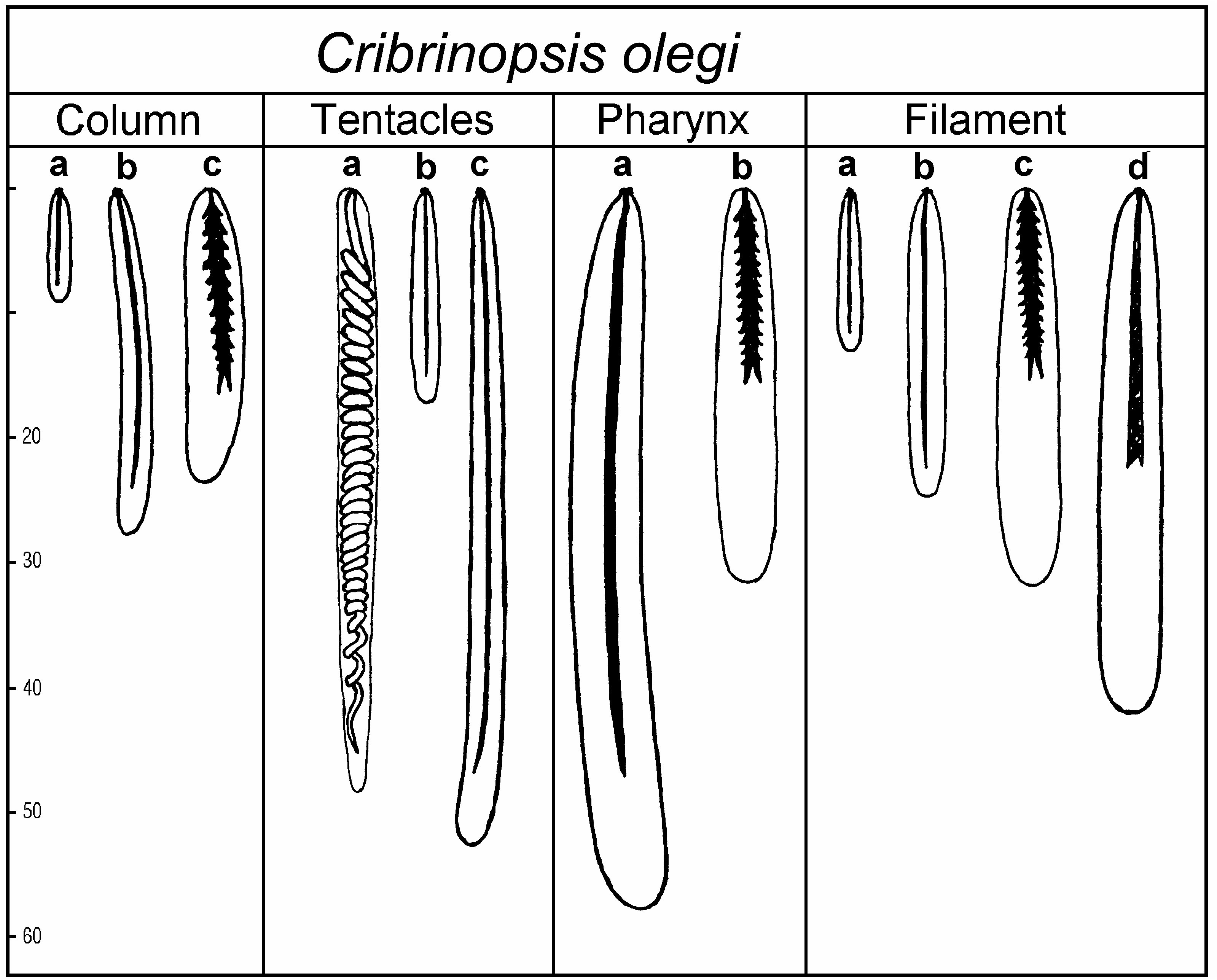
Size and distribution of cnidae (letters in brackets refer to Figure 4 View Figure 4 , all measurements in Mm; N is the proportion of examined specimens that had a particular type of cnida; distribution of all cnidae is confirmed on sections):
Column basitrichs (a): 6–12×1–2 (rare), N513/15 basitrichs (b): 13–31×2–3.5 (common), N515/15 p-mastigophores A (c): 18–29×4–6 (very rare), N58/15
Tentacles spirocysts (a): 26–62×2–3.5 (numerous), N515/15 basitrichs (b): 12–26×2–2.5 (very rare), N57/15 basitrichs (c): 32–60×2.5–3.5 (numerous), N515/15
Actinopharynx basitrichs (a): 35–70×4.5–7 (numerous), N515/15 p-mastigophores A (b): 22–38×5.5–6.5 (very rare), N59/15
Filaments basitrichs (a): 11–20×1.5–2.5 (common), N512/14 basitrichs (b): 21–37×2.5–4 (numerous), N514/14 p-mastigophores A (c): 23–36×4.5–6 (common), N514/14 p-mastigophores B (d): 34–55×4.5–7 (common), N514/14
Habitat
Several specimens of C. olegi sp. nov. were found at 6 m depth, but the majority of the specimens are from 10 to 32 m. The species is always buried in sand, gravel, or broken shell with the pedal disk always attached to buried stones so only the oral disk with the tentacles is visible on the surface. Contracted specimens are buried completely in sand. Symbiotic shrimps [probably Lebbeus grandimanus (Brazhnikov) ] were found on many specimens.
Etymology
The species is named after Oleg Vlasenko, the captain of the boat Chaika, who helped in our field work.
Remarks
Cribrinopsis olegi sp. nov. differs from all known species of Urticina and Cribrinopsis by its short thick tentacles with almost spherical expanded ends. Bunodes crassus Andres, 1884 , wrongly assigned to Cribrinopsis by Schmidt (1972), also has thick tentacles with rounded ends, but their shape is clearly different, and this warm-water Mediterranean species differs from C. olegi sp. nov. in many features, including the hexamerous arrangement of the mesenteries. Cribrinopsis albopunctata sp. nov. resembles the present species in the presence of the white verrucae on the usually red or pink column. Living specimens of C. olegi sp. nov. and C. albopunctata sp. nov. are very different in appearance and are readily distinguished by the shape and colour of the tentacles (the latter species lacking the red stripes characteristic of C. olegi sp. nov.), and by habitat ( C. olegi sp. nov., unlike C. albopunctata sp. nov., always being buried in the sand). Preserved specimens of the two species often are similar, especially when the tentacles are strongly contracted and their real shape obscured. The presence of the spirocysts in the columnar ectoderm in C. albopunctata sp. nov. and their absence in the same tissue in C. olegi sp. nov. is a good distinguishing feature confirmed in cross-sections.
Cribrinopsis similis Carlgren, 1921 View in CoL
( Figure 5 View Figure 5 )
Cribrinopsis similis Carlgren 1921, p 156 View in CoL ; 1928, p 279 (synonymy); Zhiubikas 1977, p 108.
Material examined
Vessel Pogranichnik Pertov , Sea of Okhotsk, trawl 97, 53 ° 41.49N, 154 ° 32.99E – 53 ° 39.79N, 154 ° 33.29E, 200 m, globigerina ooze, 18 July 2001, seven specimens, collector A. V. Chetvergov ( KBPIG 268 /1, 269/2) GoogleMaps .
Description
External appearance. Preserved specimens are large, cylindrical, up to 7 cm diameter and 8 cm high. Size and colour of living specimens are not known, although the specimens examined shortly after fixation showed remnants of red colour in some parts of the column and tentacles. The column is smooth, without columnar verrucae or marginal pseudospherules. The circular base is the same diameter as the column. Remnants of mud on the lower part of the column and an undamaged intact base in all specimens suggests that the specimens live unattached on soft bottoms with the lower part of the column immersed in the mud. The specimens have about 80–90 conical or cylindrical tentacles, wrinkled transversely and with pointed tips.
Internal structure. The marginal endodermal sphincter is strong, circumscribed, with not always pronounced central lamella. Longitudinal muscles of the tentacles are mesogloeal and strong.
The mesenteries are arranged decamerously in three cycles, with a few additional small mesenterial pairs in some exocoels. The number of mesenteries is the same distally and proximally, and small and large specimens have about the same number. Well-developed gonads are present on all cycles. On the younger cycles gonads are developed in the distal part of the mesenteries, while in older cycles the gonads are situated proximally, near the base.
The retractor muscles are strong, restricted, and almost reniform on younger cycles. Well-developed parietobasilar muscles form a long free pennon.
Size and distribution of cnidae (letters in brackets refer to Figure 5 View Figure 5 , all measurements in Mm; N is the proportion of examined specimens that had a particular type of cnida; distribution of all cnidae is confirmed on sections):
Column spirocysts (a): 30–70×3–4.5 (common), N53/3
basitrichs (b): 9–12×1.5–2 (common), N53/3
basitrichs (c): 21–32×2–3 (common), N53/3
p-mastigophores A (d): 30–39×5.5–6 (rare), N53/3
Tentacles spirocysts (a): 27–76×3–5 (numerous), N52/2 basitrichs (b): 14–19×2.5 (very rare), N52/2 basitrichs (c): 42–73×2.5–3.5 (numerous), N52/2
Actinopharynx basitrichs (a): 39–72×3.5–5.5 (numerous), N52/2 p-mastigophores A (b): 35–43×6–7 (common), N52/2
Filaments basitrichs (a): 11–16×2 (common), N52/2 basitrichs (b): 23–41×2–3 (numerous), N52/2 p-mastigophores A (c): 25–39×5–6.5 (common), N52/2 p-mastigophores B (d): 25–52×4–5.5 (common), N52/2
Remarks
The present specimens from the Sea of Okhotsk agree well with the original description of C. similis . This large species is characterised by the relatively small and more or less constant number of the mesenteries and tentacles. As in all species of Cribrinopsis and Urticina , the number of tentacles is the same or slightly smaller than the number of mesenteries in the middle of the column. Carlgren’s (1921, p 159) statement that ‘‘the number of mesenteries seems sometimes to be a little smaller than that of tentacles’’ is an obvious mistake, as appears from his definition of the genus and the statement that the mesenteries grow from the base upward. The colour of the living specimens of this species was recorded only by Zhiubikas (1977), who reported the body wall to be carmine-red, or yellowish with dark green or red patches. The oral disk and lips are pink. The tentacles near their bases are pale red and become dark red toward the tips. Light bands run from the lips to the tentacle bases. The mesenteries are probably always arranged decamerously, the two hexamerous specimens reported by Carlgren (1921) from the Bering Sea and Ikamiut, Greenland, being either abnormal, or, especially in the case of the Bering Sea specimen, belonging to another species.
The species is known from numerous Arctic locations including Greenland, Iceland, Faroe Islands, Spitsbergen, Barents Sea and, probably, the Bering Sea. Carlgren (1921) reported also a specimen from the Korea Strait, a location too distant from the known range. The record may be based on incorrect identification.


No known copyright restrictions apply. See Agosti, D., Egloff, W., 2009. Taxonomic information exchange and copyright: the Plazi approach. BMC Research Notes 2009, 2:53 for further explanation.
|
Kingdom |
|
|
Phylum |
|
|
Class |
|
|
Order |
|
|
Family |
|
|
Genus |
Cribrinopsis olegi
| Sanamyan, N. P. & Sanamyan, K. E. 2006 |
Cribrinopsis similis
| Zhiubikas II 1977: 108 |
| Carlgren O 1921: 156 |