
Lebertia (Pilolebertia) longiseta Bader, 1955
|
publication ID |
https://doi.org/10.11646/zootaxa.3619.5.6 |
|
publication LSID |
lsid:zoobank.org:pub:0AB793D1-D40D-47FF-9835-C83BBD94CE27 |
|
DOI |
https://doi.org/10.5281/zenodo.6160821 |
|
persistent identifier |
https://treatment.plazi.org/id/CD0C7A0F-243C-FFF4-B79E-3FC2FDFED5C8 |
|
treatment provided by |
Plazi |
|
scientific name |
Lebertia (Pilolebertia) longiseta Bader, 1955 |
| status |
|
Lebertia (Pilolebertia) longiseta Bader, 1955
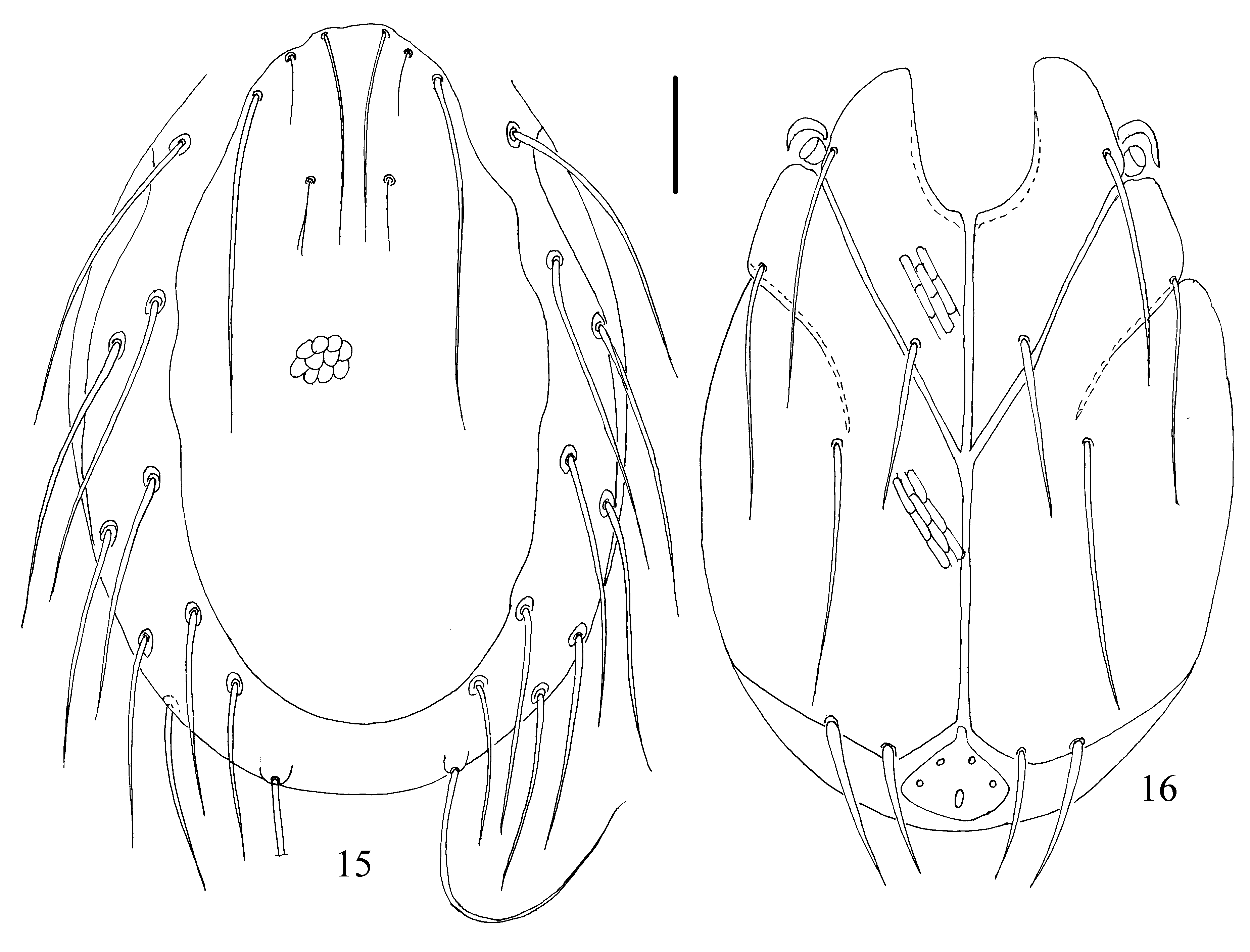
( Figs 15 View FIGURES 15 – 16 –26)
Material examined. Larvae (n = 16) were reared from four females collected in a cold brook near the village Postyltsevo, Nekouz District, Yaroslavl Province: two females were collected on 16 June 2002, one female on 2 July 2002 and one female on 8 August 2005. The duration of the embryonic period was 11–16 days.
Diagnosis. Dorsal plate elongated (L/W ratio 1.75–2.15), setae Oe, Hi, He, Sci and Sce equal in length; excretory pore plate wider than long (L/W ratio 0.75–0.95); Ae longer and thicker than Ai; chelicerae bases completely fused to each other; II–Leg-4 with a single thickened seta, III–Leg-4 with two greatly thickened setae.
Description. Idiosoma flat, dorsal plate in unengorged larvae covering large part of dorsum ( Fig. 15 View FIGURES 15 – 16 ), with slightly convex lateral margins. Anterior margin of the dorsal plate slightly convex, posterior margin rounded, with short and punctuate scale-like patterns and bearing four pairs of setae: two pairs of simple setae ( Fch, Vi) and two pairs of trichobothria ( Fp, Oi). Simple setae long and thick, anterior setae ( Fch) shorter and thinner than posterior setae ( Vi). Both pairs of trichobothria short and thin, equal in size and branched. Seven pairs of long setae ( Oe, Hi, He, Sci, Sce, Li, Le) situated on soft wrinkled membrane: Oe longest, and Li and Le shortest.
Coxal plates large, plates II–III on each side completely fused to each other, suture line between them developed only in their lateral parts ( Fig. 16 View FIGURES 15 – 16 ). Anterior coxal plates completely separated from coxal plates II and III. Setae C1 slightly shorter than other coxal setae (C2–C4) and approximately equal in length. Posterior margin of coxal plate III has two unequal setae, Pe slightly longer than Pi. Setae Ci very long, located on small tubercles and much longer than Si and Se. All coxal plates with reticulated pattern consisting of elongated cells. Urstigma lateral to the border between coxal plates I and II, covered by flaps.
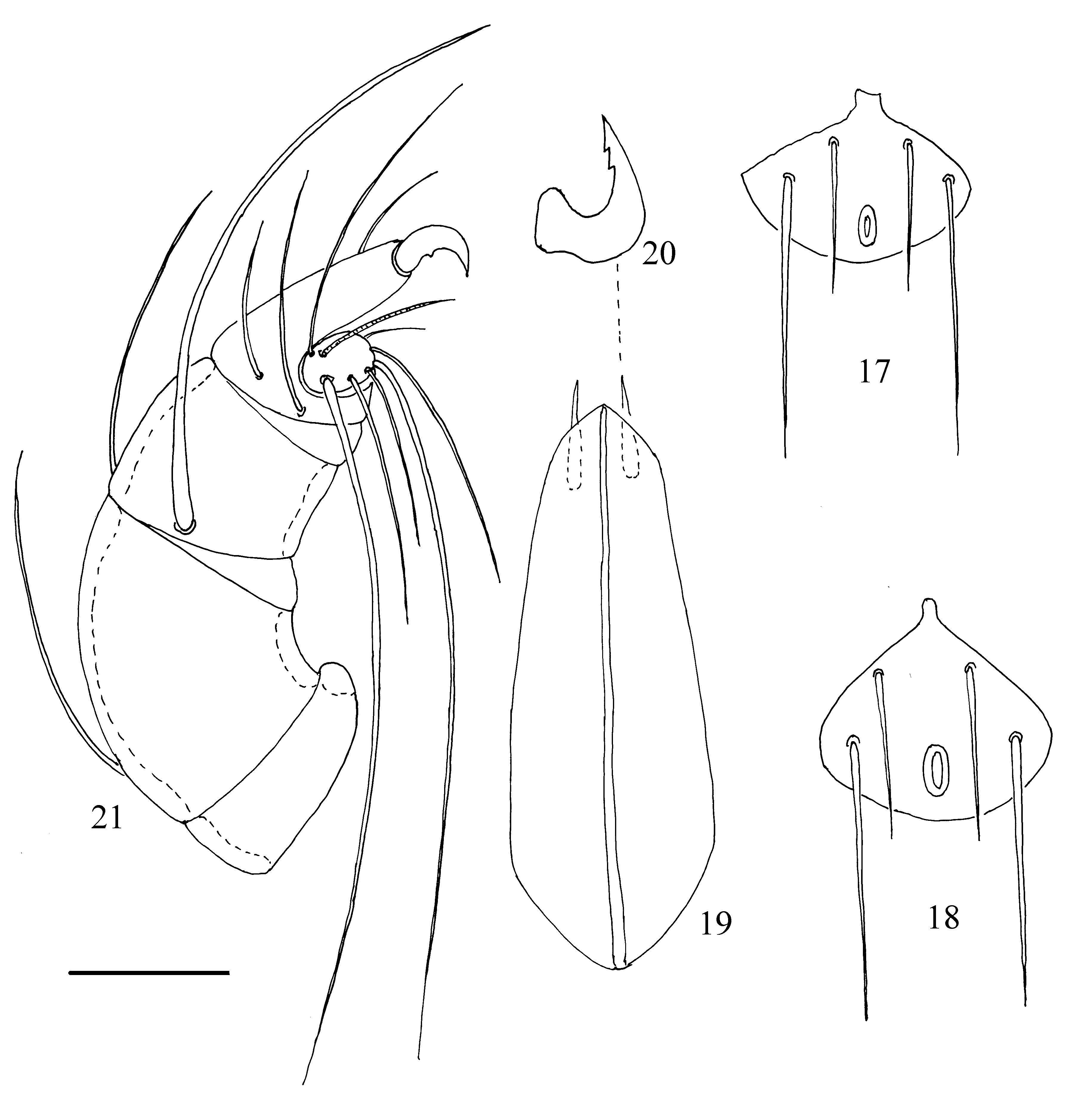
Excretory pore plate wider than long, with more or less developed anteromedial projection ( Fig. 17–18 View FIGURES 17 – 21 ), rounded posteriorly; setae Ae much longer than Ai, located laterally in the posterior half of plate. Excretory pore placed posteromedial to Ae near posterior margin of plate; distance between setae Ae–Ae almost two times longer than distance between Ai–Ai.
Chelicerae bases completely fused to each other ( Fig. 19 View FIGURES 17 – 21 ), its dorsal margins convex, ventral margins concave; cheliceral stylets relatively small, crescent–shaped, with two small subapical teeth ( Fig. 20 View FIGURES 17 – 21 ).
Pedipalps moderately developed ( Fig. 21 View FIGURES 17 – 21 ): P–1 short and glabrous; P–2 large with convex dorsal margin and a single dorsoproximal seta; P–3 with very long, thick lateroproximal seta and a relatively short, fine dorsoproximal one; P–4 with two rather long unequal lateroproximal setae, one relatively short dorsodistal seta and a massive dorsodistal claw; P–5 small, with single solenidion and seven unequal simple setae, two of them very long.
FIGURES 22–26. Lebertia longiseta Bader, 1955 , larva: 22, leg I; 23, leg II; 24, leg III; 25, claws of leg III, 26—claw of leg I. Scale bars: 22–24 = 50 μm, 25–26 = 20 μm.
Legs 5–segmented, basifemur and telofemur of all legs fused to each other, with indistinct suture line. Shape and arrangement of setae on legs segments as shown in Figs 22–24. Total number of leg setae, excluding eupathidia, is as follows (specialized setae given in parentheses): I–Leg-1–5: 1, 7, 5 (s), 10 (2s), 15 (s, ac); II–Leg- 1–5: 1, 7, 5 (s), 11 (2s), 15 (s, ac); III–Leg-1–5: 1, 6, 5 (s), 10 (s), 12 (11); proximal ventral setae on tarsus III sometimes absent. Number of thickened setae from trochanter to tarsus: I–Leg: 0, 1, 1, 1, 0; II–Leg: 0, 2, 2, 1, 0; III–Leg: 0, 2, 2, 2, 0. I–Leg-1 with relatively short seta, II–Leg-1 and III–Leg-1 each with long seta. Solenidia on genu of legs I–II and tibia III short and approximately equal in length; solenidion on genu III shorter than other legs solenidia. Both solenidia on I-leg-4 located in distal half of segments; proximal solenidion on II-leg-4 and solenidion on III-leg-4 situated proximally to middle of these segments. Acanthoid setae are present only on tarsus I and II, each bent two times. Claws of legs III (Fig. 25) larger than claws of legs I and II (Fig. 26). Lateral claws longer and thinner than heavy empodial claw: all claws are sickle shaped and provided with a lateral spur on each side.
Measurements, n=10. L of dorsal plate 255–275, W 125–155; L of setae Fch 64–70, L of setae Fp and Oi 19–23, L of setae Vi 140–150, L of setae Oe, Hi, He 110–125; L of setae, Sci and Sce 110–125; L of setae Li, Le 95–115; L of setae Si 80–85, L of setae Se 90–105, L of setae Ci 140–150, L of setae Pi 60–65, L of setae Pe 70–75, L of setae C1 65–75, L of setae C2–C4 100–115; L of medial margins of coxae I 80 –100, L of medial margins of coxae II–III 110–125; L of excretory pore plate 20–30, W 25–35; L of capitulum 89–96; L of basal segments of chelicerae 83–90, L of cheliceral stylet 12–16; L of pedipalpal segments (P–1–5): 9–13, 35–42, 29–38, 25–30, 12–16; L of legs segments: I–Leg-1–5: 28–35, 49–52, 50–55, 65–70, 64–80; II–Leg-1–5: 40–48, 48–52, 50–55, 80–83, 89–100; III–Leg-1–5: 45–55, 55–60, 57–65, 89–93, 95–115.
FIGURES 27–29. Lebertia inaequalis Koch, 1837 , larva: 27, ventral view; 28–29, excretory pore plate. Scale bars: 27 = 50 μm, 28–29 = 20 μm.
Remarks. The larva of L. longiseta is similar to the larva of L. inaequalis (Koch, 1837) , the latter described by Wainstein (1980) and Martin (2000), but the description were very different, most probably refer two different species. A comparison is made with the Yaroslavl larvae of this species, which is similar to Wainstein’s description (Figs 27–29). The idiosoma in unengorged of larva L. inaequalis has a distinct posteromedian protrusion (Fig. 27), the excretory pore plate has short setae, which are thick and nearly equal in length (Fig. 28–29). In contrast, the idiosoma in unengorged of larval L. longiseta are without posteromedian protrusion ( Fig. 16 View FIGURES 15 – 16 ), the excretory pore plate has relatively long, thin unequal setae, Ae much longer than Ai ( Figs 17–18 View FIGURES 17 – 21 ).
The morphology of the larva of L. longiseta is very similar to Martin’s larva L. inaequalis but these species are not identical. In Martin’s larva L. inaequalis III-Leg-5 with 10 setae + 1 solenidion, all segments of III-Leg without thickened setae (Martin 2000). In contrast, in the larva of L. longiseta III-Leg-5 bearing only simple setae, III-Leg- 2–4 with two thickened setae each. I believe, that is possible Martin’s larva L. inaequalis belongs to any other species of the genus Lebertia .
This species is found in Russia for the first time.
Distribution. Europe: Northern Alps and Pre-Alps (K. O. Viets 1978, Gerecke 2009); Russia, Yaroslavl Province.
Habitat. Standing and running waters.
No known copyright restrictions apply. See Agosti, D., Egloff, W., 2009. Taxonomic information exchange and copyright: the Plazi approach. BMC Research Notes 2009, 2:53 for further explanation.
|
Kingdom |
|
|
Phylum |
|
|
Class |
|
|
Order |
|
|
Family |
|
|
Genus |