
Lebertia (Mixolebertia) dubia (Thor, 1899)
|
publication ID |
https://doi.org/ 10.11646/zootaxa.3619.5.6 |
|
publication LSID |
lsid:zoobank.org:pub:0AB793D1-D40D-47FF-9835-C83BBD94CE27 |
|
DOI |
https://doi.org/10.5281/zenodo.6160819 |
|
persistent identifier |
https://treatment.plazi.org/id/CD0C7A0F-2439-FFF8-B79E-3F43FD48D683 |
|
treatment provided by |
Plazi |
|
scientific name |
Lebertia (Mixolebertia) dubia (Thor, 1899) |
| status |
|
Lebertia (Mixolebertia) dubia (Thor, 1899)
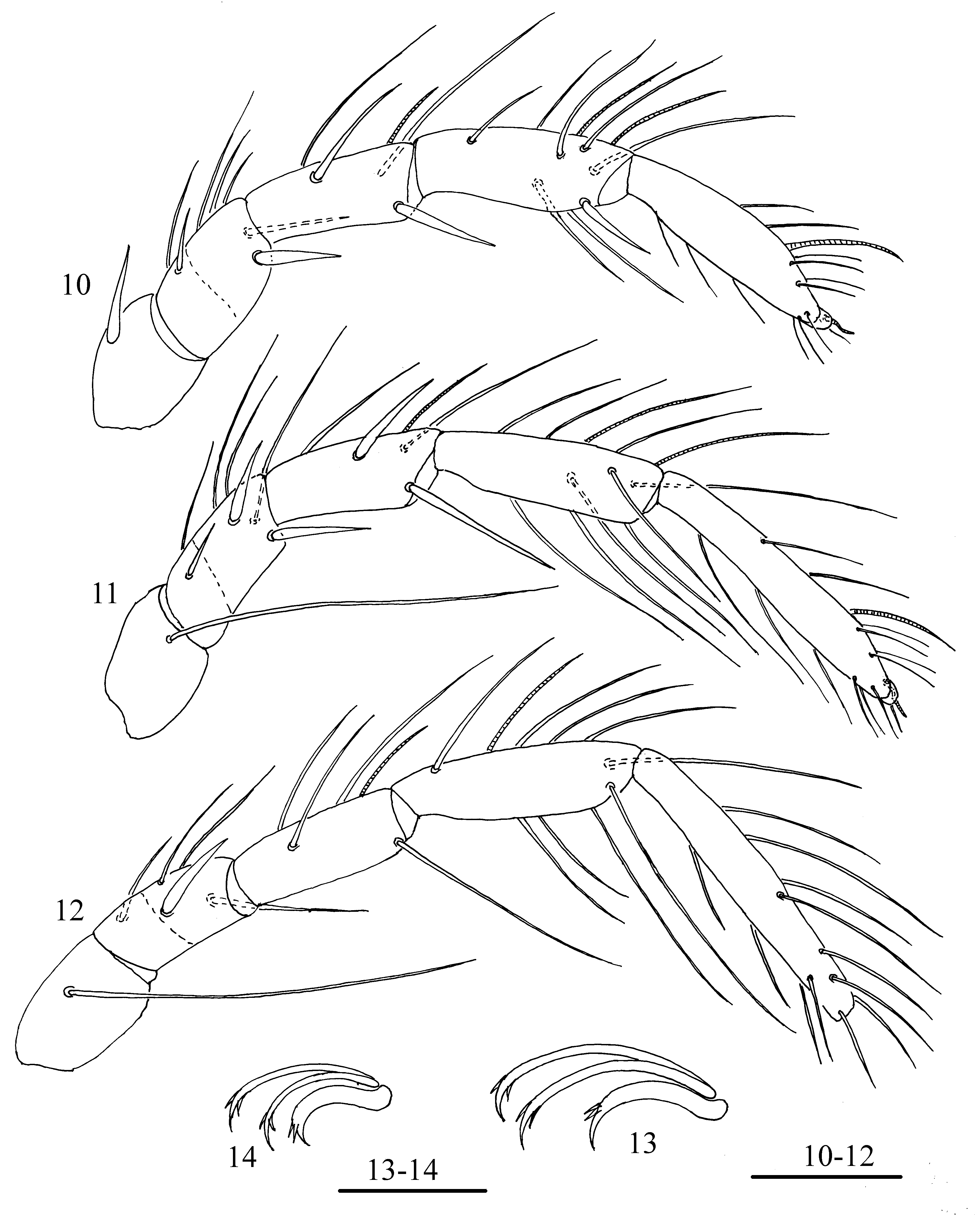
( Figs 1–14 View FIGURES 1 – 2 View FIGURES 10 – 14 )
Material examined. Larvae (n = 15) were reared from three females collected in a cold brook near the village Postyltsevo, Nekouz District, Yaroslavl Province: two females on 19 July 2004, and one female on 22 June 2005. The duration of the embryonic period was 20–90 days.
Diagnosis. Dorsal plate elongated (L/W ratio 1.3–1.4), setae Oe longer than Hi, He, Sci and Sce; trichobothria Fp and Oi branched; chelicerae bases partly fused to each other; excretory pore plate longer than wide, setae Ae longer than Ai, II–Leg-4 and III–Leg-4 without greatly thickened setae.
Description. Idiosoma flat, dorsal plate elongate in unengorged larvae covering large part of dorsum ( Fig. 1 View FIGURES 1 – 2 ), with slightly convex lateral margins. Its anterior margin slightly convex, posterior margin rounded, with short and punctuate scale-like patterns and bearing four pairs of setae: two pairs of simple setae (Fch, Vi) and two pairs of trichobothria (Fp, Oi). Simple setae long and thick, anterior setae (Fch) shorter and thinner than posterior setae (Vi). Both pairs of trichobothria short and thin, equal in sizes and branched. Seven pairs of long setae (Oe, Hi, He, Sci, Sce, Li, Le) situated on soft wrinkled membrane: Oe longest, Li and Le shorter than Hi, He, Sci and Sce.
Coxal plates large, plates II–III on each side completely fused to each other, suture line between them developed only in their lateral parts ( Fig. 2 View FIGURES 1 – 2 ). Anterior coxal plates completely separated from coxal plates II and III. Setae C1 slightly shorter than C2 and C3, which are approximately equal in length; C4 a little longer than C2 and C3. Posterior margin of coxal plate III with two unequal setae, Pe slightly longer than Pi. Setae Ci very long, located on small tubercles and much longer than Si and Se. All coxal plates have a reticulated pattern consisting of elongated cells. Urstigma lateral to border between coxal plates I and II, covered by flaps.
Excretory pore plate (Figs 3–6) longer than wide (L/W ratio 1.15–1.30), oval or rhomboidal with more or less developed anteromedian projection, rounded posteriorly; setae Ae longer and thicker than Ai, located laterally in posterior half of plate; excretory pore placed posteromedial to Ae and near posterior edge of plate; distance between setae Ae–Ae a little longer than distance between Ai–Ai.
Capitulum (Fig. 7) elongated (L/W ratio 1.20–1.25) with a wide and long base and relatively short, narrow rostrum; mouth cavity oval, anterior setae longer than posterior ones.
Chelicerae bases partly fused to each other, its dorsal margins convex, ventral margins concave; cheliceral stylets relatively small, crescent–shaped, each with two small subapical teeth (Fig. 8).
Pedipalps moderately developed (Fig. 9): P–1 short and glabrous; P–2 large with a convex dorsal margin and a single dorsal setae near the middle of segment; P–3 with a very long, thick lateroproximal seta and a relatively short, fine dorsoproximal seta; P–4 with two rather long unequal lateroproximal setae, one short dorsodistal seta and a massive dorsodistal claw; P–5 small, with single solenidion and seven unequal simple setae, two of them very long.
Legs 5–segmented, basifemur and telofemur of all legs fused to each other, with indistinct suture line. Shape and arrangement of setae on legs segments as shown in Figs 10–12 View FIGURES 10 – 14 . Total number of leg setae, excluding eupathidia, is as follows (specialized setae given in parentheses): I–Leg-1–5: 1, 7, 5 (s), 10 (2s), 15 (s, ac); II–Leg- 1–5: 1, 7, 5 (s), 11 (2s), 15 (s, ac); III–Leg-1–5: 1, 6, 5 (s), 10 (s), 11–12; proximal ventral setae on III–Leg-5 sometimes absent. Number of thickened setae from trochanter to tarsus: I–Leg: 0, 1, 1, 1, 0; II–Leg: 0, 2, 2, 0, 0; III–Leg: 0, 1, 0, 0, 0. I–Leg-1 with relatively short seta, II–Leg-1 and III–Leg-1 each with long seta. Solenidion on genu of legs I–III and tarsi I–II short; solenidia on tibia I–II relatively long and located in distal half of these segments; solenidion on tibia III situated proximally to the middle of segment. Acanthoid setae are present only on tarsus I and II, and each bent two times. Claws of legs III ( Fig.13 View FIGURES 10 – 14 ) larger than claws of legs I and II ( Fig. 14 View FIGURES 10 – 14 ). Lateral claws longer and thinner than heavy empodial claw, all claws are sickle shaped and provided with a lateral spur on each side.
FIGURES 3–9. Lebertia dubia Thor, 1899 , larva: 3–6, excretory pore plate; 7, capitulum, ventral view; 8, chelicera, lateral view; 9, pedipalp, lateral view. Scale bars: 3–6 = 25 μm, 7–9 = 20 μm.
Measurements, n=10. L of dorsal plate 250–275, W 145–190; L of setae Fch 60–70, L of setae Fp and Oi 42–48, L of setae Vi 150–160, L of setae Oe 145–160; L of setae Hi, He, Sci and Sce 130–145; L of setae Li 110–120, L of setae Le 95–115; L of setae Si 95–105, L of setae Se 75–90, L of setae Ci 145–155, L of setae Pi 48–55, L of setae Pe 55–70, L of setae C1 95–105, L of setae C2 and C 3 110–125, L of setae C4 130–145; L of medial margin of coxae I 85 –95, L of medial margins of coxae II–III 110–130; L of excretory pore plate 45–55, W 29–42; L of capitulum 76–92; L of basal segments of chelicerae 83–90, L of cheliceral stylet 16–19; L of pedipalpal segments (P–1–5): 6–9, 28–42, 28–33, 16–21, 12–15; L of legs segments: I–Leg-1–5: 38–45, 40–55, 54–58, 65–75, 80–90; II–Leg-1–5: 40–48, 50–65, 55–60, 73–85, 95–110; III–Leg-1–5: 48–55, 53–65, 55–65, 75–85, 105–122.
Remarks. The larva of the present species is similar to larva of L. oudemansi Koenike, 1898 (= syn. L. densa Koenike, 1902 after Gerecke 2009). The anterior margin of the dorsal plate in the larva of L. oudemansi is straight, the setae Ai and Ae are equal in length and the capitulum has a narrow base (L/W ratio 1.7–1.9) (Tuzovskij 2006). In contrast, the anterior margin of the dorsal plate in the larva of L. dubia is convex, setae Ai is shorter than Ae and the capitulum has a wide base (L/W ratio 1.2–1.3).

No known copyright restrictions apply. See Agosti, D., Egloff, W., 2009. Taxonomic information exchange and copyright: the Plazi approach. BMC Research Notes 2009, 2:53 for further explanation.