
Dialeurotrachelus cambodiensis Takahashi, 1942
|
publication ID |
https://doi.org/10.11646/zootaxa.4695.6.7 |
|
publication LSID |
lsid:zoobank.org:pub:027E90CF-944B-468F-B73B-49B317BBE733 |
|
DOI |
https://doi.org/10.5281/zenodo.5942879 |
|
persistent identifier |
https://treatment.plazi.org/id/CB4987B2-EE40-EE55-FF3F-AB7EC0EAFC07 |
|
treatment provided by |
Plazi |
|
scientific name |
Dialeurotrachelus cambodiensis Takahashi |
| status |
|
Dialeurotrachelus cambodiensis Takahashi View in CoL
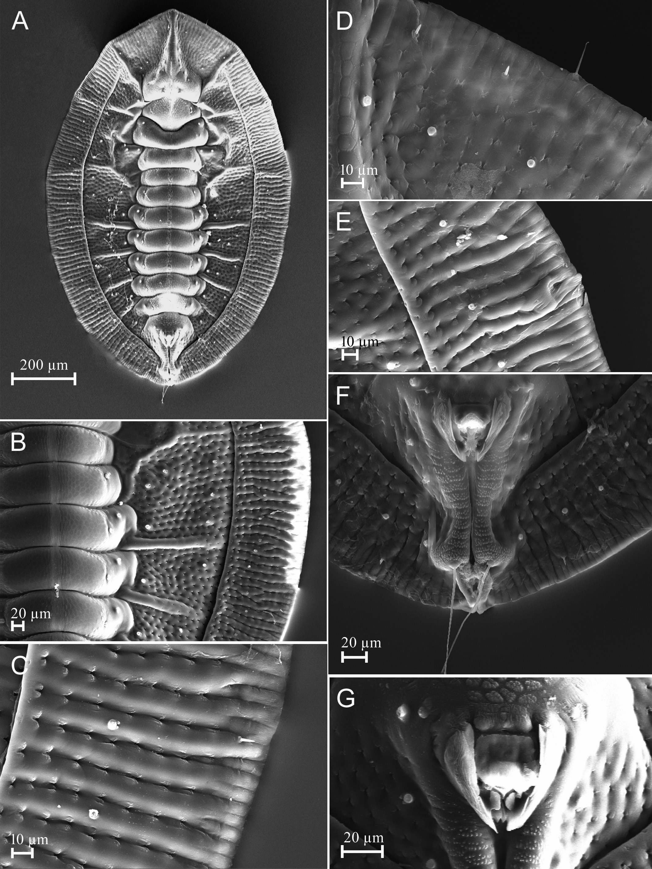
( Figs 1–6 View FIGURE 1 View FIGURE 2 View FIGURE 3 View FIGURE 4 View FIGURE 5 View FIGURE 6 )
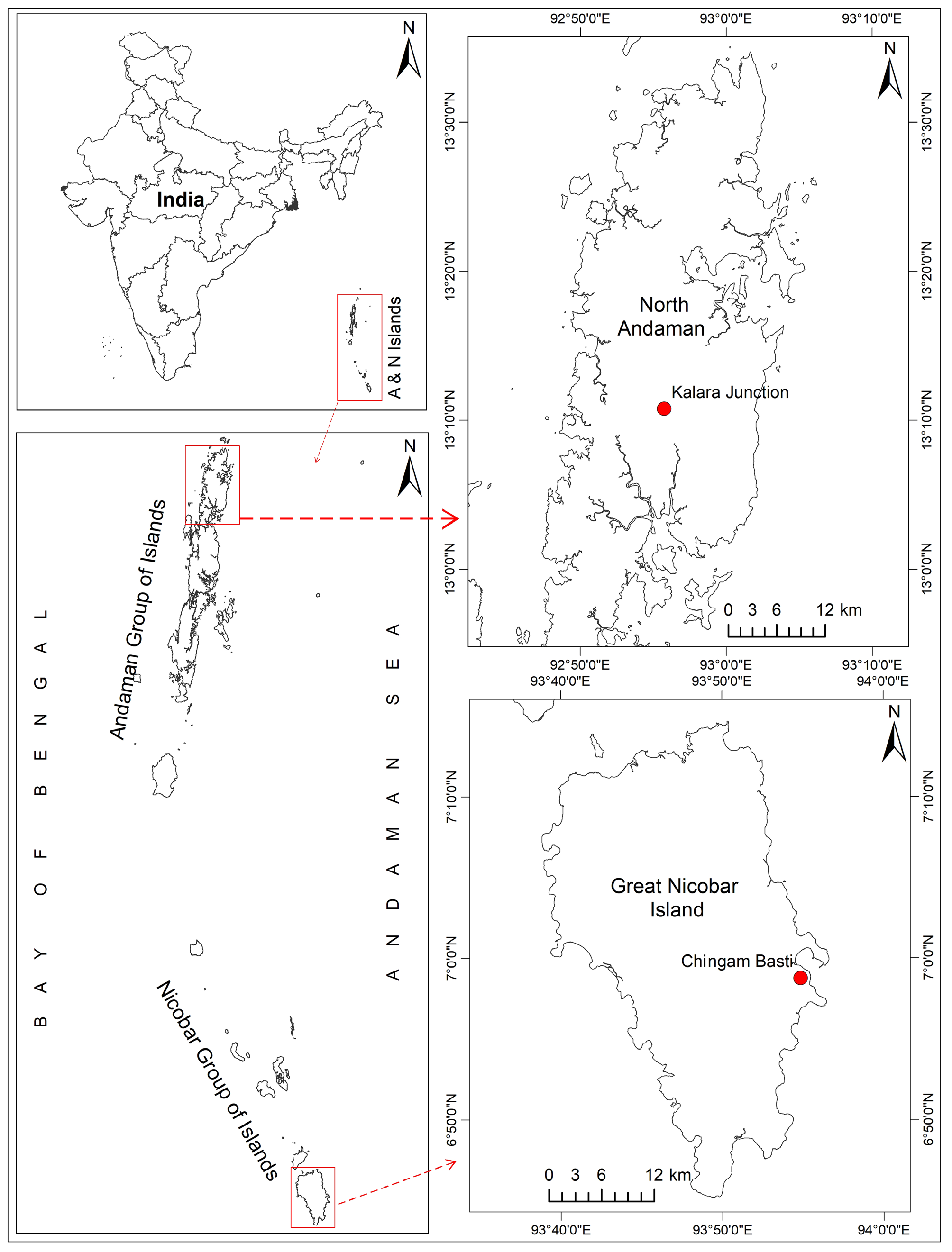
Dialeurotrachelus cambodiensis Takahashi, 1942: 102 View in CoL . Three syntypes, Cambodia, Angkor, on an unidentified tree, 23.iv.1940, R. Takahashi, apparently lost or destroyed. A neotype is designated here from India, Andaman Island, North Andaman, Diglipur, Kalra Junction, 1 puparium on slide, on Diospyros kurzii View in CoL , 17.x.2017, A. K. Dubey (ZSI in Kolkata).
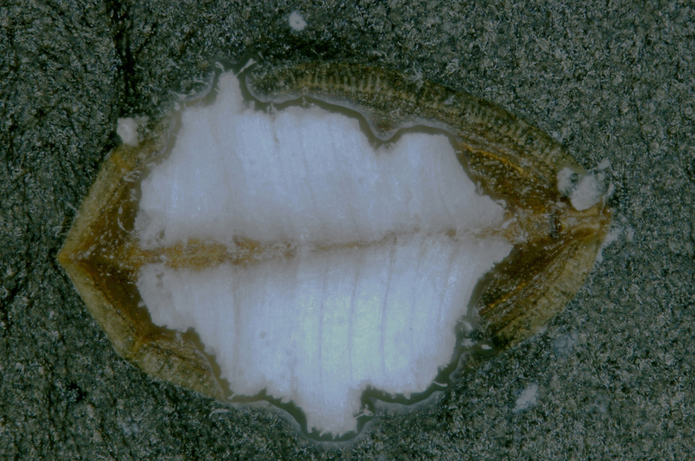
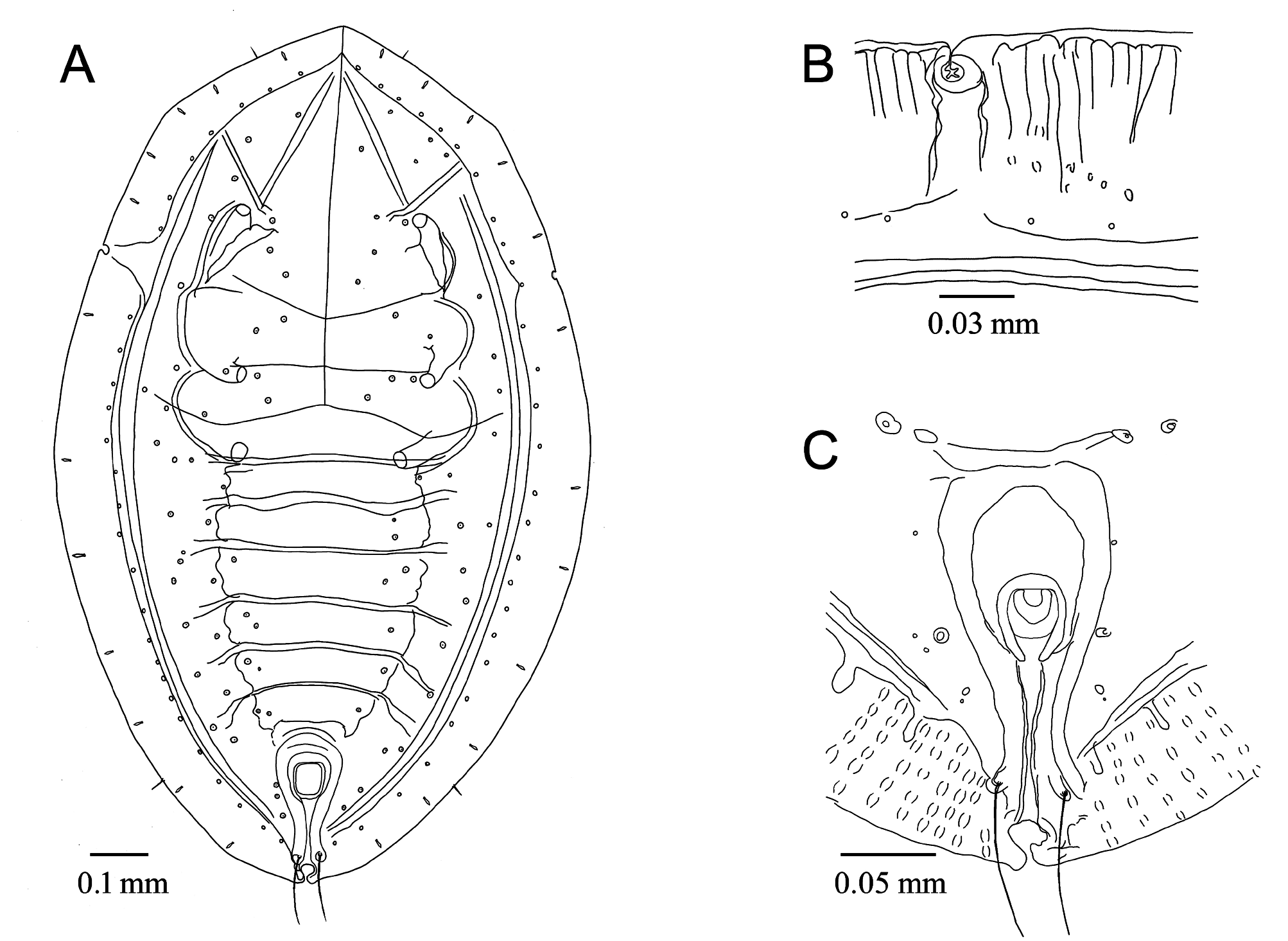
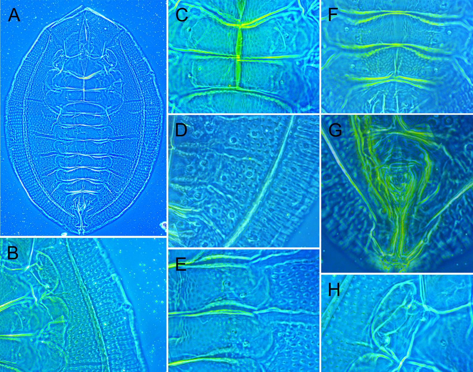
Puparium. In life, pale; median area convex; with secretion of white wax arising from submedian area in two longitudinal rows and extending to margin ( Fig. 2 View FIGURE 2 ); oblong ( Figs 3A View FIGURE 3 , 4A View FIGURE 4 , 5A View FIGURE 5 ); dimorphic, female puparia larger than male; female 1110–1220 microns long, 790–830 microns wide; male 910–1140 microns long, 550–590 microns wide; broadest at metathorax and gradually narrowing towards anterior and posterior ends.
Margin. Smooth, thoracic tracheal pores deeply invaginated from margin ( Figs 3B View FIGURE 3 , 5E View FIGURE 5 ), anterior ends overlapping; caudal tracheal pore also deeply inset from margin but apical ends widely open.
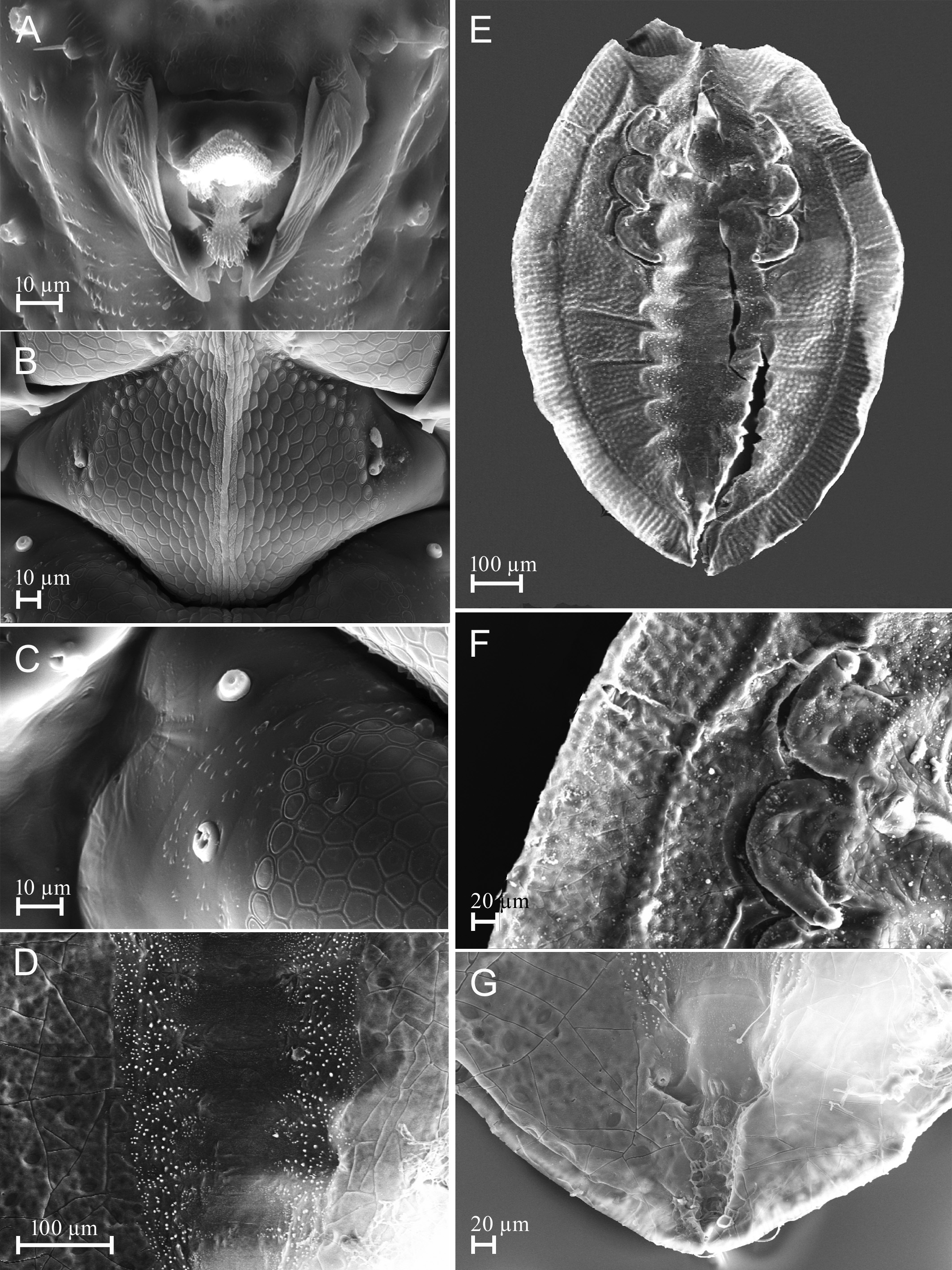
Dorsum. Cephalic median area raised, median keel present. Longitudinal moulting suture reaching margin, and transverse moulting suture reaching subdorsum near submarginal furrow. Dorsum with six pairs of rhachises, one pair located on cephalothorax and five pairs on abdomen, of these each abdominal rhachis extends along the intersegmental sutures of abdominal segments III/IV, IV/V, V/VI, VI/VII and VII/VIII. The cephalic pair of rhachises reach the margin whereas abdominal rhachises reach the submarginal furrow. Submargin differentiated from the dorsal disc by a pair of submarginal furrows except at anterior and posterior margins; submarginal furrows reaching anterior to prothorax near cephalic rhachis, and posteriorly to abdominal segment VIII, not intersecting caudal tracheal furrow. Submarginal and subdorsal area with many rows of diverging plates arranged in linear pattern ( Fig. 5B View FIGURE 5 ). Cephalic and abdominal submedian area rhachisform. The submedian area of cephalothorax and abdominal segments marked with polygonal sculpturing ( Fig. 6B View FIGURE 6 ). Intersegmental sutures prominent, reaching subdorsal area, intersegmental sutures arising from segments V–VIII reaching the submarginal furrow. The median length of cephalothorax was smaller than the abdomen; median length of cephalothorax and abdomen in female measured 480–525 microns and 675–700 microns long, respectively, and 425–440 microns and 90–520 microns long, respectively, in male. Median length of meso-, and metathorax measured 57–60 microns and 62–65 microns long, respectively, in female, and 47–50 microns and 55–60 microns long, respectively, in male. The median length of abdominal segments I–VIII measured in female as: A1: 62–70, A2: 57–70, A3: 70–75, A4: 72–75, A5: 62–72, A6: 60–65, A7: 57–62, A8: 55–57 microns long; and A1: 45–57; A2: 50–57, A3: 50–60, A4: 50–57, A5: 47–55, A6: 45–47, A7: 37–45, A8: 37–43 microns long, respectively, in male. Abdominal segments III and IV were longest and subequal in both male and female puparia (III=IV) whereas other segments (I, II, V and VI) varied little, the segment VI was the shortest or equal to segment VIII. Thoracic tracheal furrows absent. Caudal tracheal furrow prominent; caudal ridges made up of many transverse rows of fine granules, reaching half the length of submargin and bearing caudal setae at the end ( Figs 4G View FIGURE 4 , 5F View FIGURE 5 ). Geminate pores present, prominent, each occupying an area of 8 microns in length, arranged in five rows, one row on submargin along the submarginal furrow, two rows on subdorsal area, and two closely placed rows on cephalic and abdominal longitudinal rhachises, posterior to submedian pockets. Geminate pores appear raised from dorsal surface in SEM images (Fig. 23). The distance between vasiform orifice and puparial caudal margin measured 115–120 microns long in female and 85–95 microns long in male.
Vasiform orifice. Quadrangular; slightly elevated posteriorly; located anterior to caudal margin by nearly 2½ times of its own length ( Fig. 3C View FIGURE 3 ); broadest at anterior half, posteriorly narrow, divided at hind end, forming bilobed invagination; longer than wide; 45–62 microns long, 40–57 microns wide in female; 42 microns long, 40–42 microns wide in male; operculum subrectangular, much shorter, covering half the length of vasiform orifice; wider than long; 27–35 microns long, 33 microns wide in female, 25–32 microns long, 25–35 microns wide in male. Lingula apex exposed, setose; not divided apically.
Venter. Oblong; submedian area below dorsal abdominal rhachises and intersegmental sutures with prominent stipples ( Fig. 6D View FIGURE 6 ); submarginal area with imprint of dorsal submarginal groove ( Fig. 6E View FIGURE 6 ); a pair of ventral eighth abdominal setae present ( Fig. 6G View FIGURE 6 ), 27–35 microns long, 27–37 microns apart in female and 45 microns apart in male. Antennae extending through inside the prothoracic legs and reaching near base of them ( Fig. 4H View FIGURE 4 ), keel overlapping margins of the legs, 100 microns long (including keel, 7 microns long). Microsetae in middle of prothoracic legs approximately 2 microns long, and 5 microns long in middle of meso-, and metathoracic legs. Thoracic tracheal folds without stipples. Caudal tracheal fold with a few fine stipples only on subdorsal area. Spiracles and adhesive sacs visible.
Chaetotaxy. Submargin with a row of small, lanceolate setae, 5–7 microns long; located 5 pairs anterior to thoracic tracheal pores and 7 pairs posterior to it. Anterior and posterior marginal setae 7–13 microns and 13–18 microns long, respectively. Eighth abdominal setae 7–13 microns long. Caudal setae 95–113 microns long in female, 87 microns long in male; 15–30 microns apart.
Host plant: Unidentified tree ( Takahashi, 1942); Diospyros kurzii Hiern (Ebenaceae) (new record).
Distribution: Cambodia ( Takahashi, 1942); India: Andaman and Nicobar Islands (new record).
Material examined. India: Andaman and Nicobar Islands, North Andaman, Diglipur, Kalra Junction , 8 puparia on 5 slides, on Diospyros kurzii , 17.x.2017, A. K. Dubey, of these one puparium on a slide is selected as the neotype (deposited in ZSI, Kolkata) ; Nicobar Islands, Chingam Basti , 11 puparia on 6 slides, on Diospyros kurzii , 25.xii.2017, A. K. Dubey ( Indian Agricultural Research Institute, New Delhi, and Forest Research Institute, Dehradun). There are also a few puparia in 95% alcohol ( A. K. Dubey private collection) .
Remarks. Puparia of the new species were found feeding only on the lower surface of leaves and some of them were parasitized. Population of this species remains very low throughout year and puparia are barely visible on leaf surface when its wax deposits are removed by the wind or rain. Female puparia are larger in size than those of the male, the caudal furrow and vasiform orifice longer, and wider apart of ventral eighth abdominal setae. Takahashi (1942) stated that the new genus differs from its closely related genus in having a divided lingula; however SEM studies confirmed that the lingula is not divided ( Figs 5F View FIGURE 5 , 6A View FIGURE 6 ), but the posterior area of vasiform orifice is open and forms a bilobed structure, anteriorly supported by two adjoining plates ( Fig. 5G View FIGURE 5 ) where the tip of the lingula overlaps ( Fig. 5G View FIGURE 5 ); all of these characteristics together give the apical end of the lingula a divided appearance in slide mounted puparia which Takahashi noted in the original description of the genus. However, the genus Dialeurotrachelus remains valid and differs from Rhachisphora Quaintance and Baker in that the submargin is demarcated from the dorsal disc by a submarginal furrow.
No known copyright restrictions apply. See Agosti, D., Egloff, W., 2009. Taxonomic information exchange and copyright: the Plazi approach. BMC Research Notes 2009, 2:53 for further explanation.
|
Kingdom |
|
|
Phylum |
|
|
Class |
|
|
Order |
|
|
Family |
|
|
Genus |
Dialeurotrachelus cambodiensis Takahashi
| Id, Anil Kumar Dubey Orcid 2019 |
Dialeurotrachelus cambodiensis
| Takahashi, R. 1942: 102 |