
Mimosicerya williamsi Foldi
|
publication ID |
https://doi.org/10.5281/zenodo.191685 |
|
DOI |
https://doi.org/10.5281/zenodo.6217009 |
|
persistent identifier |
https://treatment.plazi.org/id/C8140C4D-FFA2-FF8C-FF41-FBB2FCA52B41 |
|
treatment provided by |
Plazi |
|
scientific name |
Mimosicerya williamsi Foldi |
| status |
sp. nov. |
Mimosicerya williamsi Foldi n. sp.
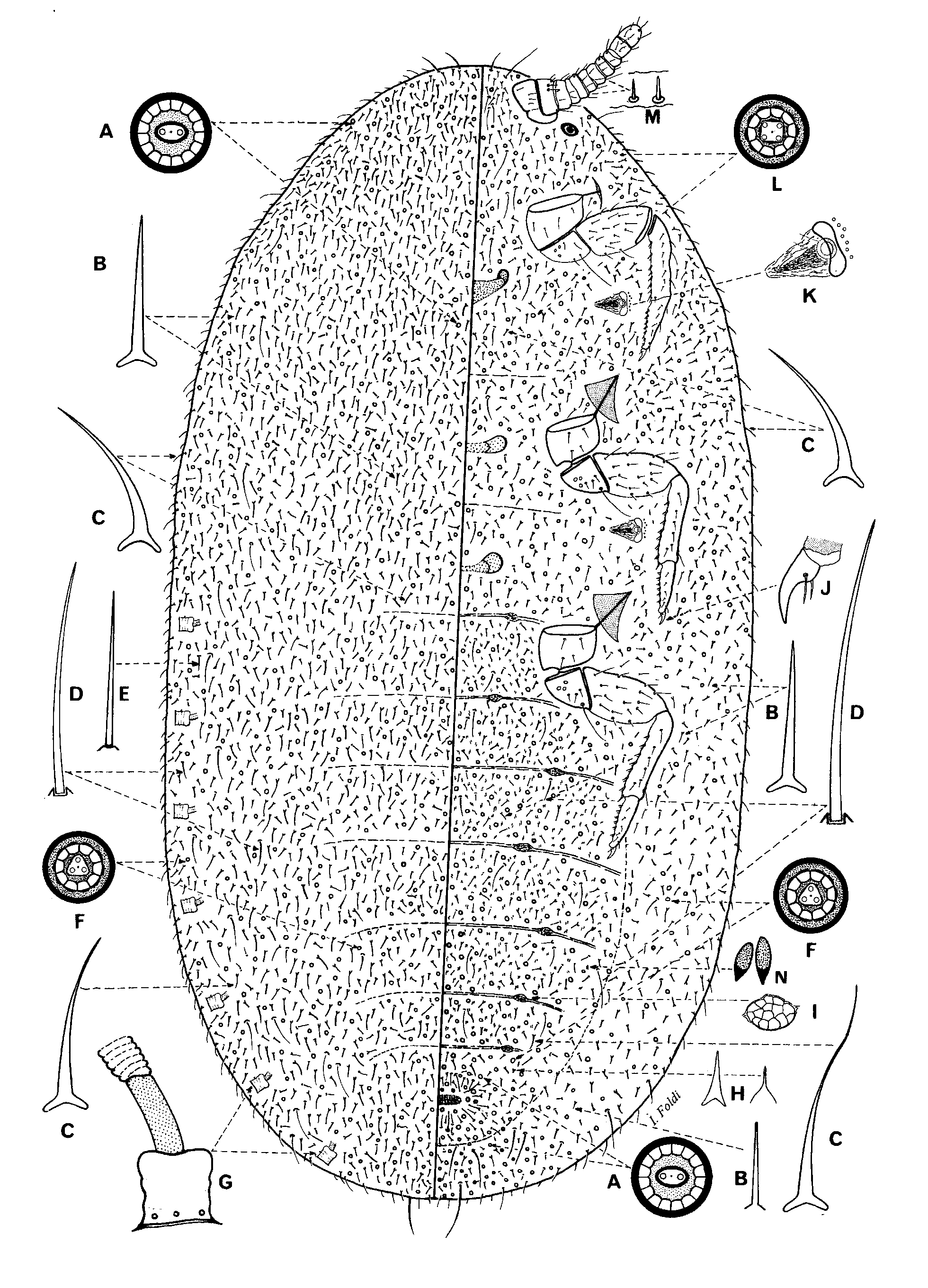
ADULT FEMALE ( Fig. 8 View FIGURE 8 )
Description based on 3 specimens in fair condition.
Material examined. Holotype adult female. VENEZUELA: around Mérida, state Mérida, 2500 m, on the young stem of Inga sp., on footpath in the forest, 24.x.1984, I. Foldi coll., MNHN. Paratypes. 2 adult females on 2 slides, 7 preadult females on 7 slides, data as for holotype, MNHN. 1 paratype of adult and 2 paratype preadult females in USNM.
Mounted material. Female broadly oval, 11–14 mm long, 9.5–11 mm wide ( Holotype 14 mm long, 10 mm wide), characterised by a strongly sclerotised dermal zone on anterior apex of body. Rest of derm very thin, membranous. Adult females extracted from strongly sclerotised preadult in which it was retained after last moult.
Sclerotised area: Anterior apex of body with a large circular sclerotised area ( Fig. 4 View FIGURE 4 A), most heavily sclerotised medially, becoming less so laterally, about 2.6–3.4 mm wide, extending from dorsally on head through to ventral mesothorax; sclerotised area with a dense pattern of minute dermal thickenings giving the appearance of a ridged and wrinkled network; sclerotised zone bearing spines, setae and multilocular pores, and enclosing antennae and eyes. Within this zone, multilocular pores densely distributed, each 9–10 µm wide, mostly with an oval centre ( Fig. 4 View FIGURE 4 C) and with 1 tiny central loculus and about 9–10 triangular-shaped outer loculi, surrounded by a large rim; also slightly triangular multilocular pores, each with trilocular centre ( Fig. 4 View FIGURE 4 B) and 9–11 outer triangular-shaped loculi, sparsely scattered among oval-centred pores? pores. Also covered in abundant spines ( Fig. 4 View FIGURE 4 D) throughout, very variable in length, from 30 to 115 µm long; central area of sclerotised zone with robust spines, each 50–65 µm long, and with fewer hair-like setae, each 60–70 µm long, scattered among pores; also bifurcated setae, each about 210–230 µm long, rare; more peripheral areas with longest spines, each up to 115 µm long, and longest setae, each 90–130 µm long, mixed with shorter spines, each 25–40 µm long.
Dorsum. Multilocular pores, each about 10–12 µm wide, with a large oval centre ( Fig. 4 View FIGURE 4 C) with 2 tiny central loculi plus about 10 triangular-shaped outer loculi; multilocular pores with circular centre ( Fig. 4 View FIGURE 4 G) with 2 tiny loculi and 12 outer loculi, and multilocular pores with triangular centre ( Fig. 4 View FIGURE 4 B) with 4 tiny loculi and 10 outer loculi, sparsely distributed throughout. Hair-like setae ( Fig. 4 View FIGURE 4 E), each 50–70 µm long, distributed throughout, mixed with slender flagellate setae ( Fig. 4 View FIGURE 4 F) each 30–60 µm long, latter most abundant on abdomen. Anal tube ( Fig. 4 View FIGURE 4 J) 550–575 µm long, with a sclerotised ring at inner end.
Venter. Multilocular pores, each about 10–12 µm in diameter, with a large oval centre ( Fig. 4 View FIGURE 4 C), with 2 tiny central loculi and about 10 triangular shaped outer loculi as on dorsum, sparsely distributed throughout, most frequent throughout abdomen, particularly around vulvar area and on posterior end of abdomen, where mixed with multilocular pores with a trilocular centre ( Fig. 4 View FIGURE 4 B) with 3 inner loculi and 7–11 outer triangularshaped loculi. Multilocular pores each with a large circular centre ( Fig. 4 View FIGURE 4 P) and 8–12 outer loculi, in a large group just laterad to each spiracular opening. Slender flagellate setae (Fig, 4F), as on dorsum, each 30–60 µm long, plus hair-like setae ( Fig. 4 View FIGURE 4 H), each 55–90 µm long, scattered throughout venter. Longest hair-like setae ( Fig. 4 View FIGURE 4 M) each 115–250 µm, in a longitudinal median band extending from thorax to anal area. Hairs ( Fig. 4 View FIGURE 4 L), each 60–80 µm long, and hair-like setae ( Fig. 4 View FIGURE 4 K), each about 80–135 µm long, plus a few short, slender flagellate setae (Fig, 4F), each 40–50 µm long, in a dense group medially on posterior segments of abdomen, including vulvar area, and extending a little way onto dorsum. Meso- and metathorax with: (i) spines ( Fig. 4 View FIGURE 4 D) medially, each 50–70 µm long, rarely with swollen apex; (ii) short spiniform setae ( Fig. 4 View FIGURE 4 S), each 25–30 µm long, each with an enlarged base, 18−21 µm wide, sparsely scattered medially on thorax; (iii) slender flagellate setae same than those on dorsum ( Fig. 4 View FIGURE 4 F), each 90–120 µm long and (iv) hairs ( Fig. 4 View FIGURE 4 L), each 45– 50 µm long, sparsely distributed throughout. Bifurcated setae rare, each about 210–230 µm long. Each thoracic spiracle surrounded by: (i) straight or curved spines ( Fig. 4 View FIGURE 4 D), each about 55–90 µm long, rarely with swollen apex, (ii) spiniform setae ( Fig. 4 View FIGURE 4 O), each 50-60 µm long, and (iii) with fewer flagellate setae, each 90–140 µm long.
Antennae 1 segmented ( Fig. 4 View FIGURE 4 R), about 120−130 µm long, 110–120 µm wide, widest at base, tapering to apex which bears several hair-like setae, each 30−60 µm long, plus one seta 80 µm long and 6–7 stout, straight fleshy setae, each 35−55 µm long. Eyespots, each about 100–130 µm wide, just posterolateral to each antenna. Mouthparts absent. Thoracic spiracles ( Fig. 4 View FIGURE 4 P) rather large, each peritreme about 450−520 µm wide, with a large group of perispiracular multilocular pores just laterad to peritreme, each pore with a large inner loculus and 8−12 outer loculi; atrium 200−250 µm wide, without pores; a broad apodeme present. Abdominal spiracles ( Fig. 4 View FIGURE 4 I) in 7 pairs; atrium 110−130 µm wide, number of atrial pores highly variable, 2−10, or apparently absent but presumably always present in some atria. Legs indistinct ( Fig. 4 View FIGURE 4 N), each apparently reduced to a small, short elongate segment, each with a group of long setae located just mesad to each leg on an area of sclerotised derm; each seta with a strongly sclerotised collar; prothoracic leg 230 µm long, 70 µm wide, setal group with about 4 setae, each 100−150 µm long, plus 6 setae each 60−80 µm long, all with strongly sclerotised collar; mesothoracic leg 180 µm long, 60 µm wide, with a long bifurcated seta about midway along length, 180−200 µm, plus group of about 5−9 setae, each 80−120 µm long; metathoracic leg 150 µm long, 40 µm wide; setal group with about 5 setae, each 50-100 µm long, plus a long seta, 180 µm long. Cicatrices absent.
Distribution. Venezuela.
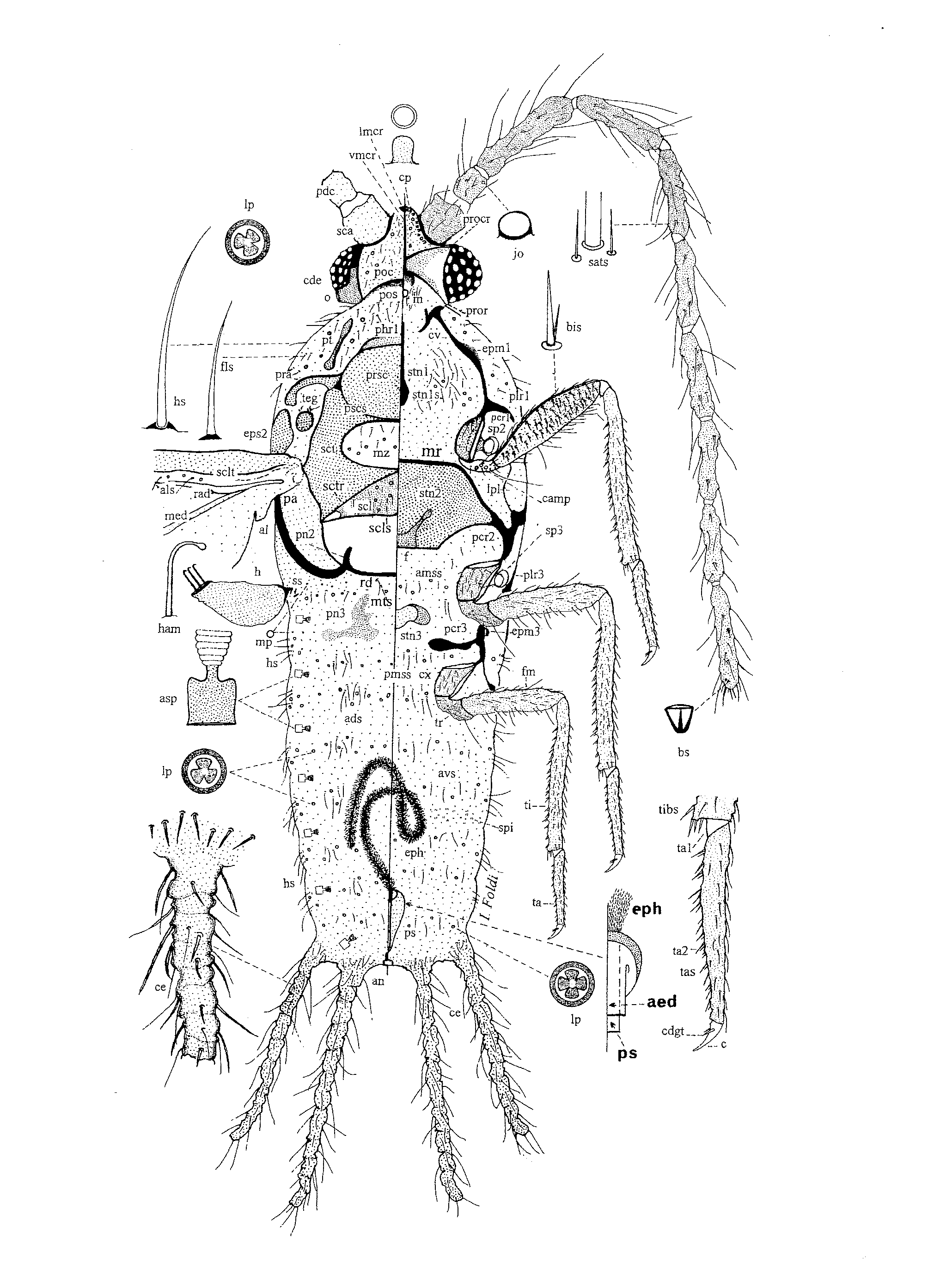
PREADULT FEMALE ( Fig. 9 View FIGURE 9 )
Description based on 4 specimens in fair condition.
Live material. Collected from a large colony on young branches. Dorsum covered by a coat of white filamentous secretion; venter with a thick coat of solidified amorphous secretion that appeared to provide a strong attachement to host-plant. Body broadly oval, dark-brown. After last moult, adult female remains within exuviae of preadult.
Mounted material. Body broadly oval to almost spherical, 7.2–11 mm long, 6.2–10.5 mm wide.
Dorsum Derm rather thick. Multilocular pores, each 10–11 µm wide, of 2 types: (i) with triangular centre ( Fig. 5 View FIGURE 5 L) with 3 inner loculi and about 12 outer loculi; densely scattered throughout, and (ii) pore with a quadrate centre ( Fig. 5 View FIGURE 5 H) with 4 inner loculi and 12 outer loculi, rare, sparsely scattered on over dorsum.
Raised pores ( Fig. 5 View FIGURE 5 A): circular, convex closed pores, each about 18–22 µm in diameter, with a narrower base about 8–10 µm wide, located in small cavity; very densely distributed in irregular transverse rows throughout. Long hair-like setae ( Fig. 5 View FIGURE 5 B), each 80–120 µm long, scattered throughout among slender and shorter flagellate setae ( Fig. 5 View FIGURE 5 F), latter each about 30–50 µm long, frequent throughout. Spiniform setae ( Fig. 5 View FIGURE 5 E), each 25–35 µm long, with a sharp apex and a well-developed basal socket; rather scattered in a large submarginal band ( Fig. 5 View FIGURE 5 E). Stout conical spines ( Fig. 5 View FIGURE 5 C), each 30–90 µm long, densely distributed throughout, but least frequent medially on abdomen. Hairs ( Fig. 5 View FIGURE 5 G), each 60–75 µm long, scattered. Anal opening dorso-apical; anal tube 530–560 µm long, 190–200 µm wide, with a wide collar of polygonal wax pores at inner end, plus 3 or 4 rows of multilocular disc-pores, each with a trilocular centre, present nearer opening ( Fig. 5 View FIGURE 5 J). Anal area and posterior end of abdomen with: (i) dense covering of raised pores ( Fig. 5 View FIGURE 5 M) mixed with hairs ( Fig.5 View FIGURE 5 G); (ii) hair-like setae ( Fig. 5 View FIGURE 5 B), which may be up 240 µm long, and (iii) multilocular pores, each with a triangular ( Fig. 5 View FIGURE 5 L) or quadrate centre ( Fig. 5 View FIGURE 5 H) with 3 or 4 inner loculi and 8 or 9 outer loculi.
Venter. Derm thick-membranous, except strongly sclerotised from head to metathorax in a wide band including submedian and submarginal areas, extending to anterior abdominal segments or even, in old specimens, to end of body where it can form a circular sclerotised zone; submargin rarely sclerotised. Sclerotised zone on head and thorax with multilocular pores, raised pores, hair-like setae and spines.
Multilocular pores in this zone each 10–12 µm in diameter, mostly with trilocular centres ( Fig. 5 View FIGURE 5 S) but occasionally with quadrilocular centres as on dorsum ( Fig. 5 View FIGURE 5 H), and with 8 (rarely 9) outer loculi; densely distributed on head, thorax, and on submargin of abdomen. Raised pores ( Figs. 5 View FIGURE 5 A, 5M), as described for dorsum, abundant on head and thorax, distributed also in irregular transverse rows, each 2–5 pores wide, throughout venter. Tubular pores ( Fig. 5 View FIGURE 5 U) quite large, each 16–20 µm wide, 18–20 µm deep, with large thickened rim and thickened base, sparsely scattered on thorax, and in transverse rows one pore wide on abdomen. Setae and spines of several types present: (i) hair-like setae ( Fig. 5 View FIGURE 5 B), each 170–195 µm long with a high collar, present on anterior end of body; also with shorter hair-like setae, each 100–130 µm long, around anal area but a few occasionally up to 240 µm long, and with few setae, each about 350 µm long on posterior end of abdomen. (ii) hairs ( Fig. 5 View FIGURE 5 G), each 160–270 µm, flexible, with expanded bases and mostly with a swollen apex ( Fig. 5 View FIGURE 5 X), very densely distributed medially and submedially on thorax; also with shorter hairs, each 70–90 µm long, numerous on posterior abdominal segments; (iii) slender flagellate setae ( Fig. 5 View FIGURE 5 F), each 30–60 µm long, present on abdomen, and on anterior end of body and on thorax; (iv) spines of various lengths present across all segments of body, each 10–14 wide at base, of two main types: (a) stout conical spines ( Fig. 5 View FIGURE 5 Y), each 60–130 µm long, sparsely distributed throughout venter, densest on head and around anal area where mixed with numerous long hair-like setae; and (b) shorter spines, each 20–50 µm long, rather frequent on margin and submargin of abdomen, but less frequent on rest of venter.
Antennae 9 segmented ( Fig. 5 View FIGURE 5 D), each 380–420 µm long, with 7 ring-like segments, each segment with a few hair-like setae up to 130 µm long; scape with 6 setae; apical segment with a rounded apex, 180–200 µm long, 80–90 µm greatest width, with many hair-like setae, each 40–90 µm long, plus 4 or 5 fleshy setae, each 40–55 µm long. Eyespot 135 µm wide, lens 70 µm in diameter. Thoracic spiracles large ( Fig. 5 View FIGURE 5 Z), each with a large apodeme, each anterior peritreme 145–160 µm wide, each posterior peritreme 155–170 µm wide, each spiracle with a distinct group of about 40–50 multilocular pores laterad to opening, each pore mainly with a circular centre with 3 inner loculi and 10 outer loculi; plus a small group of 6–9 simple pores; atria without pores. Abdominal spiracles ( Fig. 5 View FIGURE 5 O) 7 pairs; each with peritreme 90–100 µm wide, outer half of atrium with about 32–36 multilocular pores, each with a circular centre with a central elongate loculus plus a circular loculus on each side, plus 12 outer loculi; small cruciform pores ( Fig. 5 View FIGURE 5 P) scattered amongst these multilocular pores; external surface of atrium covered with minute spiniform setae ( Fig. 5 View FIGURE 5 N). Mouthparts present; clypeolabral shield proportionately small; labium narrow, apparently 2 segmented, apical segment with about 18–22 sensory setae, each 60–75 µm long, plus 2–4 long, slender hair-like setae, each 160–180 µm long. Legs short but robust with few setae. Metathoracic legs: coxa: 260 µm long, 500 µm wide, with few hair-like setae, each 8–12 µm long; trochanter + femur 950 µm long; trochanter with 2 or 3 campaniform sensilla on each side, and with a long distal trochanteral seta, 360 µm long; femur 340 µm greatest width, with many hair-like setae, each 100–130 µm long; tibia 670 µm long, 150 µm greatest width, with spur-like setae along ventral surface, each 45–55 µm long; tarsus ( Fig. 5 View FIGURE 5 W) 360 µm long, with spur-like setae ventrally, each 50–60 µm long; distally with 2 tarsal digitules, each 90 µm long; claws curved, each with 1 pair of acute, setose digitules, each 60–65 µm long. Spinules ( Fig. 5 View FIGURE 5 T) very densely distributed on thorax, and on anterior abdominal segments. Cicatrices ( Fig. 5 View FIGURE 5 V) circular, each 35–45 µm in diameter, with a large rim and a rather weakly convex surface; distributed in irregular, transverse rows 2–4 cicatrices wide, on abdomen.
Derivatio nominis. The new species “ williamsi ” is named in honour of Dr. Douglas J. Williams (The Natural History Museum, London, UK) for his considerable contribution to our knowledge of scale insects of the world.
Comment. The adult females of M. williamsi share several major features with M. hempeli which also have: strongly sclerotised zone on head; antennae reduced to 1 segment; mouthparts absent; spines and spiniform setae on dorsum and venter; multilocular pores with oval or triangular centre present; and adult females retained within the exuviae of preadult. However, M. williamsi differs from M. hempeli as follows (character states on M. hempeli in parentheses): multilocular pores with an oval centre more abundant across all abdominal segments and also across meso– and metathorax (fewer, rather restricted to abdomen); absence of pores with 5 or 6 loculi (present), long hair-like setae in a median line on abdomen (absent); legs each reduced to a protuberance (legs present but slender with segments considerably reduced in size); submedian apodemes on ventral abdomen absent (present, 2 apodemes per abdominal segment in submedial lines); abdominal spiracles opening at derm surface, with a few atrial pores (opening on a raised derm and without atrial pores). The preadult female of M. williamsi differs from that of M. hempeli due to the presence of: raised pores on the dorsum and venter; large tubular pores on venter; spines on head and thorax, and small minute spiniform setae on atria of abdominal spiracles.


No known copyright restrictions apply. See Agosti, D., Egloff, W., 2009. Taxonomic information exchange and copyright: the Plazi approach. BMC Research Notes 2009, 2:53 for further explanation.
|
Kingdom |
|
|
Phylum |
|
|
Class |
|
|
Order |
|
|
SuperFamily |
Coccoidea |
|
Family |
|
|
Genus |