
Gnathia pilosus, Hadfield, Kerry A., Smit, Nico J. & Avenant-Oldewage, Annemarié, 2008
|
publication ID |
https://doi.org/ 10.5281/zenodo.184379 |
|
DOI |
https://doi.org/10.5281/zenodo.5661695 |
|
persistent identifier |
https://treatment.plazi.org/id/C708879F-FFA2-AF4A-AECE-996CFB0C76C8 |
|
treatment provided by |
Plazi |
|
scientific name |
Gnathia pilosus |
| status |
sp. nov. |
Gnathia pilosus View in CoL sp. nov.
Material examined. Holotype. Male, 1.9 mm, 14 September 2006, Sheffield Beach (29°29’09.63”S, 31°15’24.64”E), South African Museum, Cape Town ( SAM A45580 View Materials ). Paratypes. 7 males, 5 females, 5 larvae, 14 September 2006, Sheffield Beach (29°29’09.63”S, 31°15’24.64”E), South African Museum, Cape Town ( SAM A45581). Other material. 4 males, 17 females, 50 larvae, 16 February 2007, Tinley Manor (29°27’09.25”S, 31°17’11.11”E), in the collection of the author.
Type host. Scartella emarginata (Günther, 1861) . Other hosts. Abudefduf sordidus (Forsskål, 1775) , Acanthurus triostegus (Linnaeus, 1758) , Antennablennis bifilum (Günther, 1861), Diplodus sargus capensis (Smith, 1844) , Epinephelus marginatus (Lowe, 1834) , Halichoeres nebulosus (Valenciennes, 1839) , Istiblennius dussumieri (Valenciennes, 1836) , Istiblennius edentulus (Forster & Schneider, 1801) , Omobranchus banditus Smith, 1959 , Plectroglyphidodon leucozoncus (Bleeker, 1859) , Psammogobius knysnaensis Smith, 1935 , Pterois miles (Bennett, 1828) , Terapon jarbua (Forsskål, 1775) , and Thalassoma purpureum (Forsskål, 1775) .
14 Diplodus sargus capensis (Smith, 1844) 54–109 (87.8) 4 1 25 2 ± 1 (0–2)
15 Rhabdosargus sarba (Forsskål, 1775) 210 1 0 0 0 Terapontidae
16 Terapon jarbua (Forsskål, 1775) 59–109 (86.8) 6 3 50 3.7 ± 2 (0–2) Diagnosis. Eyes large, bulbous. Slightly produced frontal border, superior frontolateral process and inferior mediofrontal process. Mandibles short, curved inwards with dentate blade. Numerous tubercules and setae over cephalosome and pereon.
Male description ( Figs 1–3 View FIGURE 1 View FIGURE 2 View FIGURE 3 , 10 View FIGURE 10 )
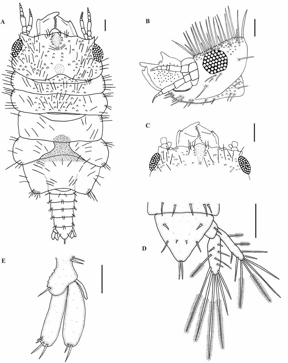
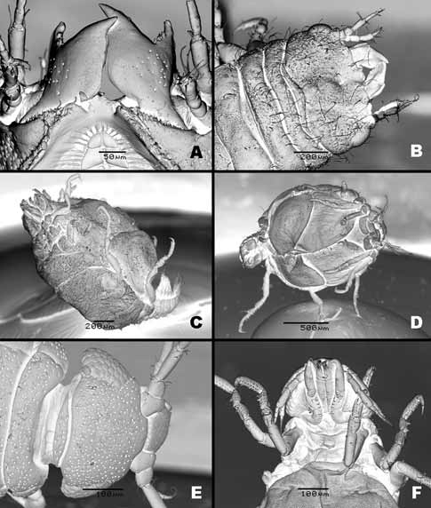
Description. Body ( Fig. 1 View FIGURE 1 A) length 1.6–2 mm, approximately 2.2 times as long as wide. Cephalosome ( Fig. 1 View FIGURE 1 A) rectangular, twice as wide as long, deep dorsal sulcus, narrower than width of median process, extending almost half length of cephalosome, lateral margins convex, densely covered in tubercles and short simple setae on dorsal and lateral surfaces as well as numerous small sensory pits covering entire cephalosome surface ( Fig. 10 View FIGURE 10 A), posterior margin concave. Well developed oval-shaped, bulbous, sessile compound eyes on lateral margin of cephalosome, length of eye slightly more than two thirds of cephalosome. Short simple setae around eye and no para-ocular tubercles. Numerous simple setae covering dorsal cephalosome ( Fig. 1 View FIGURE 1 B). Frontal border ( Fig. 1 View FIGURE 1 C) slightly produced, small rounded superior frontolateral process, with three long simple setae on outer border and two on inner border. Row of tubercles extends from superior frontolateral process, posteriorly along edge of dorsal sulcus ( Fig. 10 View FIGURE 10 B). Mediofrontal process inferior forming one distinct rounded lobe, no frontolateral process. Lamina dentata visible. External scissura shallow. Supraocular lobe slightly produced. Pereon ( Fig. 1 View FIGURE 1 A) one and two thirds times as long as wide, short simple setae on all pereonites, densely covered with small sensory pits. Pereonite 1 fused with cephalosome, dorsally visible, not reaching lateral margins, anterior border convex, posterior margin slightly concave, numerous tubercles covering dorsal surface. Pereonite 2 and 3 of similar shape and length but pereonite 3 slightly wider, lateral margins pointing anteriorly, numerous tubercles covering dorsal surface. Pereonite 4 anterior border convex with an anterior constriction medially separating it from pereonite 3, median groove present, longer than pereonite 3 and narrower then previous pereonites. Pereonite 5 with areae laterales and dorsal sulcus as thick groove, widest part of body. Pereonite 5 and 6 not fused. Pereonite 6 at least twice as long as other pereonites, narrowest part of body, anterior border concave, posterior margin deeply concave, with lobi laterals with at least five simple setae and pectinate scales on posterior point, no lobuii. Pereonite 6 almost separates pereonite 5 into two triangular sections. Pereonite 7 dorsally visible, small with rounded posterior margin, overlapping first pleonite. Pleon and pleotelson less than half of total length ( Fig. 1 View FIGURE 1 A). Pleonites subequal, epimera dorsally visible, short simple setae and pectinate scales on all pleonites.
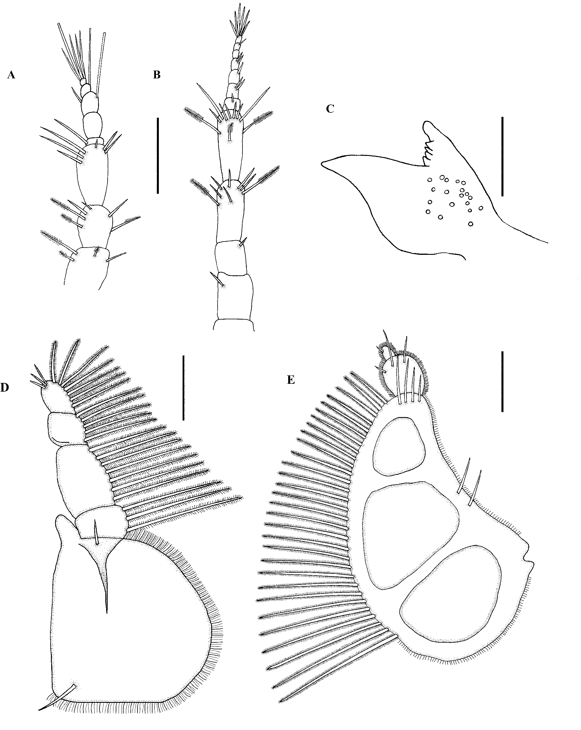
Antenna 1 ( Fig. 2 View FIGURE 2 A) with first two peduncle articles similar in shape and size both with plumose setae, first article with pectinate scales and simple setae. Flagellum with article 3 largest, article 3 and 4 each with one aesthetasc and one simple seta on article 3, article 5 terminating in one aesthetasc and four simple setae, no setae on second article.
Antenna 2 ( Fig. 2 View FIGURE 2 B) slightly longer than antenna 1. Antenna 2 with first article base covered with pectinate scales, article 3 and 4 largest with several simple and at least two plumose setae and six or seven simple setae on each, flagellum with seven articles, article 1 largest, article 7 terminating in six simple setae.
Mandible ( Fig. 2 View FIGURE 2 C, 10A) short, half the length of cephalosome, twice as long as wide, broad basal neck, curved inwards with dentate blade and short tufts of setae between teeth. Apex conical with rounded point, distally slightly raised in lateral view. Incisor present with tubercles present on posterior area. Single simple mandibular seta present. Carina armed, forming ridge on lateral margin extending from basal neck to a third along mandible. Small sensory pits on dorsal and ventral surface of blade.
Maxilliped ( Fig. 2 View FIGURE 2 D) five-articled, proximal article largest with two simple setae and mediodistal endite reaching article 3. Outer margin of proximal article densely setose. Distal four articles bearing plumose setae on lateral margins in order of 3–7–5–7. Distal article with three short simple setae. Palp 1.2 times as long as wide.
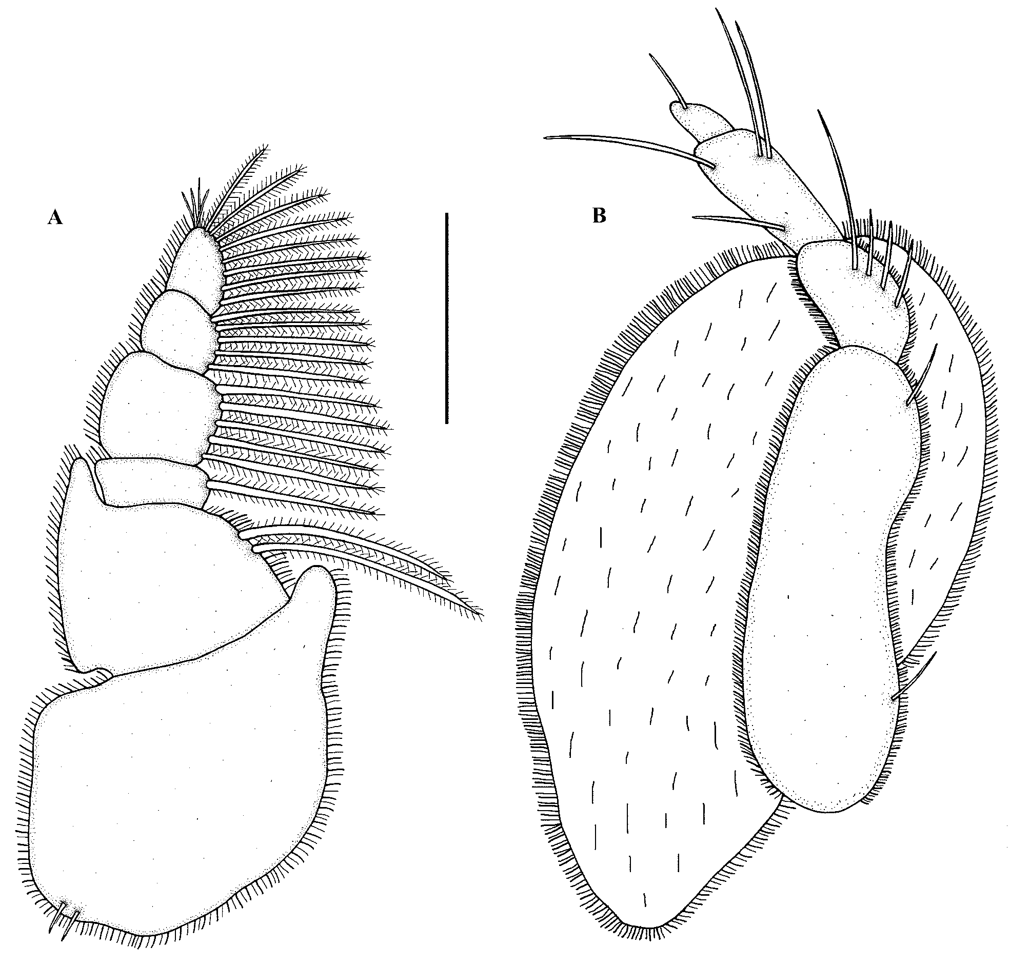
Pylopod ( Fig. 2 View FIGURE 2 E) with three articles, slightly convex and overlapping. First article greatly enlarged, convex mesial border fringed with 28–33 plumose setae, fine setae on other borders, a pair of simple setae near lateral border and four short simple setae distally on posterior surface. Three large areolae. Second article, as long as wide, margins setose, three simple setae distally on posterior surface. Third article small with fringing setae and single simple seta, twice as long as wide.
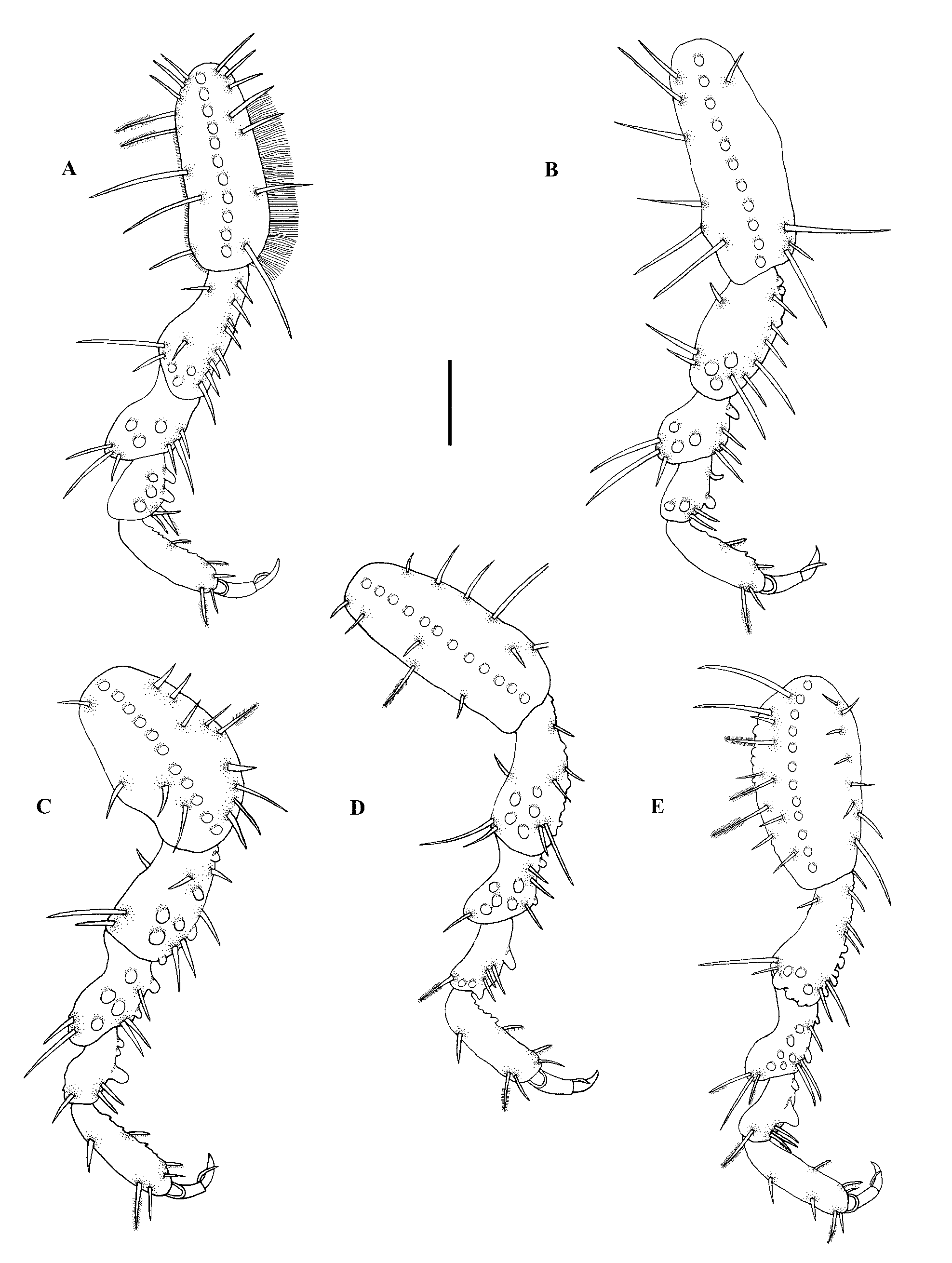
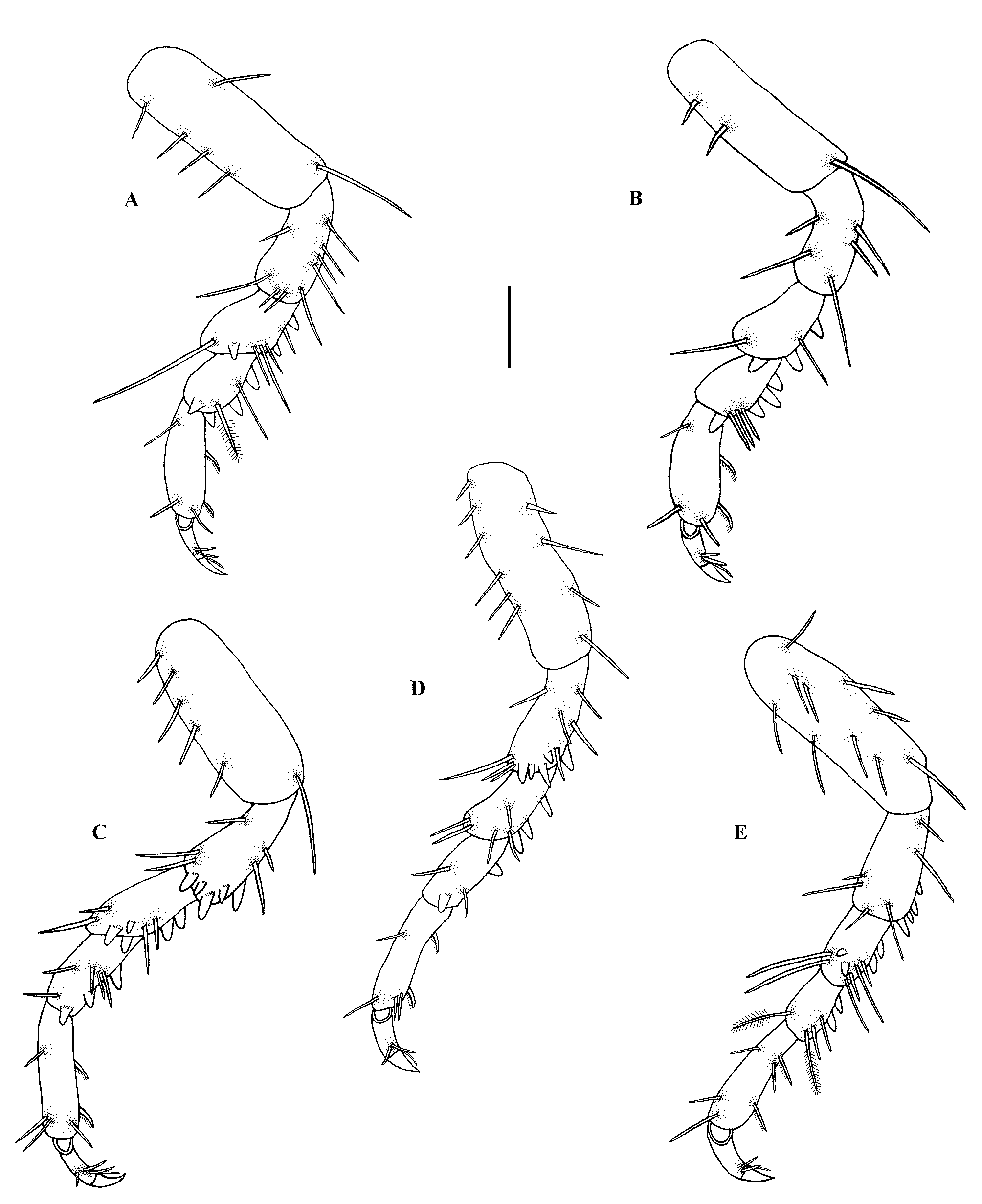
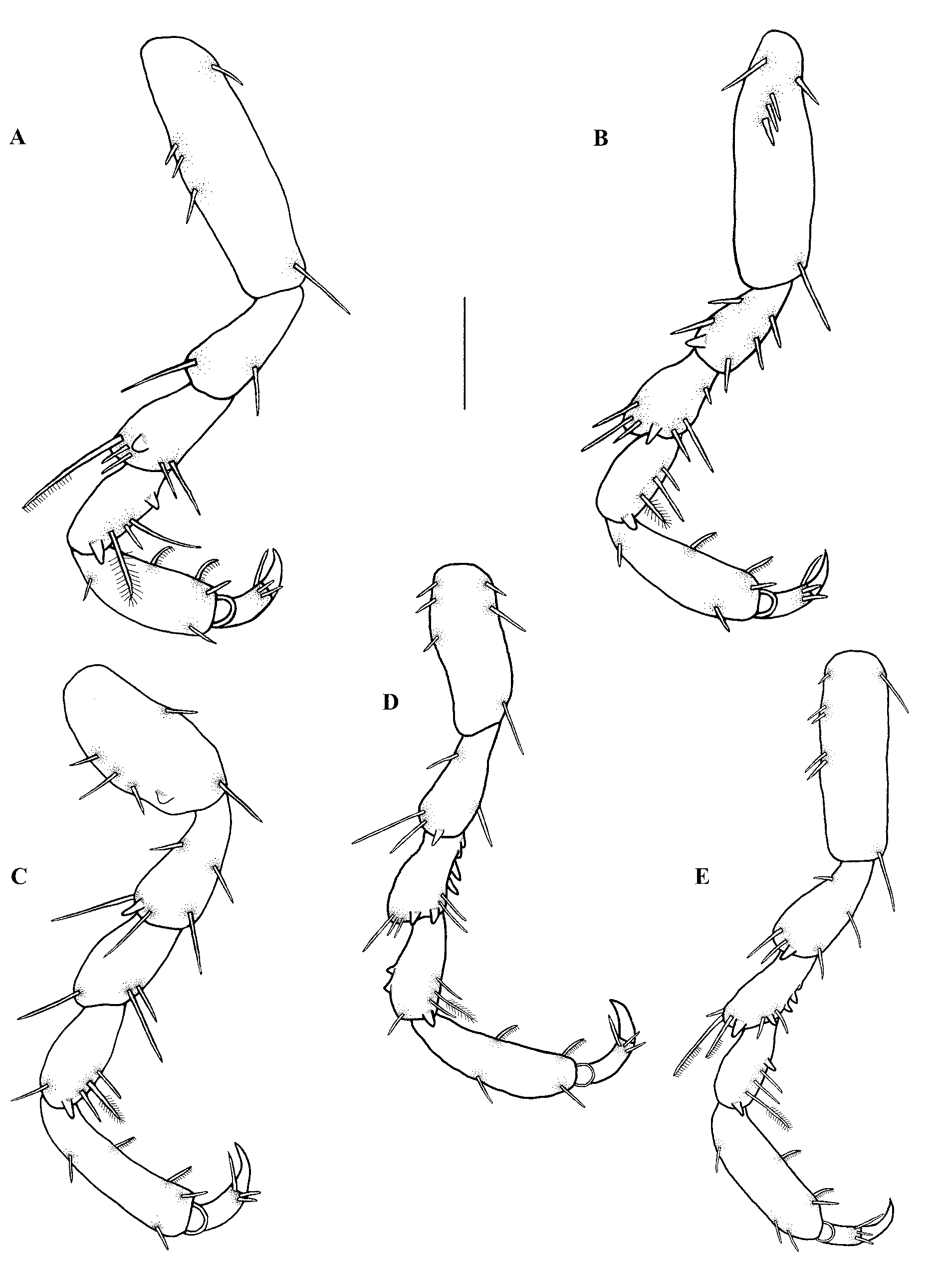
Pereopod 2 ( Fig. 3 View FIGURE 3 A) basis elongated with a strip of distinct tooth-like tubercles running the length of the basis, oval-shaped with 10 to 13 simple setae and two plumose setae on posterior margin, outer section of anterior margin densely covered with short setae and posterior margin densely covered with longer setae, pectinate scales on anterior border. Ischium with tooth-like tubercles on distal part, up to 12 simple setae scattered over the segment, pectinate scales on anterior border. Merus with anterior bulbous protrusion, three simple setae and tubercles on bulbous protrusion, posterior margin with simple setae, pectinate scales covering anterior border. Carpus with slight anterior bulbous protrusion, posterior margin with tooth-shaped tubercles, simple setae and an elongated denticulated compound spine, pectinate scales covering anterior border. Propodus with two tooth-shaped tubercles on proximal posterior margin terminating in a sharp point, only a few short simple setae anteriorly with one plumose seta distally, pectinate scales covering posterior margin. Dactylus terminates in sharp posterior pointing unguis, prominent spine on posterior side proximal to unguis ( Fig. 3 View FIGURE 3 A). Pereopods 3 to 6 ( Figs 3 View FIGURE 3 B–E) basic shape similar to pereopod 2, but setation as well as distribution of tubercles differ. Dorsal surface of all pereopods covered with pectinate scales which are not shown in illustrations.
Pleopod 2 ( Fig. 1 View FIGURE 1 E) with endopod slightly longer than exopod. Endopod ending distally in at least two short simple setae and exopod distally fringed with at least four simple setae, two short simple setae on interior posterior margin of sympodite and one simple setae on front lateral margin. Sympodite with retinaculae on medial margin. Basis with pectinate scales on lateral margin. Pleopod 2 endopod with appendix masculina, appendix masculina one third length of rami. Other pleopods similar to pleopod 2 but without appendix masculina.
Pleotelson ( Fig. 1 View FIGURE 1 D) triangular, base slightly wider than long, lateral margins slightly concave, dorsal surface with three pairs of short simple setae and many pectinate scales, distal apex terminating in pair of simple setae.
Uropodal ( Fig. 1 View FIGURE 1 D) rami extending beyond apex of pleotelson, endopod slightly longer and wider than exopod, pectinate scales covering both uropods. Four plumose setae and six simple setae on exopod, five plumose and seven simple setae on endopod. Uropodal basis covered with pectinate scales and single simple seta.
Penis prominent with two contiguous papillae, wider than long.
Pigmentation light brown in live specimens, occurring between eyes on anterior-dorsal surface of the cephalosome and in a T-shape over the medial dorsal sulcus. Pereonites 4, 5 and 6 have dark brown pigmentation with pereonite 4 being the darkest of the pereonites. Pleon also coloured with dark brown randomly distributed pigmentation.
Adult female ( Figs 4–6 View FIGURE 4 View FIGURE 5 View FIGURE 6 , 10 View FIGURE 10 )
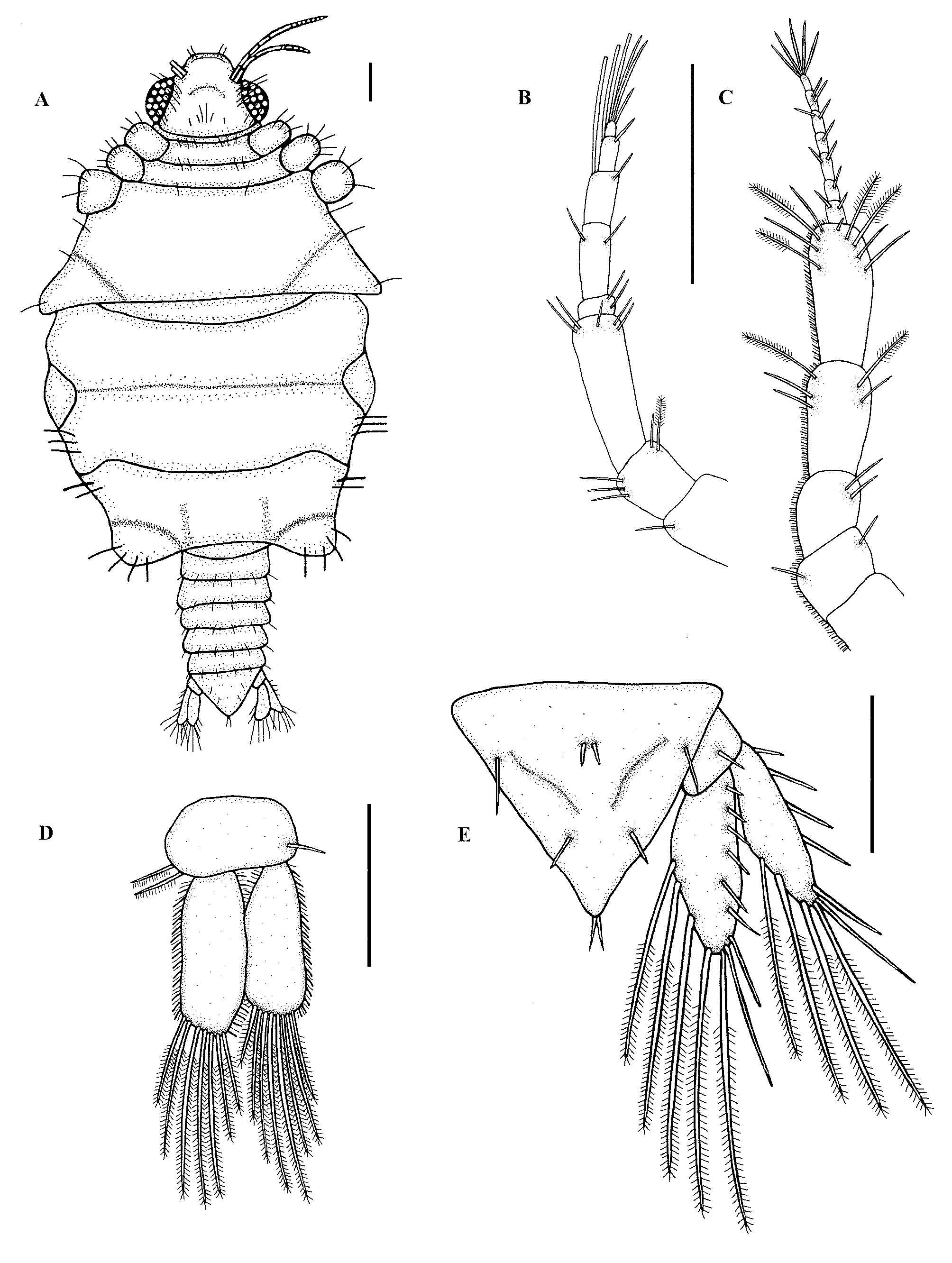
Description. Body ( Fig. 4 View FIGURE 4 A) length 1.8–2.5 mm, approximately 1.2 times as long as wide. Cephalosome ( Fig. 4 View FIGURE 4 A) broadened and short. Rectangular, 1.2 times as wide as long, short simple setae on dorsal, lateral and ventral cephalosome, posterior margin straight. Well developed oval-shaped, bulbous, compound eyes on lateral margin of cephalosome, length of eye two thirds of cephalosome. No paraocular ornamentation, only four to six short simple setae. Entire cephalosome covered with fine setae ( Fig. 10 View FIGURE 10 C). Frontal border ( Fig. 4 View FIGURE 4 A) broadly rounded, produced, slightly concave anteriorly, covered with pectinate scales. Pereon ( Fig. 4 View FIGURE 4 A) swollen round, sutures between pereonites 5–7, 1.2 times as long as wide, wider than cephalosome, short simple setae covering the entire dorsal surface and margins. Pereonites 5–7 form thin plate-like oostegites, enclose brood pouch, oostegites overlapping ( Fig. 10 View FIGURE 10 D). Pereonite 7 dorsally visible, small with rounded posterior margin, overlapping first pleonite. Ventral area of pereonite 6 with slit which appears to be genital opening. Pleon ( Fig. 4 View FIGURE 4 A) and pleotelson less than a third of total length. Pleonites subequal, epimera not distinct, short setae and pectinate scales covering pleonites. Single simple seta on each posterior lateral side of pleonite and two pairs of simple setae on the posterior margins of each pleonite.
Antenna 1 ( Fig. 4 View FIGURE 4 B) with three peduncle articles increasing in length distally with third article as long as first and second articles combined. Few short simple setae on distal end of articles 1 and 2 with a single plumose seta on distal end of article 2, two to five short simple setae on article 3. Flagellum with five articles, article 2 largest, articles 1 and 2 with short simple setae, articles 3 and 4 with one aesthetasc seta and one simple seta each, article 5 terminating in one aesthetasc and three simple setae.
Antenna 2 ( Fig. 4 View FIGURE 4 C) longer than antenna 1. Antenna 2 with five peduncle articles, article 5 largest, short simple setae on distal end of articles 1 to 3, three to four short simple setae on distal ends of with two and four plumose seta on articles 4 and 5 respectively. Flagellum with seven articles, short simple setae on distal end of each article, article 7 terminating in three to five simple setae. Peduncle articles of both antennae covered with pectinate scales and very short, fine simple setae on the peduncle of antenna 2.
Mandible and maxillae absent.
Maxilliped ( Fig. 5 View FIGURE 5 A) consists of basis, oostegite and four articled palp. Endite long, setose, reaching article 2 of palp. Lateral margins of basis fringed with two long plumose setae. Palp bearing plumose setae on lateral margins in order of 1–5–5–8. Distal article of palp with three to four short simple setae. Oostegite broader and almost as long as palp, with two simple setae on posterior border. All borders of basis, palp and oostegite densely setose.
Pylopod ( Fig. 5 View FIGURE 5 B) with four articles, articles 1 and 2 not fused. Article 1 broad, robust, curved anteriorly, with two simple setae on lateral border. Article 2 with four to five simple setae distally. Article 3 with two to four simple setae distally. Article 4 small with one to two simple setae. Surface of articles 2 and 3 covered with pectinate scales and lateral borders with short fine simple setae. Oval-shaped oostegite, 2 times longer than broad, covers mouthparts ventrally, not surpassing frontal border, lateral borders and surface of oostegite with short fine simple setae. Article 1 and 2 covered with pectinate scales and short fine simple setae.
Pereopod 2 ( Fig. 6 View FIGURE 6 A) basis elongated oval shaped with four to five short simple setae anteriorly, two to three posterior simple setae. Ischium two to five anterior short simple setae, five short simple setae posteriorly. Merus with anterior bulbous protrusion, one long simple seta on bulbous protrusion, posterior margin with two to three large tooth-shaped tubercles as well as three short and a single long simple seta. Carpus without anterior bulbous protrusion, posterior margin with three to four large tooth-shaped tubercles, single simple setae and a single plumose seta. Propodus with two elongated denticulated compound spines ending in sharp points situated on middle and distal part of posterior margin respectively, two simple setae anterio-distally. Dactylus terminates in sharp posterior pointing unguis, prominent spine on posterior side proximal to unguis, two simple setae on dorsal and ventral sides of spine ( Fig. 6 View FIGURE 6 A). Pereopods 3 to 6 similar to pereopod 2 in basic form, differ in setation, shape and number of tubercles ( Figs 6 View FIGURE 6 B–E). Dorsal surface of ischium, merus, carpus and propodus of all pereopods covered with pectinate scales but not shown in illustrations.
Pleopod 2 ( Fig. 4 View FIGURE 4 D) with endopod longer and wider than exopod. Both fringed distally with seven to eight long plumose setae and short simple fine setae on all borders. Sympodite with retinacula, single simple seta on lateral margin. Other pleopods similar to pleopod 2.
Pleotelson ( Fig. 4 View FIGURE 4 E) triangular, base slightly wider than length, lateral margins slightly concave, dorsal surface with three pairs of simple setae and pectinate scales, distal apex terminating in pair of short simple setae.
Uropodal ( Fig. 4 View FIGURE 4 E) rami extending beyond apex of pleotelson, endopod longer and wider than exopod, both with long simple setae, pectinate scales on dorsal area of uropods and basis. Endopod with six to eight simple setae on dorsal and posterior surfaces and five plumose setae on posterior margin. Exopod with four plumose setae and six long simple setae. A single short simple seta on uropodal basis.
Pigmentation lightly coloured, dark brown speckles distributed over the cephalosome, pleon and pereonites with denser pigmentation around pereonite 4. Prominent red / brown eyes.
Third-stage praniza (P3) ( Figs 7–10 View FIGURE 7 View FIGURE 8 View FIGURE 9 View FIGURE 10 )
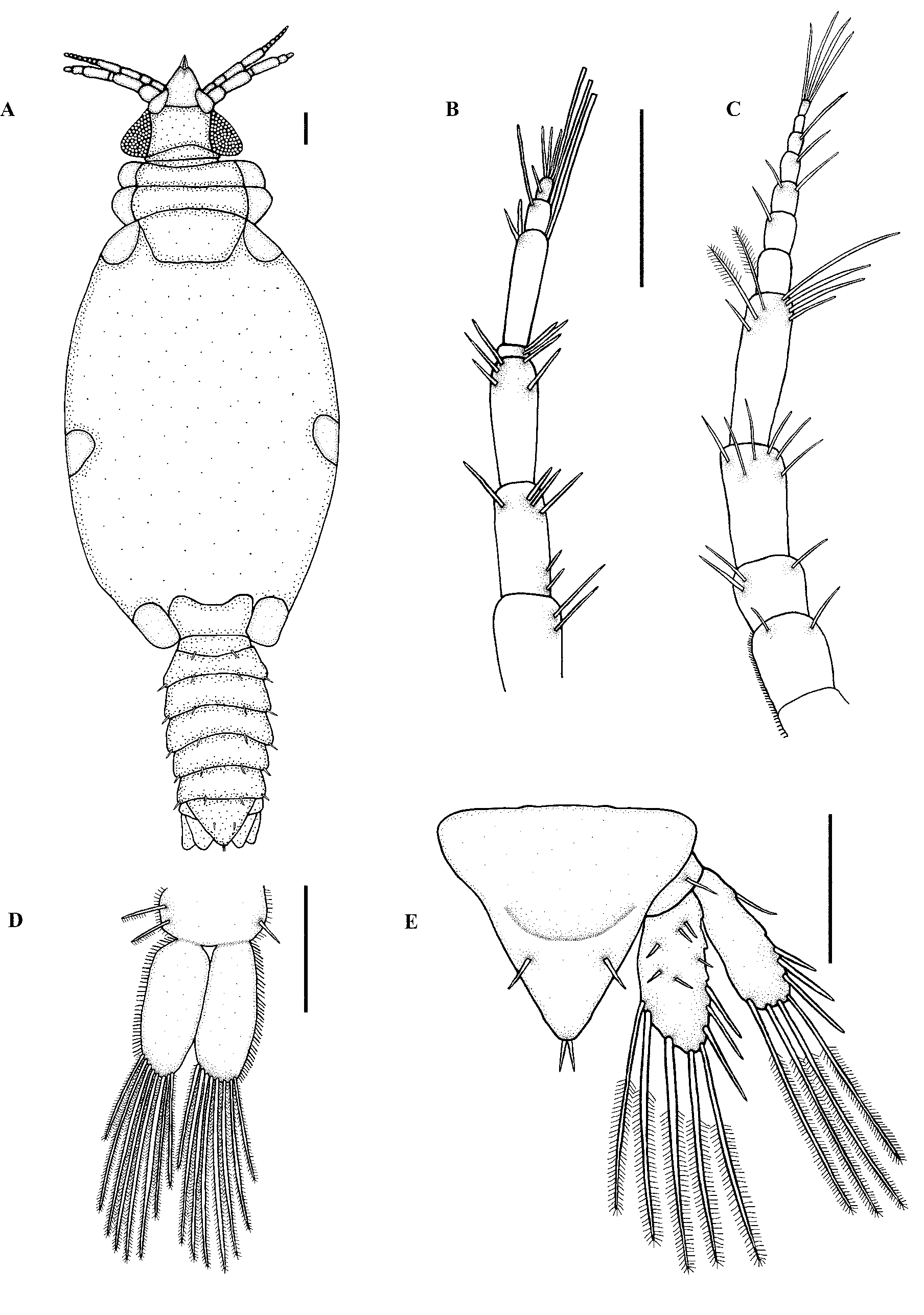
Body length of first praniza 1–1.4 mm, second praniza 1.3–1.9 mm, third praniza 1.8–3 mm. Cephalosome ( Fig. 7 View FIGURE 7 A, 10E) posterior margin concave, wider than anterior margin, wider at the base, lateral margins convex, few setae on dorsal posterior cephalon, posterior margin straight, triangular-shaped cephalosome. Many sensory pits distributed randomly over dorsal surface of cephalosome ( Fig. 10 View FIGURE 10 E), two pairs of short simple setae on eye margins. Compound eyes large, triangular-shaped, bulbous, length of eye almost same as cephalosome. Medio-anterior margin of cephalosome straight with lateral concave excavations to accommodate first articles of antennae ( Fig. 10 View FIGURE 10 E). Labrum ( Figs 7 View FIGURE 7 A, 10E) prominent, almost same length as cephalosome, triangular shaped with apical process, truncated posterior margin, anterior margin concave. Ventral part of labrum gutter-like with central groove, covers mandibles dorsally and laterally. Pereon ( Fig. 7 View FIGURE 7 A) almost twice as long as wide, wider than cephalosome. Pereonite 1 fused with cephalosome, dorsally visible, anterior border convex, posterior border straight. Pereonite 2 with anterior constriction separating it medially from pereonite 1. Pereonite 4 twice as wide as long, lateral sides tapering towards rounded posterior margin, posterior margin stretching over pereonite 5, lateral shields at leg attachment. Pereonite 6 rectangular, posterior margin slightly concave, lateral shields at leg attachment. Pereonite 7 dorsally visible, small with rounded posterior margin, overlapping first pleonite. Pleon ( Fig. 7 View FIGURE 7 A) and pleotelson half length of pereon. Pair of short simple setae in the middle of each pleonite and single short simple setae on each posterior lateral side of each pleonite. Many sensory pits distributed over pereonites and pleonites.
Antenna 1 ( Fig. 7 View FIGURE 7 B) with third article largest, short simple setae on both anterior and posterior borders and pectinate scales on articles 1 and 2. Flagellum first article with two short simple setae, article 2 largest, articles 2 and 3 with one aesthetascs setae each, article 4 terminating in one aesthetascs and three simple setae, few setae on each article.
Antenna 2 ( Fig. 7 View FIGURE 7 C) longer than antenna 1. Antenna 2 with article 4 largest and short simple setae on bor- der and pectinate scales on articles 1 and 2. Flagellum with article 1 largest, article 7 terminating in three to four simple setae, few setae on distal end of each article.
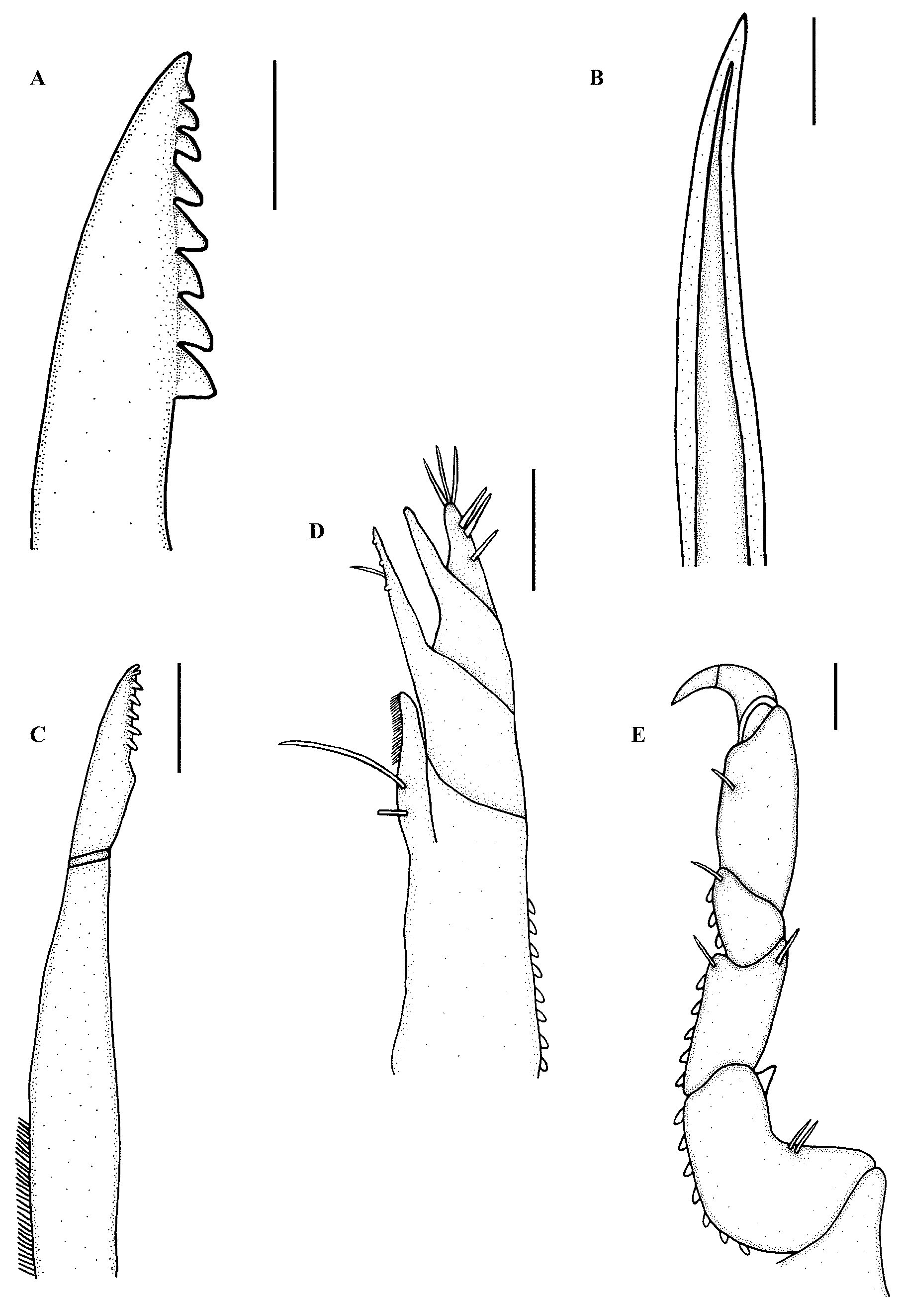
Mandible ( Fig. 8 View FIGURE 8 A) stout, swollen at base, distal margin styliform with eight teeth on inner mesial margin, triangular and backwardly directed, increasing in size from anterior to posterior.
Paragnath ( Fig. 8 View FIGURE 8 B) elongated, gutter-like, terminates in sharp point, no teeth.
Maxillule ( Fig. 8 View FIGURE 8 C) long, slender, swollen base, stretching past distal margin of labrum. Seven to eight teeth on distal inner margin. Maxilla not visible.
Maxilliped ( Figs 8 View FIGURE 8 D, 10F) large, cylindrical, elongated base, endite almost reaching palp with a single long simple seta and coupling hook. Palp with three articles, first article acute with three to four small teeth and a single simple seta mesially, articles 3 with five to six simple setae.
Gnathopod ( Figs 8 View FIGURE 8 E, 10F) smaller than pereopods, seven articles fused, dactylus strongly hooked, only few simple setae, many pectinate scales on inner and lateral sides.
Pereopod 2 ( Fig. 9 View FIGURE 9 A) basis elongated with pectinate scales and three to four simple setae anterior, one or two simple seta posteriorly. Ischium with single simple setae anteriorly and posteriorly. Merus with anterior bulbous protrusion, single serrated spine, tubercules and two simple setae on bulbous protrusion, posterior margin with simple setae. Carpus without anterior bulbous, tubercles present and single plumose seta on posterior margin. Propodus with two short denticulated spines ending in sharp points situated on middle and distal part of posterior side respectively, only a simple setae anteriorly. Dactylus terminates in sharp posterior pointing unguis, prominent spine on posterior side proximal to unguis, few simple setae on dorsal and ventral sides of spine ( Fig. 9 View FIGURE 9 A). Pereopods 3 to 6 ( Figs 9 View FIGURE 9 B–E) similar to pereopod 2 in basic shape, differ in setation and number and presence of spines and tubercules. Pereopods also differ in direction, pereopods 4 to 6 directed posteriorly and pereopods 2 and 3 anteriorly. All articles of pereopods with pectinate scales and short fine setae and dominant pointed tubercules when present.
Pleopod 2 ( Fig. 7 View FIGURE 7 D) with exopod and endopod almost the same size and covered in pectinate scales. Both fringed distally with eight to nine long plumose setae, short fine setae on all margins. Plumose setae almost as long as pleopod. Sympodite with retinacula, single simple seta on lateral margin and pectinate scales. Other pleopods similar to pleopod 2.
Pleotelson ( Fig. 7 View FIGURE 7 E) triangular, base slightly wider than length, anterior half of lateral margins slightly convex, posterior half straight. A pair of simple setae on posterior dorsal surface, distal apex terminating in pair of simple setae, dorsal surface covered with pectinate scales, slight depression medially.
Uropodal ( Fig. 7 View FIGURE 7 E) endopod extending beyond apex of pleotelson, exopod reaching pleotelson apex. Endopod longer and wider than exopod, endopod with distal five setae plumose, exopod with distal four setae plumose, remainder of setae simple. Uropodal basis with single simple setae and pectinate scales. Both endopod and exopod covered by pectinate scales.
Pigmentation slight with few markings. Zuphea larvae with dark brown pereon, specifically pereonites 4 and 5, with pereonites 1 to 3 light with light brown speckles randomly distributed. Pleon and cephalosome of zuphea with light brown pigmentation, dense on cephalosome. Praniza larvae with light brown pigmentation on pleon and cephalosome. Cephalosome densely pigmented with darker borders on the anterior margins near the mouthparts. Brown colouration on lateral margins of the pereon and dense around the anterior pereonite 4, lateral margins of pereonite 5 and the posterior margin on pereonite 6.
Etymology. The species name is derived from the Latin word, pilosus , meaning “hairy”, in reference to the large quantity of setae covering the body and especially the cephalosome of the male giving it a hairy appearance.
Remarks. The broad, three articled pylopods, frontal border processes, a straight frontal border and nonelongate mandibles with dentate blades place this new species in the genus Gnathia . Gnathia pilosus sp. nov. can clearly be distinguished from other South African species in having numerous tubercles and setae covering the cephalosome and pereon which is not as pronounced in the other species. Other distinguishable characteristics include the short, stout mandible (0.5 times length of cephalosome) and large eyes (0.7 times length of cephalosome).
Gnathia pilosus View in CoL sp. nov. is relatively similar to three species from Australia, namely G. asperifrons Holdich and Harrison, 1980 View in CoL , G. iridomyrmex Cohen and Poore, 1994 View in CoL , and G. variobranchia Holdich and Harrison, 1980 View in CoL ( Holdich & Harrison 1980; Cohen & Poore 1994). Gnathia pilosus View in CoL is similar to G. asperifrons View in CoL in having numerous minute tubercles covering the cephalosome surface, but differs from the latter species which has pereonites 5 and 6 as the widest part of the body, only 2 small areolae on the pylopod and a crenulate mandibular blade ( Holdich & Harrison 1980). Gnathia iridomyrmex View in CoL has a shallow dorsal sulcus, smaller eyes, acute superior frontolateral process and an irregularly crenulated mandibular blade compared with G. pilosus ( Cohen & Poore 1994) View in CoL . Gnathia variobranchia View in CoL differs from the new species in having a broad but shallow dorsal sulcus, a low ridge running posteriomedially from the rear of each eye on the cephalosome and pleopods which differ in size and setae from pleopod 1 to 5 ( Holdich & Harrison 1980).
Gnathia pilosus View in CoL is also similar to G. lignophila Müller, 1993 View in CoL , in being hirsute with large number of tubercles, termed granules in Müller’s (1993) work, and narrower pereonites 4 and 6 which are similar to the new species. However, differences are apparent with the presence of a pair of granular, rounded para-ocular tubercules, a shallow dorsal sulcus and two frontolateral processes and two shorter mediofrontal processes in G. lignophila View in CoL but absent from G. p i l o s u s.
There are only two gnathiid females described from South Africa, those of G. africana View in CoL and G. pantherina View in CoL . The female of G. pilosus View in CoL sp. nov. is easily distinguished from these in having long plumose setae on the pleopod, plumose setae on the lateral margins of the maxillipedal palp articles being in the order 1–5–5–8 (from proximal to distal), and that it is covered by numerous fine simple setae, especially dense on the posterior portion of the pereon ( Fig. 10 View FIGURE 10 C).
As with the females, the larvae of only two South African gnathiids have been described, namely G. a f r i - cana and G. pantherina . The larvae of G. pilosus sp. nov. differs from these in number of teeth on the first palp article of the maxilliped, triangular-shaped cephalosome as compared to the usual oval or round shape observed in the other species, and the numerous sensory pits which are distributed from the cephalosome to the pleotelson.
Distribution. Intertidal zone of Sheffield Beach and Tinley Manor, North coast of Kwazulu-Natal Province, South Africa.
Discussion. A number of morphologically important characteristics were identified for each phase of G. pilosus ; however, there are only a few characteristics which link these different stages. One such characteristic is the shape of the pleotelson which seems to remain constant during all stages. Other linking characteristics are the plumose setae on the pleopods and uropods, as well as the tooth-like tubercles on the pereopods. These characteristics could prove helpful in linking the life stages of this species if collected separately. This paper adds a new species of gnathiid to the South African isopod fauna, bringing the total number of known gnathiid species, from this region, to eight. This is the first gnathiid found in the warmer East Coast waters of South Africa and the larvae were found to parasitise a large number of common intertidal fishes.
| SAM |
South African Museum |
No known copyright restrictions apply. See Agosti, D., Egloff, W., 2009. Taxonomic information exchange and copyright: the Plazi approach. BMC Research Notes 2009, 2:53 for further explanation.
|
Kingdom |
|
|
Phylum |
|
|
Class |
|
|
Order |
|
|
Family |
|
|
Genus |
Gnathia pilosus
| Hadfield, Kerry A., Smit, Nico J. & Avenant-Oldewage, Annemarié 2008 |
G. iridomyrmex
| Cohen and Poore 1994 |
G. pilosus (
| Cohen & Poore 1994 |
G. lignophila Müller, 1993
| Muller 1993 |
G. asperifrons
| Holdich and Harrison 1980 |
G. variobranchia
| Holdich and Harrison 1980 |