
Parapolybia indica
|
publication ID |
https://doi.org/10.11646/zootaxa.3947.2.5 |
|
publication LSID |
lsid:zoobank.org:pub:36A90396-5654-45AD-90B0-4653BB98851B |
|
DOI |
https://doi.org/10.5281/zenodo.6097432 |
|
persistent identifier |
https://treatment.plazi.org/id/C423CC1F-6F78-FFD4-25A2-FB43FB85F85E |
|
treatment provided by |
Plazi |
|
scientific name |
Parapolybia indica |
| status |
|
Key to species of Parapolybia indica View in CoL species-group
Unless the sexes are mentioned, the characters given in the following key are of females.
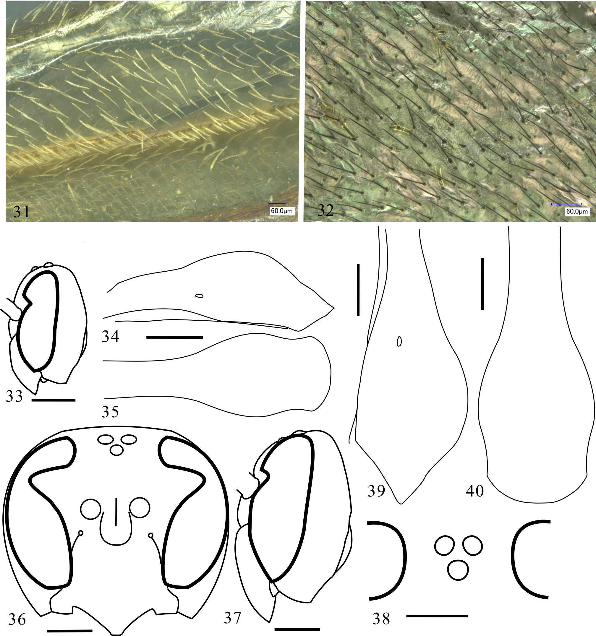
1. Body ground color brown to dark brown ( Fig. 79 View FIGURES 76 – 84. 76 ); mesoscutum more than 4 mm wide; propodeum with strongly striate; T1 strongly swollen in posterior half ( Figs 39–40 View FIGURES 31 – 40 )......................................... P. tinctipennis ( Cameron)
- Body ground color, at least of head and mesosoma, much less dark, ivory white, yellow to light brown or orange. Mesoscutum less than 3.5 mm wide; T1 weakly to moderately swollen posteriorly........................................... 2
2. Body ground color orange ( Fig. 77 View FIGURES 76 – 84. 76 ); wings yellow tinged (covered with yellow setae) ( Fig. 31 View FIGURES 31 – 40 )..... P. fulvinerva ( Cameron)
- Body ground color ivory white or yellow to light brown, with brown to black markings ( Figs 3, 5 View FIGURES 1 – 6 , 76, 78, 80–81 View FIGURES 76 – 84. 76 ); wings with black setae ( Fig. 32 View FIGURES 31 – 40 ).................................................................................. 3
3. Metasomal segments 2 and 3 much darker than mesosoma, nearly black, T2 and T3 respectively with large and paired small yellow spots ( Fig. 76 View FIGURES 76 – 84. 76 ); antenna and legs, especially in male, prominently elongated........... P. bioculata (van der Vecht)
- Metasomal segments 2 and 3 more or less same colored as mesosoma, with or without darker markings; antenna and legs not prominently elongated................................................................................. 4
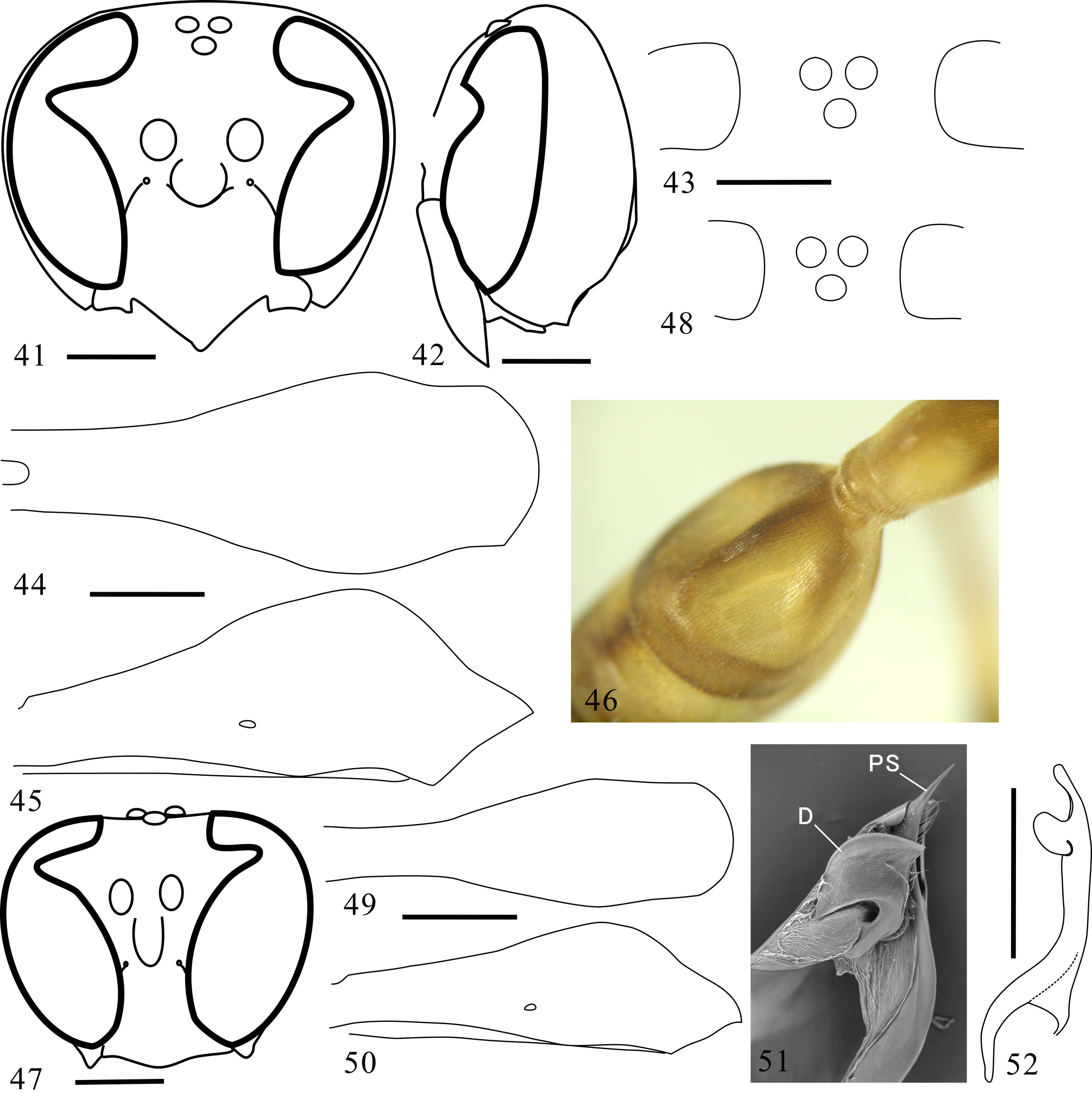
4. In both sexes, T2 distinctly concave sublaterally ( Fig. 46 View FIGURES 41 – 52 ). Gena developed, visible in frontal view of head ( Fig. 41 View FIGURES 41 – 52 ), in lateral view slightly wider than eye ( Fig. 42 View FIGURES 41 – 52 )......................................................... P. flava sp. nov.
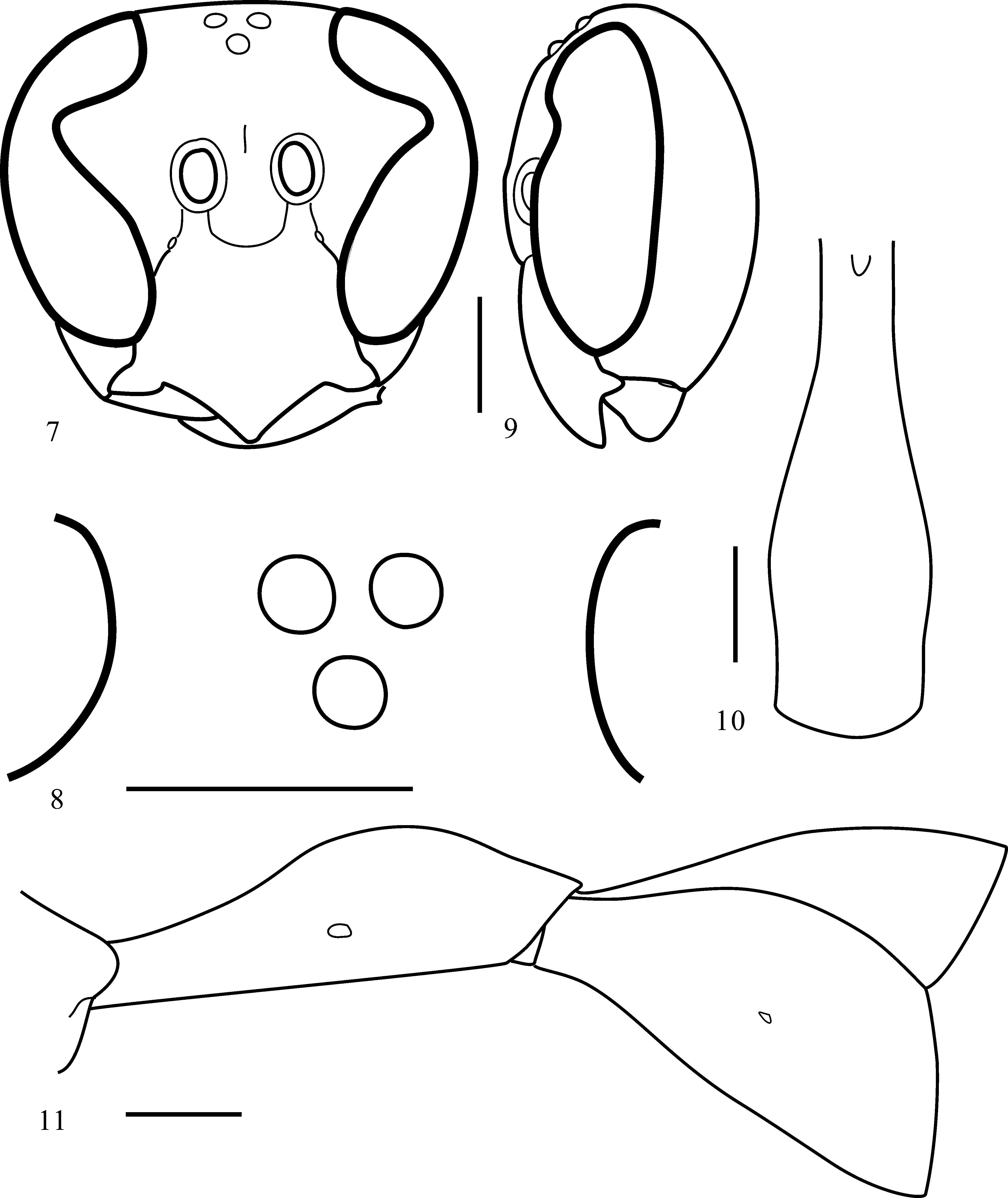
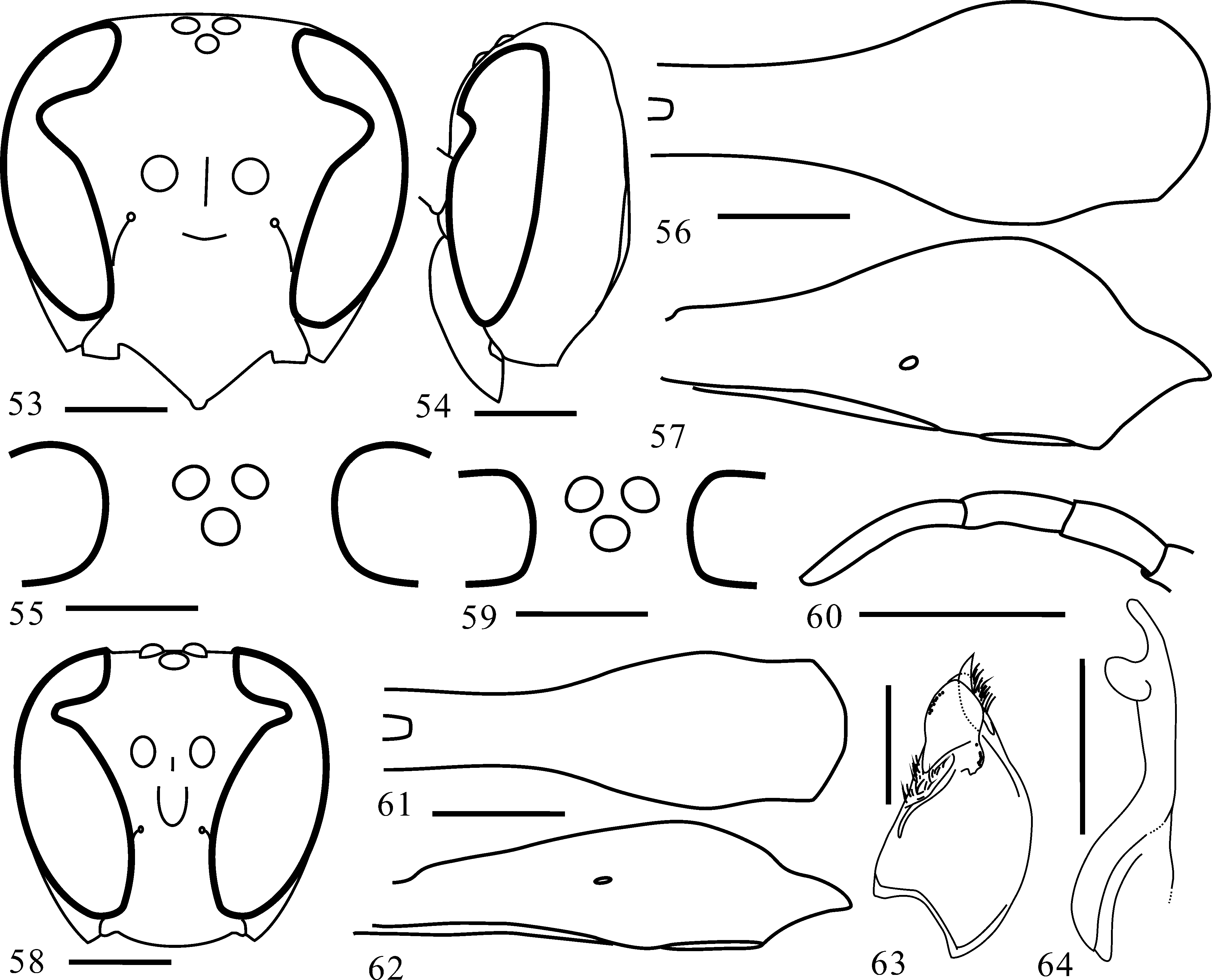
- In both sexes, T2 barely or only weakly concave sublaterally. Gena not strongly swollen laterally, invisible in frontal view of head ( Figs 7 View FIGURES 7 – 11 , 53 View FIGURES 53 – 64 , 65, 71 View FIGURES 65 – 75 ), in lateral view as wide as or narrower than eye ( Figs 9 View FIGURES 7 – 11 , 33 View FIGURES 31 – 40 , 54 View FIGURES 53 – 64 , 66, 72 View FIGURES 65 – 75 )....................... 5
5. Body ground color ivory white. Clypeus with distinct paired dark brown spots ( Fig. 70 View FIGURES 65 – 75 )................ P. albida sp. nov.
- Body ground color yellow to light brown. Clypeus without dark spots........................................... 6
6. T 1 in lateral view nodulated posteriorly ( Fig. 34 View FIGURES 31 – 40 )............................................ P. takasagona Sonan
- T 1 in lateral view more or less smoothly swollen dorsally toward level of spiracle ( Figs 11 View FIGURES 7 – 11 , 57 View FIGURES 53 – 64 , 68 View FIGURES 65 – 75 ).................... 7
7. Flat area of vertex behind posterior ocelli narrow ( Fig. 82 View FIGURES 76 – 84. 76 )....................................... P. nana sp. nov.
- Flat area of vertex behing posterior ocelli wide ( Figs 83–84 View FIGURES 76 – 84. 76 ).................................................. 8
8. Paired yellow longitudinal lines on mesoscutum and spots on T2 absent or obscure ( Fig. 5 View FIGURES 1 – 6 )......... P. indica View in CoL (de Saussure)
- Paired yellow longitudinal lines on mesoscutum and spots on T2 distinct ( Fig. 3 View FIGURES 1 – 6 )..................... P. crocea sp. nov.


No known copyright restrictions apply. See Agosti, D., Egloff, W., 2009. Taxonomic information exchange and copyright: the Plazi approach. BMC Research Notes 2009, 2:53 for further explanation.
|
Kingdom |
|
|
Phylum |
|
|
Class |
|
|
Order |
|
|
Family |
|
|
Genus |