
Zoothamnium paraentzii Song, 1991
|
publication ID |
https://doi.org/ 10.5281/zenodo.170316 |
|
DOI |
https://doi.org/10.5281/zenodo.5662214 |
|
persistent identifier |
https://treatment.plazi.org/id/BF1C87AE-FFF9-FFEF-FE9B-FEFAFC873E0F |
|
treatment provided by |
Plazi |
|
scientific name |
Zoothamnium paraentzii Song, 1991 |
| status |
|
Redescription of Zoothamnium paraentzii Song, 1991 (Figs. 3 & 4; Table 1 View TABLE 1 )
Zoothamnium paraentzii was originally found in shrimpfarming waters from the Yellow Sea, China ( Song 1991a). As the original report was made in Chinese and descriptions of oral structures were not supplied in sufficient details, a complementary redescription is thus supplied based on the present population.
Improved diagnosis: Marine Zoothamnium irregularly dichotomously branched with zooids of the same branch born on the same side. Body highly variable in shape but usually elongate, measuring about 50–80 × 25–45 µm. Peristomial lip single layered; contractile vacuole apically located; macronucleus generally Cshaped and horizontally orientated; number of transverse lines from oral area to aboral trochal band 75–83, from aboral trochal band to scopula, 28–33; inner row of peniculus 3 displaced from the other two and converged with peniculus 1 at aboral end.
Voucher slides: Two permanent slides (registration numbers: 0 4040601, 04040602) with silver nitrate and protargol prepared material are deposited at the Laboratory of Protozoology, OUC, China.
Morphology: Body flexible and highly variable, usually slender vaseshaped (Figs. 3A, 3D; 4B, 4D), widest at oral border, and slightly constricted below peristomial lip. Fully extended zooid ca. 60 × 40 µm in size, ratio of length to width mostly about 3:2. Peristomial disk moderately elevated (Fig. 4D). Singlelayered peristomial lip relatively thin (Figs. 3A, 3D; 4B). Pellicle smooth at low magnification, striations detectable at higher magnification (×400 or higher).
Cytoplasm colourless and transparent, usually becoming denser and more granular in aboral part of cell owing to presence of numerous tiny granules (ca. 2–6 µm in diameter) and several food vacuoles (Figs. 3A, 3D; 4B, 4D). Macronucleus relatively short, Cshaped and horizontally orientated (Fig. 4I). Single large contractile vacuole apically located (Figs. 3A; 4B).
Stalk robust, about 8–12 µm thick, with many drapes on surface (Figs. 3F, arrows; 4C, arrows). Colony irregularly dichotomously branched with zooids of the same branch born on the same side and reaching a total length up to 1 mm (Figs. 3E; 4A). Stalk myoneme conspicuous with numerous grayish thecoplasmic granules which measuring about 0.5 µm in diameter (Figs. 3F; 4C).
Oral apparatus typical of genus. Haplokinety and polykinety circling about one and half turns around peristomial disc and accomplishing a further turn after plunging into vestibulum (Fig. 3H). Near distal end of haplo and polykinety, always one short kinety fragment recognizable (Figs. 3H, arrow; 4H, arrow). P1 composed of three rows terminating at different levels with shortest (inner) row next to P3 and longest row opposite. P2 terminates distinctly above and between P1 and P3, row neighbouring P1 usually short than the other two rows (Figs. 3H; 4E). P3 also with three rows, and always terminating slightly above P1; the inner row slightly displaced to the other two and converged with P1 at its aboral end (Fig. 3H). Germinal kinety located parallel to haplokinety (Figs. 3H; 4G, double arrowhead). Epistomial membrane short, near the opening of vestibulum (Figs. 3H, double arrowhead; 4I, arrow). Aboral trochal band appears as a fine zigzag row of kinetosomes which encircle cell in posterior region (Fig. 4H, arrowheads).
FIGURE 3. Morphology of Zoothamnium paraentzii from live cells (A–F), after silver nitrate (G) and protargol (H, I) impregnations. (A) A general view of a typical zooid. (B) Colony form (from Song 1991). (C) Zooid at low magnification (from Song 1991). (D) Zooid at low magnification. (E) Colony form. (F) Detail of stalk, with conspicuous thecoplasmic granules in spasmoneme and arrows show drapes on the stalk. (G) Silverline system. (H) General oral infraciliature, arrow notes the distal kinety fragment, double arrowhead marks the epistomial membrane. (I) Oral structure (from Song 1991). ATB = aboral trochal band; G = germinal kinety; H = haplokinety; P1–3 = peniculus 1–3; Po = polykinety. Scale bars in (A) = 20 µm, in (E) = 200 µm.
Silverline system consisting of many parallel, transverse rows, numbering 75–83 (mean 79) from oral area to aboral trochal band, 28–33 (mean 31) from aboral trochal band to scopula with sparsely distributed pellicular pores (Figs. 3G; 4F).
FIGURE 4. Photomicrographs of Zoothamnium paraentzii from live cells (A–D), after protargol (E, G–I) and silver nitrate (F) impregnation. (A) Colony form. (B) A typical zooid, arrow to mark the contractile vacuole. (C) Stalk and spasmoneme, arrows note drapes on the surface of stalk. (D) Zooids at 400 × magnification, demonstrating flexible body. (E) Oral peniculi. (F) Silverline system. (G) Lateral view, double arrowhead marks germinal membrane. (H) Lateral view, arrow notes the oral distal fragment and arrowheads mark the aboral trochal band. (I) Lateral view, arrow to show the epistomial membrane. Ma = macronucleus; P1–3 = peniculus 1–3. Scale bars in (A) = 300 µm, in (B, D) = 30 µm.
Comparison and discussion
The special arrangement of individuals, irregularly dichotomously branching style, variable body shape, apically located contractile vacuole, high number of silverlines and marine habitat of the current population matches well to the holotype, which was found as an ectocommensal organism on the shrimp ( Song 1991a) (Figs. 3B, 3C). In original description, the P 3 was described as having only two rows of kineties (Fig. 3I). Based on a reexamination on the holotype, the peniculus 3 is composed of three rows which matched hence the present form.
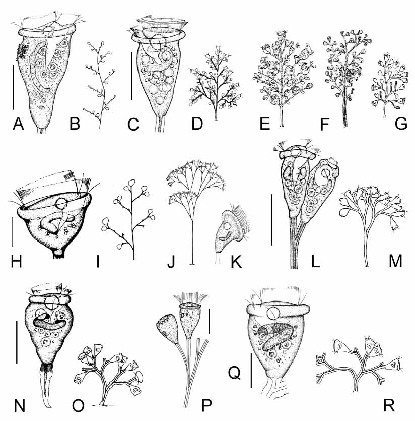
Considering the living appearances and the marine habitat, some Zoothamnium species with dichotomously branched stalk and singlelayered peristomial lip should be compared with the current organism: Z. dichotomum WrightKent, 1882 ; Z. intermedium Precht, 1935 ; Z. paragammari Song, 1991 ; Z. marinum Mereschkowski, 1879 .
Morphologically, Zoothamnium paraentzii shares many similarities with Z. dichotomum WrightKent, 1882 ( Figs. 5 View FIGURE 5 J, 5K) (Table 2). Although the oral apparatus and silverline system, even the body size of Z. dichotomum remains unknown, it can be separated from Z. paraentzii by the regularly dichotomously branching form with zooids located in pairs (vs. irregularly dichotomous branching style with zooids of the same branch born on the same side) and clearly more aboral position of the contractile vacuole (below peristomial lip vs. apically positioned) ( Kahl 1935).
Zoothamnium paraentzii can be clearly distinguished from Z. intermedium Precht, 1935 ( Figs. 5 View FIGURE 5 Q, 5R) (Table 2) ( Song 1991b) by its irregularly, dichotomously branching form and larger cell size.
Zoothamnium paragammari Song, 1991 ( Figs. 5 View FIGURE 5 N, 5 O) (Table 2) also has a similar size and body shape. However, it can be separated from Z. paraentzii by its having distinctly fewer transverse silverlines (57 vs. 75–83) ( Song 1991b) from the peristome to the trochal band. In addition, the structure of peniculus 3 is completely different. In terms of body shape and position of contractile vacuole, Zoothamnium marinum Mereschkowski, 1879 ( Fig. 5 View FIGURE 5 P) (Table 2) is also similar to Z. paraentzii . As the silverline system and infraciliature of Z. marinum remains unknown, the conspicuously larger body size (100 vs. 34 µm after fixation) could be the solely useful distinguishing character ( Kahl 1935).
Another species, Zoothamnopsis mengi Song, 1997 ( Figs. 5 View FIGURE 5 L, 5M), might also be related to the present form in the colony form, appearance of peristomial lip and body shape (Table 2). However, it differs from Zoothamnium paraentzii at least by distinctly different silverline system ( Pseudovorticella type vs. Vorticella type) ( Song 1997).
In addition, the arrangement of the living individuals of Zoothamnium paraentzii is special (zooids of the same branch born on the same side), which distinguishes it from most other known congeners and therefore could be an easily observable feature used for identification of this taxon in ecological studies.
No known copyright restrictions apply. See Agosti, D., Egloff, W., 2009. Taxonomic information exchange and copyright: the Plazi approach. BMC Research Notes 2009, 2:53 for further explanation.
|
Kingdom |
|
|
Phylum |
|
|
Class |
|
|
Order |
|
|
Family |
|
|
Genus |
Zoothamnium paraentzii Song, 1991
| Sun, Ping, Ji, Daode & Song, Weibo 2005 |
Zoothamnopsis mengi
| Song 1997 |
Zoothamnium paragammari
| Song 1991 |
intermedium
| Precht 1935 |
Zoothamnium marinum
| Mereschkowski 1879 |