
Trichomycterus dali, Rizzato & Costa-Jr & Trajano & Bichuette, 2011
|
publication ID |
https://doi.org/10.1590/S1679-62252011000300003 |
|
persistent identifier |
https://treatment.plazi.org/id/BE35C766-D421-2349-5E62-F9856E18F8C0 |
|
treatment provided by |
Carolina |
|
scientific name |
Trichomycterus dali |
| status |
sp. nov. |
Trichomycterus dali View in CoL , new species
( Fig. 2 View Fig )
Holotype. MZUSP 106630 View Materials , 78.9 View Materials mm SL, Brazil, southwestern State of Mato Grosso do Sul, Bonito, Saracura Cave ( 21º9’14.6”S 56º43’22.7”W, elevation 792 m), rio Paraguai basin, Serra da Bodoquena karst area, 15 Jan 2006, E. P. D. Costa-Jr. GoogleMaps
Paratypes. All from Brazil, southwestern State of Mato Grosso do Sul, rio Paraguai Basin , Serra da Bodoquena karst area. MZUSP 106631 View Materials , 1 View Materials , 62.4 View Materials mm SL (c&s), Jardim , Buraco das Abelhas Cave ( 21°29’18.24”S 56°43’28.6”W, elevation 490 m), 18 Apr 1998, E. P. D. Costa-Jr. MZUSP 106632 View Materials , 1 View Materials , 49.0 mm SL, Jardim, Buraco das Abelhas Cave , 18 Apr 1998, E. P. D. Costa-Jr. MZUSP 81056 View Materials , 1 View Materials , 100.8 View Materials mm SL, Jardim, Buraco das Abelhas Cave , 19 Apr 1998, E. P. D. Costa-Jr. MZUSP 106633 View Materials , 1 View Materials , 47.4 View Materials mm SL, Bodoquena, Morro do Jericó Cave ( 20°46’21.51"S 56°44’58.73"W, elevation 193 m), 1 Jul 2008, E. Trajano, A. L. F. Guil & L. M. Cordeiro. MZUSP 106634 View Materials , 1 View Materials , 21.6 View Materials mm SL, Bodoquena, Morro do Jericó Cave , 1 Jul 2008, E. Trajano, A. L. F. Guil & L. M. Cordeiro. MZUSP 106635 View Materials , 1 View Materials , 48.2 View Materials mm SL, Bonito, Saracura Cave , 9 Jul 2006, E. P. D. Costa- Jr. MZUSP 109770 View Materials , 1 View Materials , 72.5 View Materials mm SL (c&s), Bonito, Buraco das Abelhas Cave GoogleMaps .
Diagnosis. Trichomycterus dali is readily distinguished from epigean and hypogean congeners by the presence of two conspicuous, ridge-like adipose folds lining dorsally throughout the body, one anterior (pre-dorsal) and one posterior (post-dorsal) to the dorsal fin, both distinctive autapomorphies in the genus. Other characters that easily distinguish the new species, although not exclusive, are: highly reduced skin pigmentation (except for T. gorgona and caverestricted congeners, T. chaberti , T. itacarambiensis , T. spelaeus , T. sandovali , T. santanderensis and T. uisae ); total loss of eyes, not visible externally (except for T. sandovali and T. spelaeus ); barbels long, especially the nasal (99.3- 143.5% HL) and the maxillary (97.0-131.3% HL) (except for T. longibarbatus and T. spelaeus ); scapulocoracoid with a conspicuous anterior process projected forward (except for T. sandovali , T. spelaeus and T. uisae ), with a narrow base, a wide apex and a rounded distal margin; and pectoral-fin ray count reaching I,9 (except for T. hualco ). Characters possibly exclusive, but about which many taxa lack information, are listed as complementary diagnoses: cranial fontanel unique, extending from the posterior half of supraoccipital to the posterior region of the frontal bones, with a conspicuous constriction on the meeting point of supraoccipital and the two frontal bones; supraorbital long and cylindrical, without projections, with a needle appearance; 27-29 interopercular and 16-19 opercular odontodes.
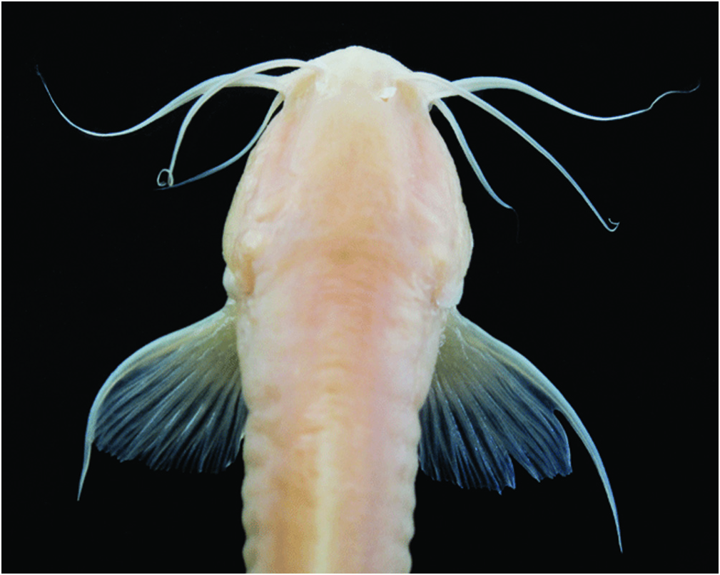
Description. Morphometric and meristic data of holotype and paratypes given in Table 1. Body elongate, semi-cylindrical, becoming compressed towards caudal fin. Dorsal profile of body slightly convex, slight and straight slope from tip of snout to anterior portion of trunk; ventral profile of body straight in lateral view. Profile of body triangular in transversal section. Dorsal portion of caudal peduncle concave at beginning of caudal fin; ventral portion of caudal peduncle straight. Dorsal lobe of caudal fin projected upward. Adipose folds well developed in adult and young specimens, ridge- like, anterior (pre-dorsal) and posterior (post-dorsal) to dorsal fin, lining dorsally throughout body, not supported by procurrent rays. Origin of pre-dorsal adipose fold posterior to pectoral-fin base, reaching dorsal-fin base; origin of postdorsal adipose fold at end of dorsal-fin base, reaching caudal fin, with higher width closer to dorsal fin than to caudal-fin ( Fig. 2 View Fig ). Urogenital and anal openings on vertical through posterior end of dorsal-fin base, between distal margin of pelvic fins.
Head relatively wide and depressed, trapezoidal in dorsal view. Eyes not visible externally ( Fig. 3 View Fig ). Anterior nostril transversally ovoid and slightly smaller than posterior one, surrounded laterally by nasal barbels. Posterior nostril rounded, surrounded anteriorly by large, laterally-folded flap of integument. Mouth slightly subterminal, convex in dorsal view, rictus laterally directed. Lips, mentum and barbels covered by many papillae ( Fig. 4 View Fig ). Barbels long, especially nasal and maxillary. Nasal barbel origin on posterolateral portion of integumentary flap around anterior nostril. When adpressed to body, maxillary barbel extending to middle of pectoral fin; nasal barbel extending to origin of pectoral fin, and submaxilary barbel extending to opercle. Opercular patch of odontodes small and circular. Interopercular odontodes forming slightly convex patch throughout ventral margin of interopercle. Opercle with two processes, upper sharp and pointed backwards, lower acute, and 27-29 opercular odontodes. Interopercle with 16-19 odontodes.
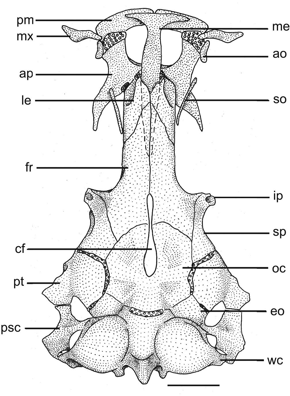
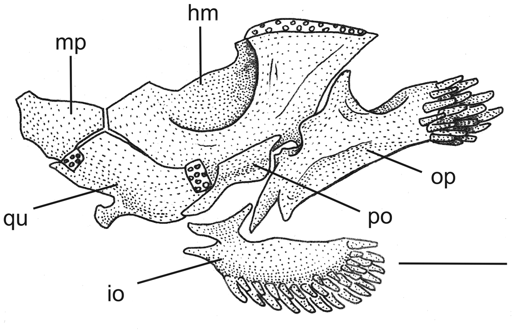
General morphology of cranium: Cranial fontanel unique, extending from posterior half of supraoccipital to posterior region of frontal bones; conspicuous constriction on meeting point of supraoccipital and two frontal bones ( Fig. 5 View Fig ). Anterior process of sphenotic and posterolateral process of frontal as conspicuous, hollow horn-like structure, inside of which emerge infraorbital sensory branches. Supraorbital long and cylindrical, without projections, and needle appearance. Palatine with convex anterior margin covered with cartilage, waist-like medial region and long, narrow posterior process, that becomes sharp on distal region and covers almost totally metapterygoid. Vomer arrow-shaped with long posterior process. Distal profile of mesethmoid straight on dorsal view, main body axis large, cornua reaching 3/4 of premaxillary length. Three to five irregular premaxillary rows of conic teeth curved backwards. Rounded proximal margin of maxilla not reaching premaxilla, covering cartilaginous margin of palatine. Lower jaw with three rows of conical teeth, curved backwards. Hyomandibular with conspicuous semicircular depression, joined tightly to metapterygoid and quadrate. Metapterygoid and quadrate united together by anterior block of cartilage ( Fig. 6 View Fig ).
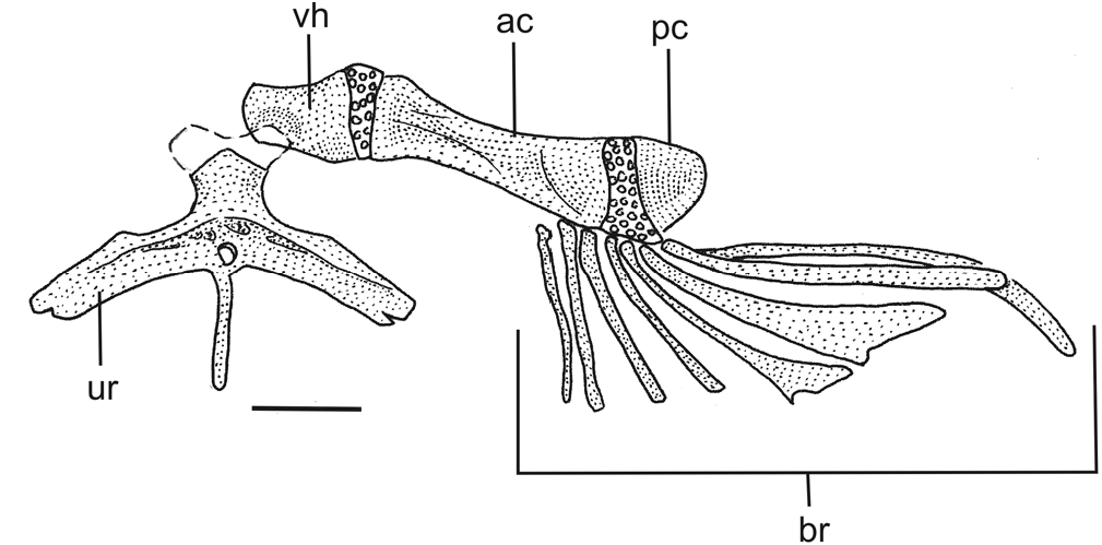
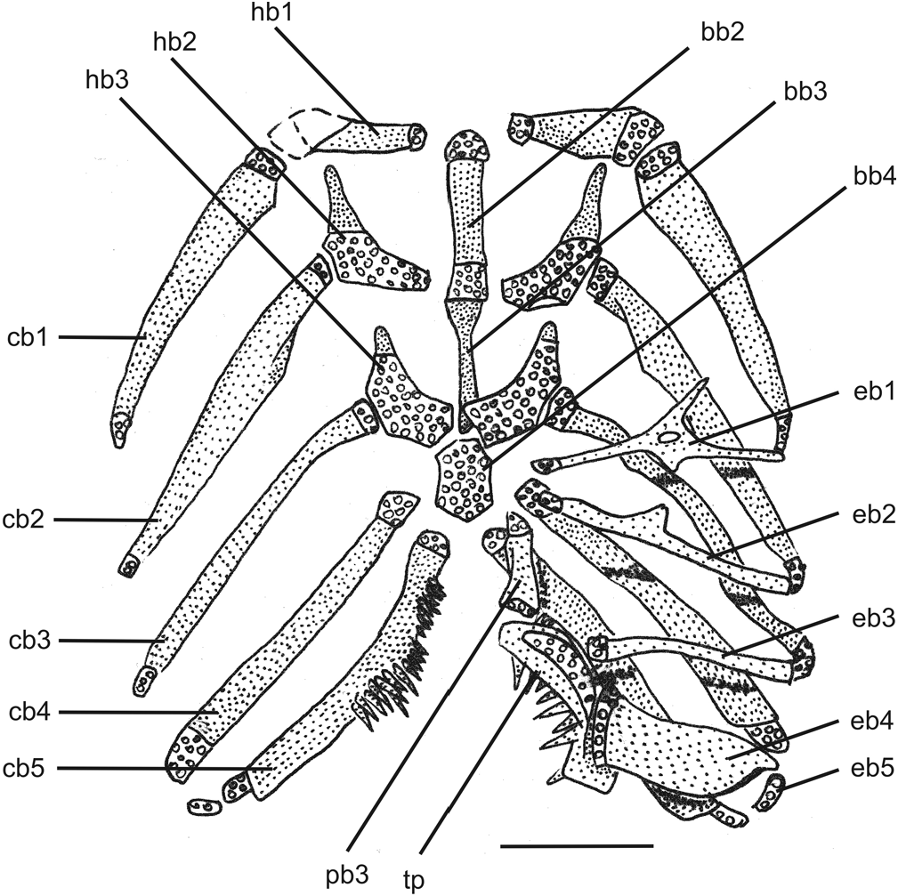
Branchial skeleton and associated structures: Branchiostegal-rays eight, rays 5, 6 and 7 with enlarged distal tip, ray 8 covered by interopercle and reaching ventral margin of opercular patch of odontodes. Urohyal with long, very narrow dorsal process, broad convex posterior margin, lateral surface with distal margins chipped, urohyal-foramen slightly ovoid. Hypohyal with depression to which articulates anterior process of urohyal. Posterior ceratohyal rounded triangular ( Fig. 7 View Fig ). Basibranchials 3, hypobranchials 3, ceratobranchials 5, epibranchials 5, pharyngobranchials 2 ( Fig. 8 View Fig ). Basibranchial 1 absent. Basibranchial 2 and 3 connected to each other by their cartilaginous tips, forming long rod. Basibranchial 3 long and narrow, rectangular on posterior half, almost triangular on anterior half, similar to chalice. Posterior tip of basibranchial 3 covered by cartilaginous posterior portion of hypobranchials 3. Basibranchial 4 completely cartilaginous, approximately hexagonal shaped. Anterior margins of basibranchial 4 bordered by cartilaginous posterior portion of hypobranchials 3, lateral and posterior margins bordered by cartilaginous anterior tips of ceratobranchial 4 and ceratobranchial 5, respectively. Hypobranchial 1 with external portion wider than internal portion, with cartilaginous tips, external cartilaginous tip two to three times wider than internal. Hypobranchial 2 boomerang shaped, posterior half cartilaginous, anterior half ossified, forming long anterior process that almost reaches external posterior margin of hipobranchial 1. Hypobranchial 3 almost completely cartilaginous, only anterior tip ossified in triangular shape, and closely joined to anterior cartilaginous tip of ceratobranchial 3. Hypobranchial 4 absent. Ceratobranchials slightly curved, with cartilaginous tips. Ceratobranchial 1 with internal tip wider than external tip. Ceratobranchial 2 with shallow concavity on its posterior margin, without defined posterior process. Ceratobranchial 3 with pronounced concavity on its posterior margin, limited posteriorly by very small process. Ceratobranchial 5 slightly enlarged, bearing patch of small, narrow conical teeth pointed dorsally on its anterior half, and connected to epibranchial 5 only by upper half of posterior margin. Ceratobranchials 3, 4 and 5 bearing one row of conical, very sharp teeth, on posterior margin of ceratobranchial 3, on both margins of ceratobranchial 4, and on anterior margin of ceratobranchial 5. Epibranchials 1, 2 and 3 narrow, rod-like, with cartilaginous tips. Epibranchial 1 with long, narrow and sharp anterior process, pointed outwards in acute angle. Epibranchial 2 with small, acute process, not uncinate. Epibranchial 3 with conspicuous, posteriorly directed, large uncinate process. Epibranchial 4 large, curved, with wide dorsal margin slightly convex joined to posterior half of tooth plate, covered with cartilage, and with ventral margin very narrow, ovoid and cartilaginous, joined to posterior cartilaginous tip of ceratobranchial 4. Epibranchial 5 very small, curved and completely cartilaginous. Pharyngobranchials 1 and 2 absent. Pharyngobranchial 3 elongate, rod-like, slightly depressed, cartilaginous tips. Pharyngobranchial 4 ossified, curved, ventral margin cartilaginous, joined tightly to anterodorsal half of tooth plate. Tooth plate well developed, curved, one row of long, conic, internally curved teeth, anteriormost of which smaller, posteriormost gradually larger.
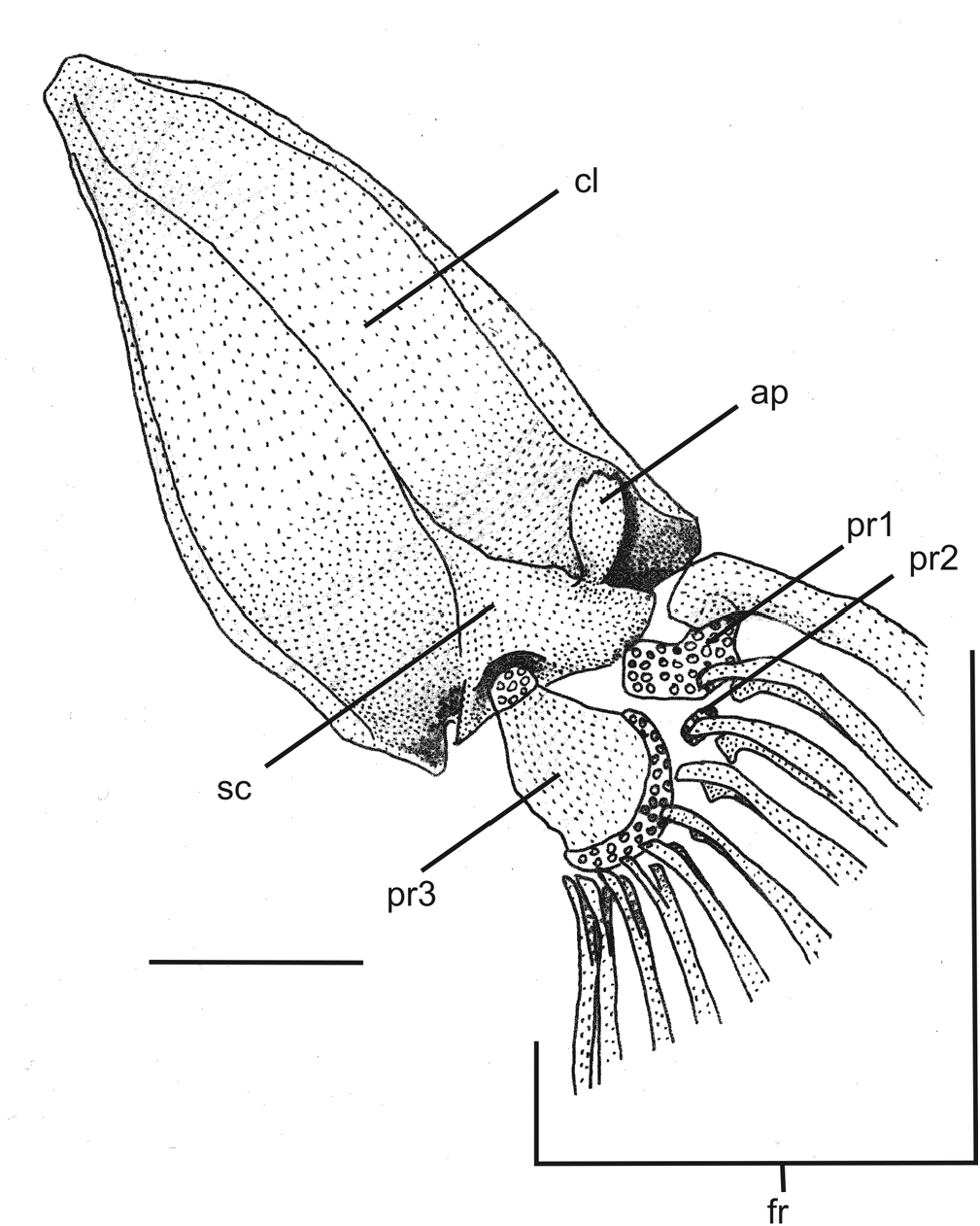
Postcranial skeleton. Total vertebrae 34-35, 10-11 pairs of ribs. First ribs thicker, posteriormost gradually thinner. Pectoral girdle with wide cleithrum, becoming narrower at anterior half, joined together by very narrow anterolateral margin. Posterior process of pectoral girdle reaching lower surface of posttemporosupracleithrum. Scapulocoracoid with conspicuous anterior process projected forward, with narrow base, wide apex and rounded distal margin. Proximal radial 1 cartilaginous, joined to first and second pectoral-fin rays. Proximal radial 2 very small, cartilaginous, joined to third pectoral-fin ray. Proximal radial 3 large and well ossified, with semicircular cartilaginous posterior margin joined to fourth to tenth pectoral-fin rays, and small, ovoid anterior cartilaginous margin, joined to scapulocoracoid ( Fig. 9 View Fig ). Pelvic girdle with external anterior process narrower and slightly longer than internal anterior process. A short medial process may occur ( Fig. 10 View Fig ). Epural absent. Neural spine (n = 1) of preural centrum reduced to almost one-fourth of uroneural length, rounded tip projected upward. Hemal spine of penultimate vertebrae with cartilaginous tip and bearing one procurrent ray. Parhypural partially fused to hypural 1+2, forming trapezoidal lower hypural plate. Hypural 3 not fused to Hypural 4+5. Uroneural with acute distal tip, not fused to Hypural 4+5, long, reaching distal margin of upper hypural plate. Upper hypural plate triangular. Lower apophysis of preural centrum projected forward, with acute tip ( Fig. 11 View Fig ).
Pectoral fin triangular in dorsal view, with wide base and ten rays, first ray longer, unbranched and filamentous (I,9). On one type C&S specimen, left pectoral fin counted I,8. Dorsal fin approximately semicircular in lateral view, with nine rays, the two first unbranched; in addition, two micro rays anterior to first ray (ii,II,7). Pterygiophores with eight narrow basal radials on dorsal fin, curved on basal portion, with espatulate distal region curved backwards and cartilaginous distal tip. First basal radial has two folds backwards and narrow cartilaginous distal margin pointing backwards. Last basal radial with laminar posterior expansion on its distal fold, joined to two last dorsal-fin rays. Each basal radial, except for first, joined to small, half cartilaginous half ossified distal radial, between two basal branches of each ray. Anal fin approximately rectangular in lateral view, distal margin straight, with seven rays; in addition two micro rays anterior to first ray (ii,II,5). On one type C&S specimen, ray count varied, and presented as one micro ray, I,6, being the micro ray branched ( Table 1). Pterygiophores with six narrow basal radials on anal fin, morphologically similar to dorsal pterygiophores: with espatulate, curved distal region, cartilaginous tips, first with two folds, last with laminar posterior expansion holding two last rays, each basal radial except first joined to small distal radial half ossified half cartilaginous between two basal branches of each ray. Pelvic fin rectangular in ventral view, separated from each other, with five rays, first ray unbranched (I,4). Origin of pelvic-fin base on vertical line through origin of dorsal-fin base. Margin of caudal fin straight, 14 principal rays (six in upper lobe, first branched or unbranched; eight in lower lobe, two first unbranched). On upper lobe, first three (uppermost) principal rays joined to hypural 4+5, next three joined to hypural 3. On lower lobe, first (lowermost) principal ray begins in gap between hemal spine of penultimate vertebrae and parhypural, second joined to parhypural and other joined to hypural 1+2. Six dorsal procurrent rays; ventral procurrent rays varying from 4 to 8 (counts of c&s paratypes, see Table 1).
Color in alcohol. Body generally pale yellowish, dorsal ridge (anterior and posterior to dorsal fin) brighter than body and tending to translucent. Mouth and barbels light yellowish pale to white. Pectoral, dorsal, pelvic, anal and caudal fins hyaline ( Fig. 2 View Fig ).
Color in life. Body generally white, tending to translucent, dorsal ridge darker than body, with light yellow color. Internal organs seen by transparency ( Fig. 12 View Fig ).
Etymology. The specific name (a noun in apposition) is an allusion to the Spanish artist Salvador Dali, in reference to his famously long moustache (or whisker).
Distribution. Trichomycterus dali is known exclusively from subterranean waters in at least three caves in Serra da Bodoquena karst area: Buraco das Abelhas, Saracura and Morro do Jericó caves.
Notes on habitat, ecology and behavior. The Buraco das Abelhas Cave is an underwater cave, with more than 2000 m of explored conduits and maximum depths of 55 m, located in the southern portion of Serra da Bodoquena National Park, in the rio Miranda lowlands. The only cave entrance so far known opens into a large room with a lake, whose level increases during rain seasons, completely flooding the saloon and only allowing access through cave diving from the entrance. The cave continues as a large, 1200 m long flooded conduit, called tunnel A. Two hundred meters from the entrance, on the right side, the tunnel A branches into tunnel B, 700 meters long and depth varying from 40 meters to up to 12 meters at the distal explored end of the conduit. Eight hundred meters far from the cave entrance, again on the right side of tunnel A, opens tunnel C; 200 meters inside tunnel C, on the left, tunnel D starts, going to a depth of up to 12 meters. Non-troglomorphic fish, such as the catfish Rhamdia quelen and tetra characins, Astyanax spp. , were observed regularly in the entrance room, and farther in tunnel A until 200 m from the cave entrance, reaching a depth of 49 m. At 100 meters from the cave entrance, 12 m deep, these fish become syntopic with T. dali .
The Saracura Cave small entrance opens into a 230 m long dry conduit with a travertine lake at its distal end accessing a large underwater cave, not totally explored so far. The underwater portion starts with a nearly vertical drop into a 4 m diameter tunnel, becoming horizontal at the depth of 60 m. The last exploratory dive took 130 meters of guidelines and ended at a depth of 65 m in a huge horizontal tunnel still to be explored.
The Jericó Cave has three small entrances opening in the Salobra canyon. It is predominantly horizontal, with dry conduits about 15 m high and 2 m wide. Trichomycterus dali specimens were collected in a large pool at the distal end of the lower conduit, which is partially filled by sandy sediments. During the collections (mid-dry season), maximum depth in the pool was about 1 m, width was 2.5 m and extension, 4 m; laboratory in São Paulo. Three specimens collected in 2000 and 2001 were still alive in 2010.
water flowed slowly transverly to the conduit (L. M. Cordeiro, pers. comm.).
Trichomycterus dali catfishes can be found in the permanently dark areas zone, calmly swimming in the water column and close to the rocky substrate (bottom and walls). In Buraco das Abelhas, individuals were observed from 12 up to 55 meters deep (maximum depth of the cave system); in Saracura cave, fish swim from near the surface up to a depth of 65 meters, where the main tunnel stops dropping down and become horizontal. The light from halogen underwater spotlights, 50W, used at the time the first specimens were collected, as well as from 10 to 20W HID spotlights used more recently, did not trigger any visible reaction in these fishes. Cave divers also seem not to disturb the fishes, but, as one try to catch them, they start to swim very fast towards the deeper parts of the caves, against the current, near the bottom and into small crevices. Sometimes they were observed digging the soft bottom of Buraco das Abelhas.
After being hand netted at depths of up to 65 m, the specimens brought to the surface did not exhibit any reactions related to pressure reduction. Several specimens were kept alive in small water boxes, aired with battery powered air pumps during at least a week and, then, traveled 1,200 km until the


No known copyright restrictions apply. See Agosti, D., Egloff, W., 2009. Taxonomic information exchange and copyright: the Plazi approach. BMC Research Notes 2009, 2:53 for further explanation.
|
Kingdom |
|
|
Phylum |
|
|
Class |
|
|
Order |
|
|
Family |
|
|
Genus |