
Chococtenus, Dupérré, Nadine, 2015
|
publication ID |
https://doi.org/10.11646/zootaxa.4028.4.1 |
|
publication LSID |
lsid:zoobank.org:pub:DE76F18B-422D-4D97-93FD-F211F691F591 |
|
DOI |
https://doi.org/10.5281/zenodo.6097677 |
|
persistent identifier |
https://treatment.plazi.org/id/BC658789-6A6F-FFBB-7397-FF3BFDEA30D1 |
|
treatment provided by |
Plazi (2016-04-22 01:08:06, last updated 2024-11-24 20:05:04) |
|
scientific name |
Chococtenus |
| status |
gen. nov. |
Chococtenus View in CoL View at ENA new genus
Type species. Chococtenus otonga n. sp.
Etymology. The generic name is a contraction of “Chocó” and “ Ctenus ” in reference to the hotspot corridor running from Panama to Peru where the type species was first discovered. The gender is masculine.
Diagnosis. The genus Chococtenus is diagnosed from other Acantheinae by the presence of unique ventral and a spine-like retrolateral tibial apophysis ( Fig. 2 View FIGURES 1 – 5 ). Males Chococtenus are differentiated from Enoploctenus Simon 1897 ( Dupérré 2014: fig. 7) and Phymatoctenus Simon 1897 by their unique, prominent ventral tibial apophysis, absent in the later genera. Furthermore males of Enoploctenus are distinguished by the presence of a retroapical tibial notch on the palpal tibia ( Dupérré 2014: fig. 8), absent in Chococtenus ( Figs 3 View FIGURES 1 – 5 , 7 View FIGURES 6 – 9 ). Females are distinguished by the presence of lateral internal pockets of the epigynum ( Figs 5 View FIGURES 1 – 5 , 9 View FIGURES 6 – 9 , 24 View FIGURES 20 – 24 ). From Enoploctenus , Phymatoctenus and Tuticanus Simon 1897 by the particularly wide median sector of the epigynum, longer than high ( Figs 12 View FIGURES 10 – 13 , 23 View FIGURES 20 – 24 , 27 View FIGURES 25 – 28 ); narrow, higher than long in Enoploctenus ( Dupérré 2014: fig. 4), Phymatoctenus and Tuticanus .
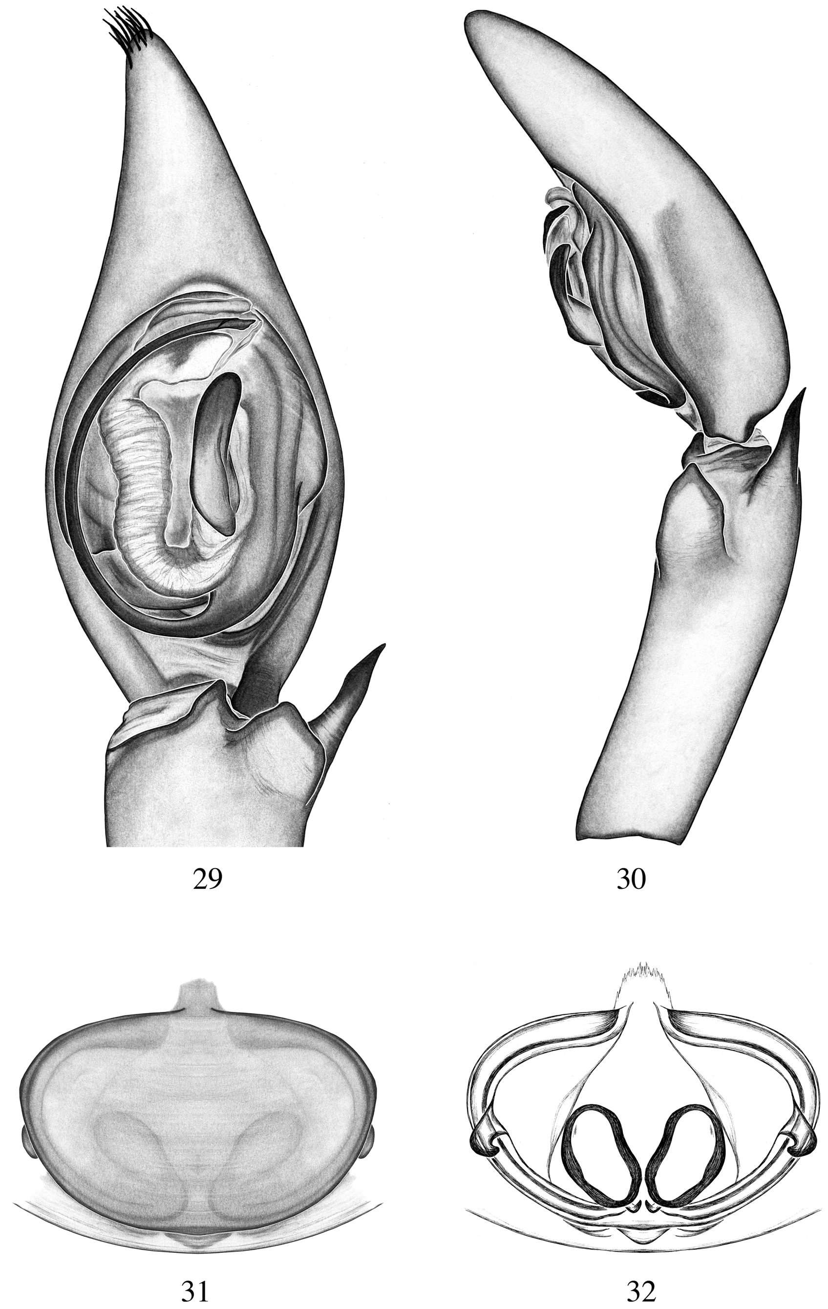
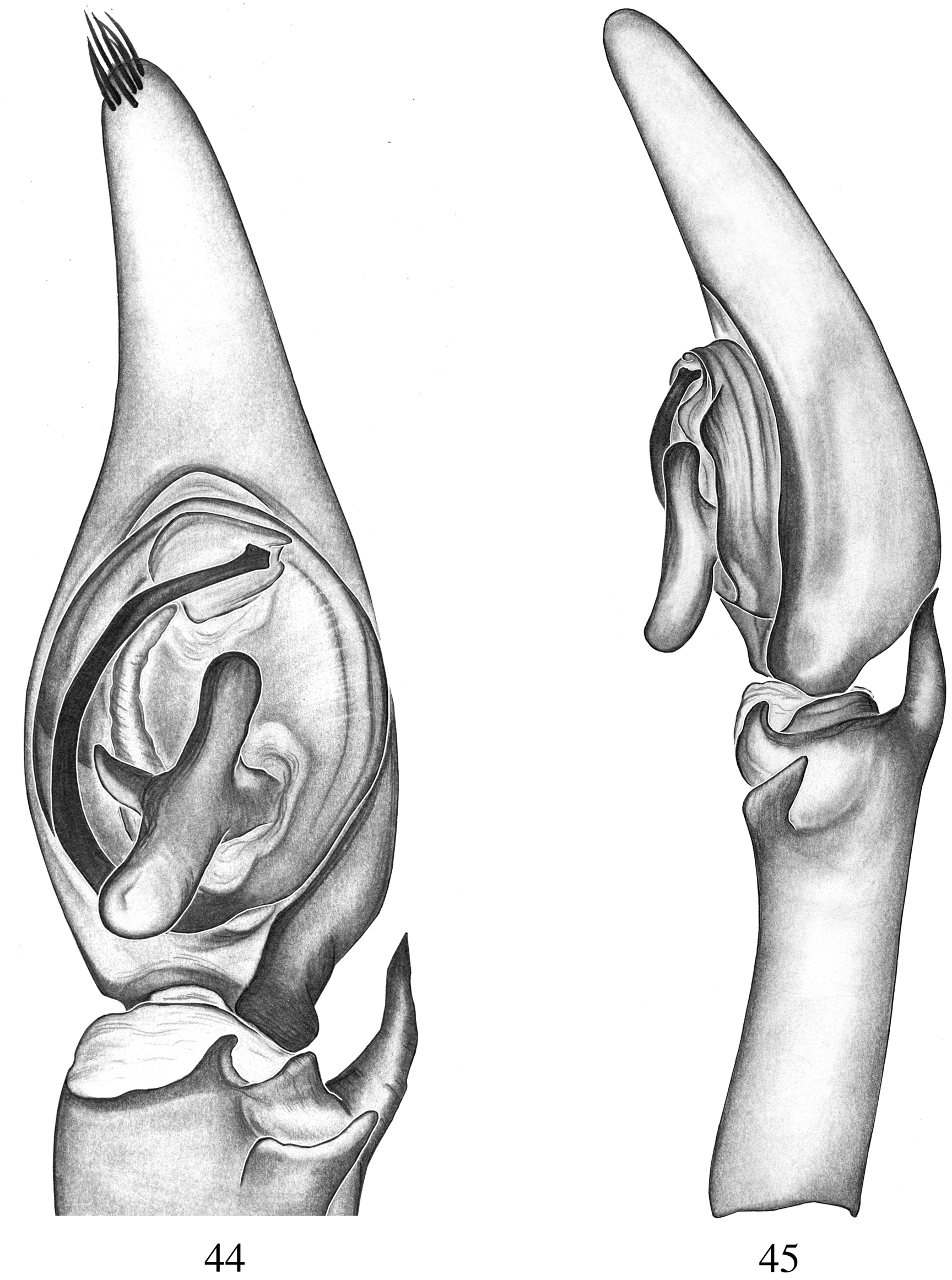
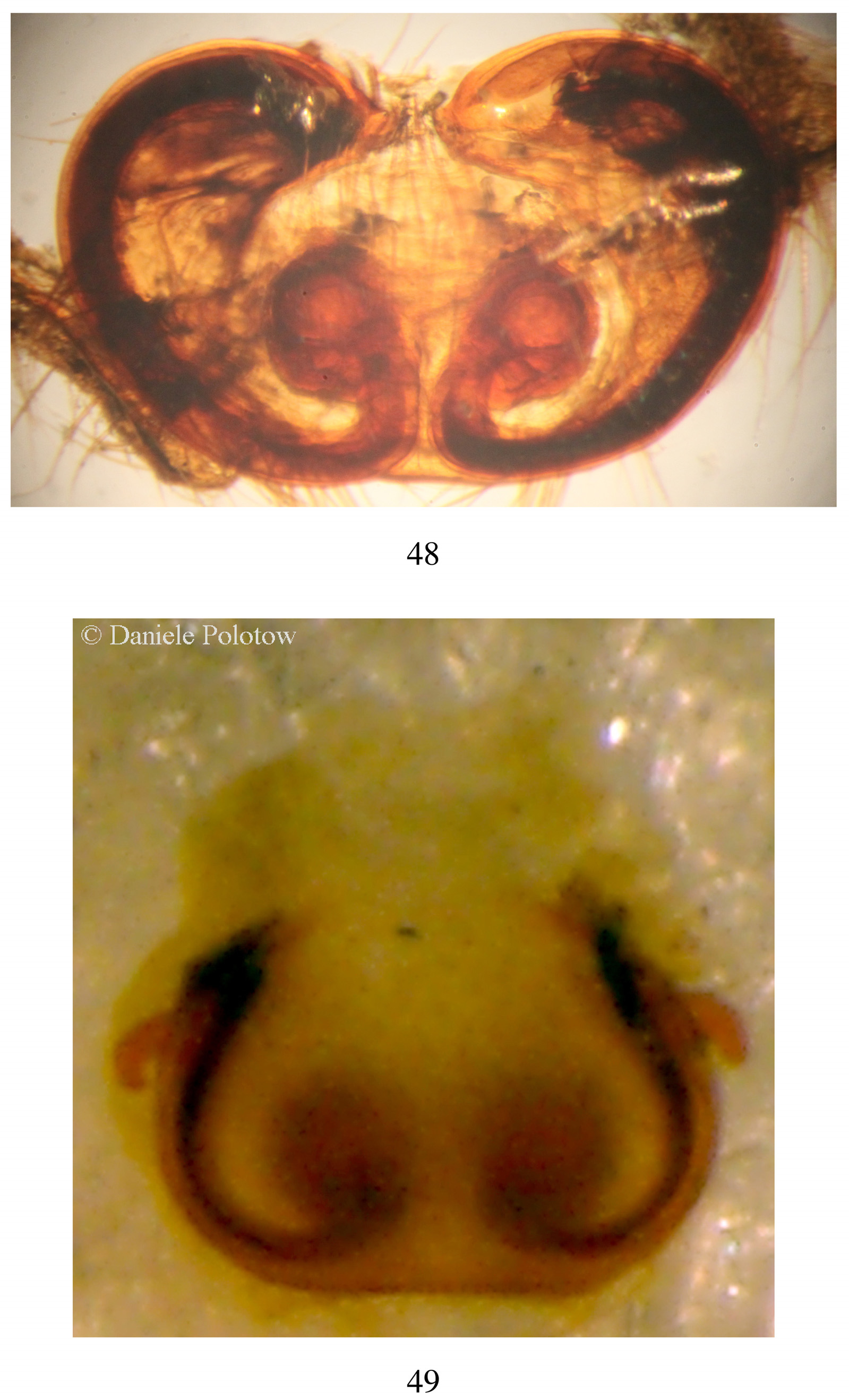
Description. Male. Small to medium (3.3–9.5mm) size spiders. CARAPACE: Piriform, slightly longer than wide, two types of colour pattern: as C. otonga ( Figs 1 View FIGURES 1 – 5 , 55 View FIGURES 55, 56. 55 ) or as C. cappuccino and C. otongachi ( Figs 20 View FIGURES 20 – 24 , 33 View FIGURES 33 – 37 ); shallow thoracic groove; fovea longitudinal. Eyes pattern 2-4-2, rounded; AME, ALE the smallest, PME, PLE the biggest ( Figs 53, 54 View FIGURES 53, 54. 53 ). Clypeus low, 1/ 2x AME ( Figs 53, 54 View FIGURES 53, 54. 53 ). Chelicerae promargin with 3 teeth, retromargin with 4 teeth; with two black stripes ( Figs 53, 54 View FIGURES 53, 54. 53 ). Endites longer than wide. Labium rectangular. Sternum flat, as long as wide. LEGS: Evenly coloured or with black bands on femur, tibiae and metatarsi. Leg formula 4123. Trochanters notched. Ventral scopula on tarsi and metatarsi I. Femur with 3 dorsal and no ventral spines. Tibia I and II with five pairs of ventral spines, metatarsi I and II with three pairs of ventral spines. Tarsal claw unipectinate, with 7–10 teeth; with dense claw tufts. ABDOMEN: oval, with median half-length pale band, uniformly greyish-brown colour ( Figs 1 View FIGURES 1 – 5 , 55, 56 View FIGURES 55, 56. 55 ) or with a pattern of darker patches. GENITALIA: Cymbium pointed apically with modified setae, as long as tibia; dorsal scopula present; with basal cymbial retrolateral keel ( Figs 2 View FIGURES 1 – 5 , 6 View FIGURES 6 – 9 , 25 View FIGURES 25 – 28 ). Tibia as long as the cymbium; two prolateral spines, one retrolateral; retrolateral tibial apophysis well developed, spine-like ( Figs 3 View FIGURES 1 – 5 , 15 View FIGURES 14 – 17 , 22 View FIGURES 20 – 24 ) or slightly curved ( Fig. 43 View FIGURES 42, 43 ); ventral tibial apophysis variable, somewhat transparent apically, curved ( Fig. 2 View FIGURES 1 – 5 ), elongated ( Figs 6 View FIGURES 6 – 9 , 10 View FIGURES 10 – 13 ), rectangular ( Figs 14 View FIGURES 14 – 17 , 18 View FIGURES 18 – 19 ) or rounded ( Fig. 42 View FIGURES 42, 43 ). Tegulum oval ( Figs 2 View FIGURES 1 – 5 , 21 View FIGURES 20 – 24 , 38 View FIGURES 38 – 41 ) sometimes with a retrolateral swelling ( Figs 6 View FIGURES 6 – 9 , 14 View FIGURES 14 – 17 , 29 View FIGURES 29 – 32 ); with a hyaline sector at the base of the median apophysis. Median apophysis variable, elongated ( Fig. 6 View FIGURES 6 – 9 ), curved ( Fig. 10 View FIGURES 10 – 13 ), straight ( Fig. 14 View FIGURES 14 – 17 ) or with basal projection ( Figs 25 View FIGURES 25 – 28 , 38 View FIGURES 38 – 41 , 42 View FIGURES 42, 43 , 44 View FIGURES 44, 45 , 46 View FIGURES 46, 47 ). Membranous tegular process originating at the base of the median apophysis and the embolus, elongated, hyaline, ribbed ( Fig. 2 View FIGURES 1 – 5 ), long ( Fig. 14 View FIGURES 14 – 17 ) or short ( Fig. 10 View FIGURES 10 – 13 ), sometimes hidden behind the embolus ( Fig. 38 View FIGURES 38 – 41 ). Conductor positioned at tip of embolus, hyaline, variably shaped, sometimes folded ( Fig. 21 View FIGURES 20 – 24 ). Embolus base sometimes twisted ( Fig. 2 View FIGURES 1 – 5 ) or straight ( Figs 6 View FIGURES 6 – 9 , 10 View FIGURES 10 – 13 ). Embolus filiform, thin ( Figs 6 View FIGURES 6 – 9 , 14 View FIGURES 14 – 17 , 21 View FIGURES 20 – 24 ) or broad ( Figs 38 View FIGURES 38 – 41 , 42 View FIGURES 42, 43 ), tip of different shapes ( Figs 21 View FIGURES 20 – 24 , 34 View FIGURES 33 – 37 ).
Female. Small to medium (3.3–9.5mm) size spiders. Somatic morphology as in male, colouration somewhat darker and more contrasted than in males. LEGS: As in males, sometimes with pattern of yellow-orange to dark brown or blackish bands; with less leg spines; legs shorter than males compared to body length. Tarsal claw unipectinate, with 4 teeth and dense claw tufts. Pedipalp claw unipectinate with 4 short teeth. GENITALIA: Epigynum with strongly sclerotized median sector, either pentagonal ( Fig. 4 View FIGURES 1 – 5 ), triangular ( Fig. 8 View FIGURES 6 – 9 ), rounded ( Fig. 16 View FIGURES 14 – 17 ), elliptical ( Figs 23 View FIGURES 20 – 24 , 31 View FIGURES 29 – 32 , 36 View FIGURES 33 – 37 ) or heart-shaped ( Figs 12 View FIGURES 10 – 13 , 27 View FIGURES 25 – 28 ); unsclerotized median sector can be wide ( Fig. 4 View FIGURES 1 – 5 ), medium ( Fig. 16 View FIGURES 14 – 17 ), narrow ( Fig. 23 View FIGURES 20 – 24 ) or absent ( Fig. 12 View FIGURES 10 – 13 ); slightly concave ( Fig. 4 View FIGURES 1 – 5 ) or flat ( Fig. 23 View FIGURES 20 – 24 ); lateral processes elongated ( Figs 4 View FIGURES 1 – 5 , 8 View FIGURES 6 – 9 ) or short ( Figs 12 View FIGURES 10 – 13 , 16 View FIGURES 14 – 17 ), positioned basally ( Figs 23 View FIGURES 20 – 24 , 27 View FIGURES 25 – 28 ) or medially ( Figs 36 View FIGURES 33 – 37 , 40 View FIGURES 38 – 41 ). Internal genitalia with copulatory ducts long, curved, positioned laterally ( Fig. 5 View FIGURES 1 – 5 ) or short and positioned more ventrally ( Fig. 13 View FIGURES 10 – 13 ) in internal lateral pockets ( Figs 5 View FIGURES 1 – 5 , 9 View FIGURES 6 – 9 , 24 View FIGURES 20 – 24 ); spermathecae rounded ( Figs 5 View FIGURES 1 – 5 , 48 View FIGURES 48, 49. 48 ) to oval ( Fig. 17 View FIGURES 14 – 17 ); fertilization ducts short positioned at the base of the spermathecae, directed medially ( Fig. 5 View FIGURES 1 – 5 ).
Composition. Fifteen species: C. acanthoctenoides ( Schmidt 1956) n. comb., C. cappuccino n. sp., C. kashakara n. sp., C. cuchilla n. sp., C. duendecito n. sp., C. fantasma n. sp., C. lasdamas n. sp., C. luchoi n. sp., C. miserabilis ( Strand 1916) , C. neblina n. sp., C. otonga n. sp., C. otongachi n. sp., C. piemontana n. sp., C. suffuscus n. sp. and C. waitti n. sp.
Distribution. Colombia and Ecuador.
Duperre, N. (2014) Demystifying three species of Ctenidae (Arachnida: Araneae) described by Embrik Strand. Part I, Ecuador. Zootaxa, 3784 (1), 67 - 73. http: // dx. doi. org / 10.11646 / zootaxa. 3784.1.3
Schmidt, G. (1956) Genus- und Speziesdiagnosen neuer, mit Bananen eingeschleppter Spinnen nebst Mitteilung uber das Auffinden der Mannchen zweier Spinnenarten. Zoologischer Anzeiger, 157, 24 - 31.
Strand, E. (1916) Systematische-faunistische Studien uber palaarktische, afrikanische und amerikanische Spinnen des Senckenbergischen Museums. Archiv fur Naturgeschichte, 81 (A 9), 1 - 153.

FIGURES 1 – 5. Chococtenus otonga new species. Male. 1 – 3. Female. 4, 5. 1. Habitus, dorsal view. 2. Palp, ventral view. 3. Palp, retrolateral view. 4. Epigynum, ventral view. 5. Internal genitalia, dorsal view. Abbreviations: Male genitalia: c, conductor; ck, cymbial keel; e, embolus; eb, embolus base; ma, median apophysis; mtp, membranous tegular process; rta, retrolateral tibial apophysis; te, tegulum; vta, ventral tibial apophysis; vtp, ventral tibial process. Female genitalia: bs, base of spermathecae; cd, copulatory ducts; fd, fertilization ducts; hs, head of spermathecae; ilp, internal lateral pockets; lp, lateral processes; ms, median sector; ums, unsclerotized median sector.
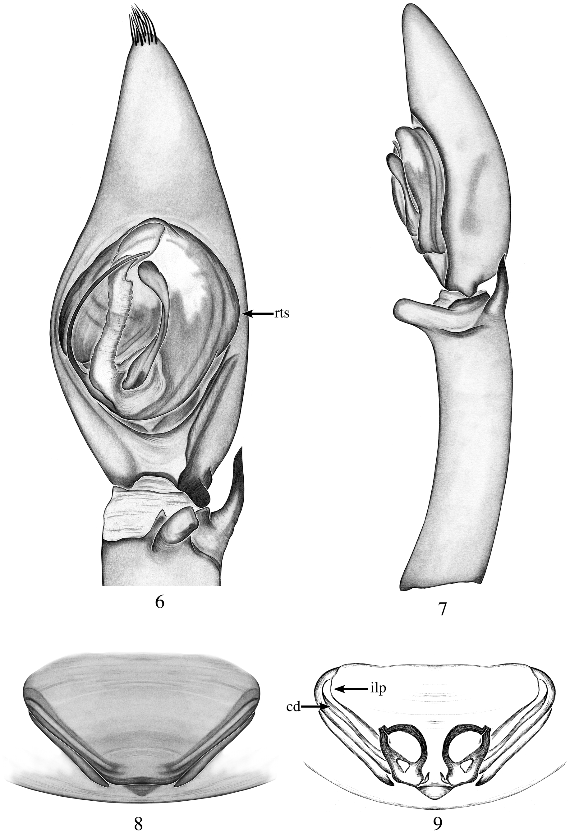
FIGURES 6 – 9. Chococtenus neblina new species. Male. 6, 7. Female. 8, 9. 6. Palp, ventral view. 7. Palp, retrolateral view. 8. Epigynum, ventral view. 9. Internal genitalia, dorsal view. Abbreviations: Male genitalia: rts, retrolateral tegular swelling. Female genitalia: cd, copulatory ducts; ilp, internal lateral pockets.

FIGURES 20 – 24. Chococtenus cappuccino new species. Male. 20 – 22. Female. 23, 24. 20. Carapace, dorsal view. 21. Palp, ventral view. 22. Palp, retrolateral view. 23. Epigynum, ventral view. 24. Internal genitalia, dorsal view. Abbreviations: Female genitalia: ilp, internal lateral pockets; ums, unsclerotized median sector.
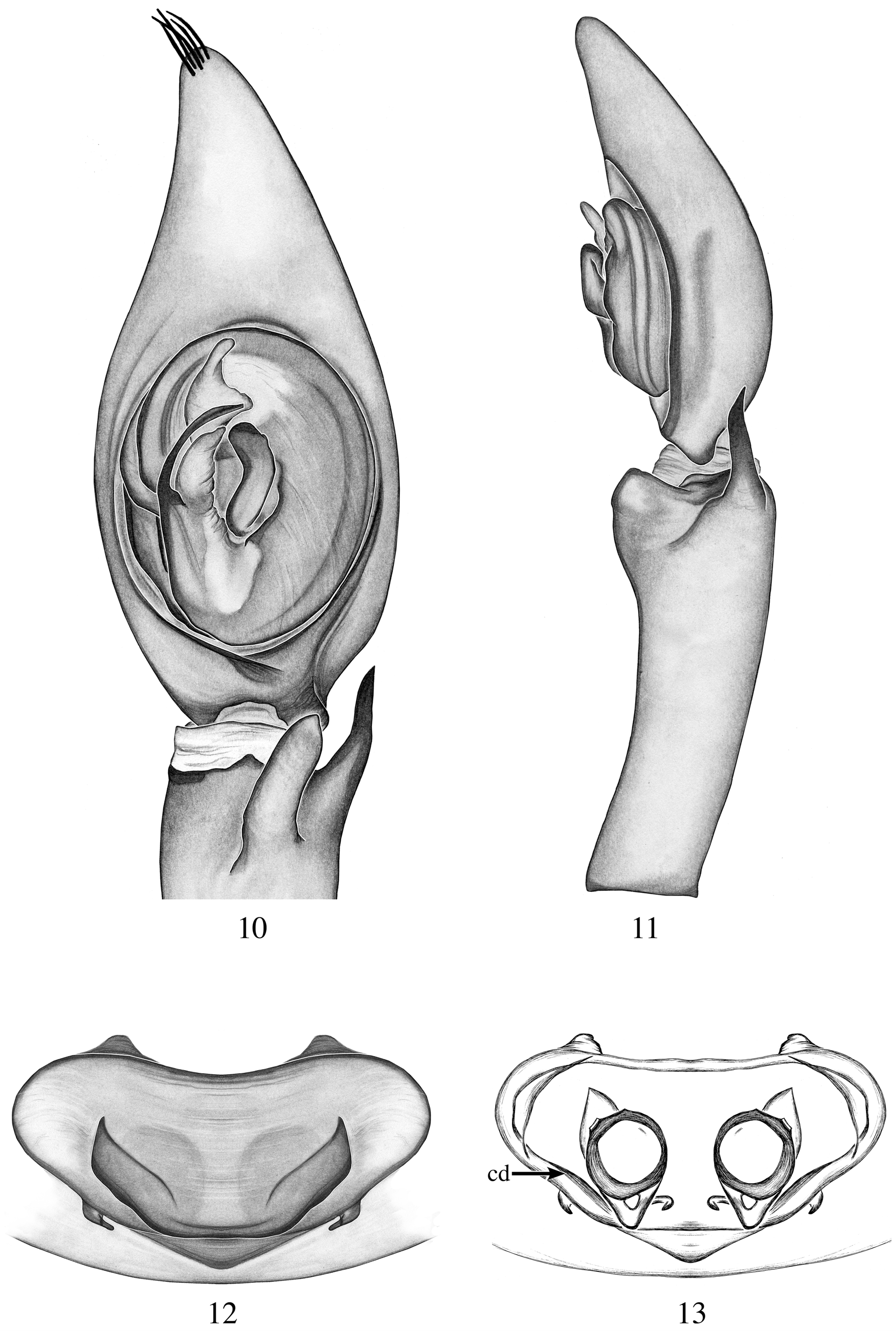
FIGURES 10 – 13. Chococtenus cuchilla new species. Male. 10, 11. Female. 12, 13. 10. Palp, ventral view. 11. Palp, retrolateral view. 12. Epigynum, ventral view. 14. Internal genitalia, dorsal view. Abbreviations: Female genitalia: cd, copulatory ducts.

FIGURES 25 – 28. Chococtenus lasdamas new species. Male. 25, 26. Female. 27, 28. 25. Palp, ventral view. 26. Palp, retrolateral view. 27. Epigynum, ventral view. 28. Internal genitalia, dorsal view.

FIGURES 55, 56. 55. Male Chococtenus otonga new species. 56. Female Chococtenus neblina new species.
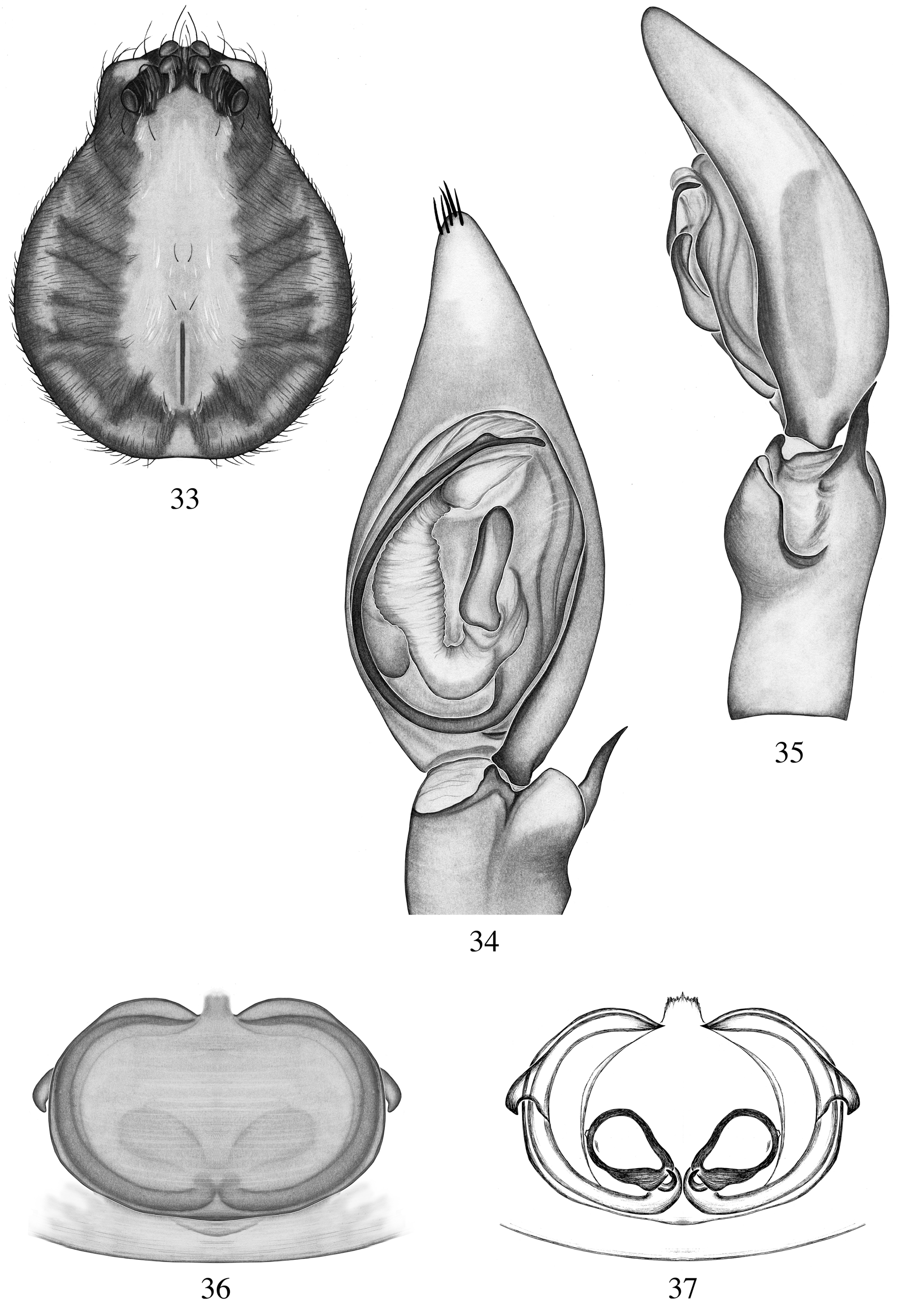
FIGURES 33 – 37. Chococtenus otongachi new species. Male. 33 – 35. Female. 36, 37. 33. Carapace, dorsal view. 34. Palp, ventral view. 35. Palp, retrolateral view. 36. Epigynum, ventral view. 37. Internal genitalia, dorsal view.
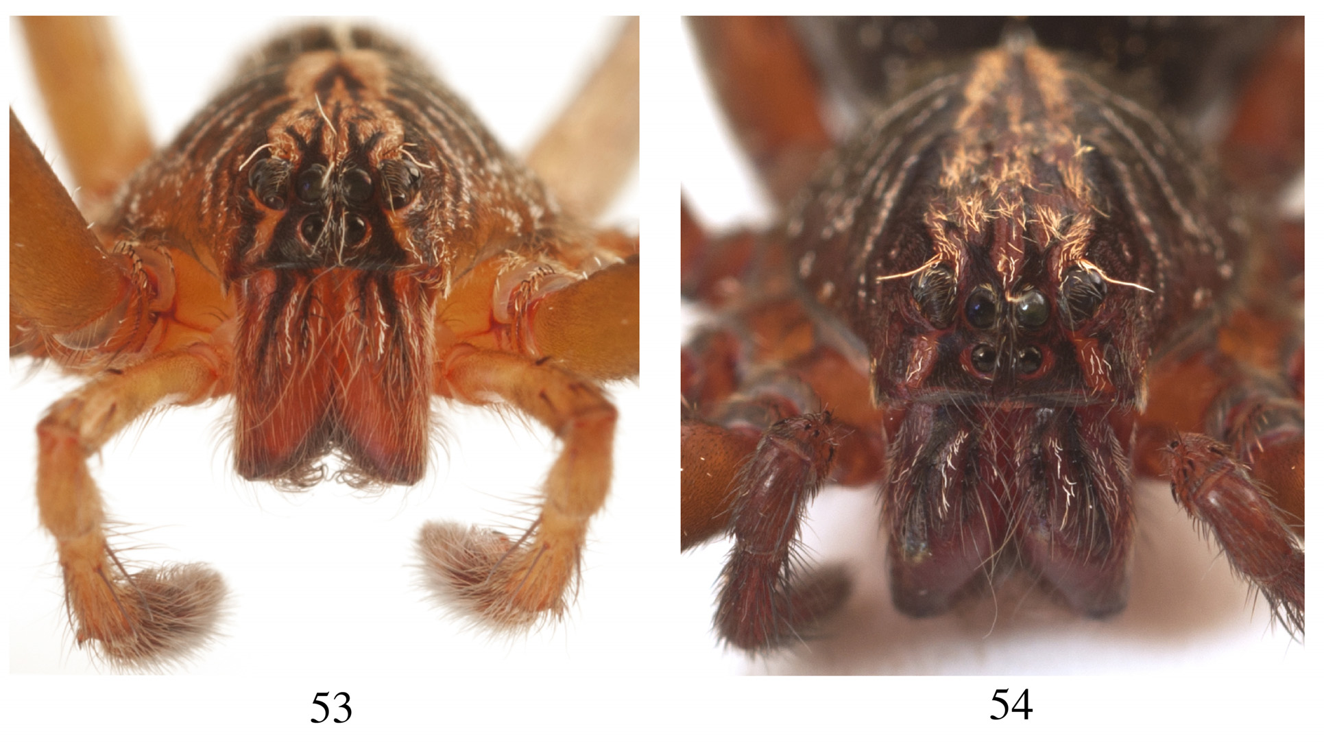
FIGURES 53, 54. 53. Male Chococtenus otonga new species, frontal view. 54. Female Chococtenus neblina new species, frontal view.

FIGURES 14 – 17. Chococtenus fantasma new species. Male. 14, 15. Female. 16, 17. 14. Palp, ventral view. 15. Palp, retrolateral view. 16. Epigynum, ventral view. 17. Internal genitalia, dorsal view. Abbreviations: Female genitalia: cd, copulatory ducts; ums, unclerotized median sector.
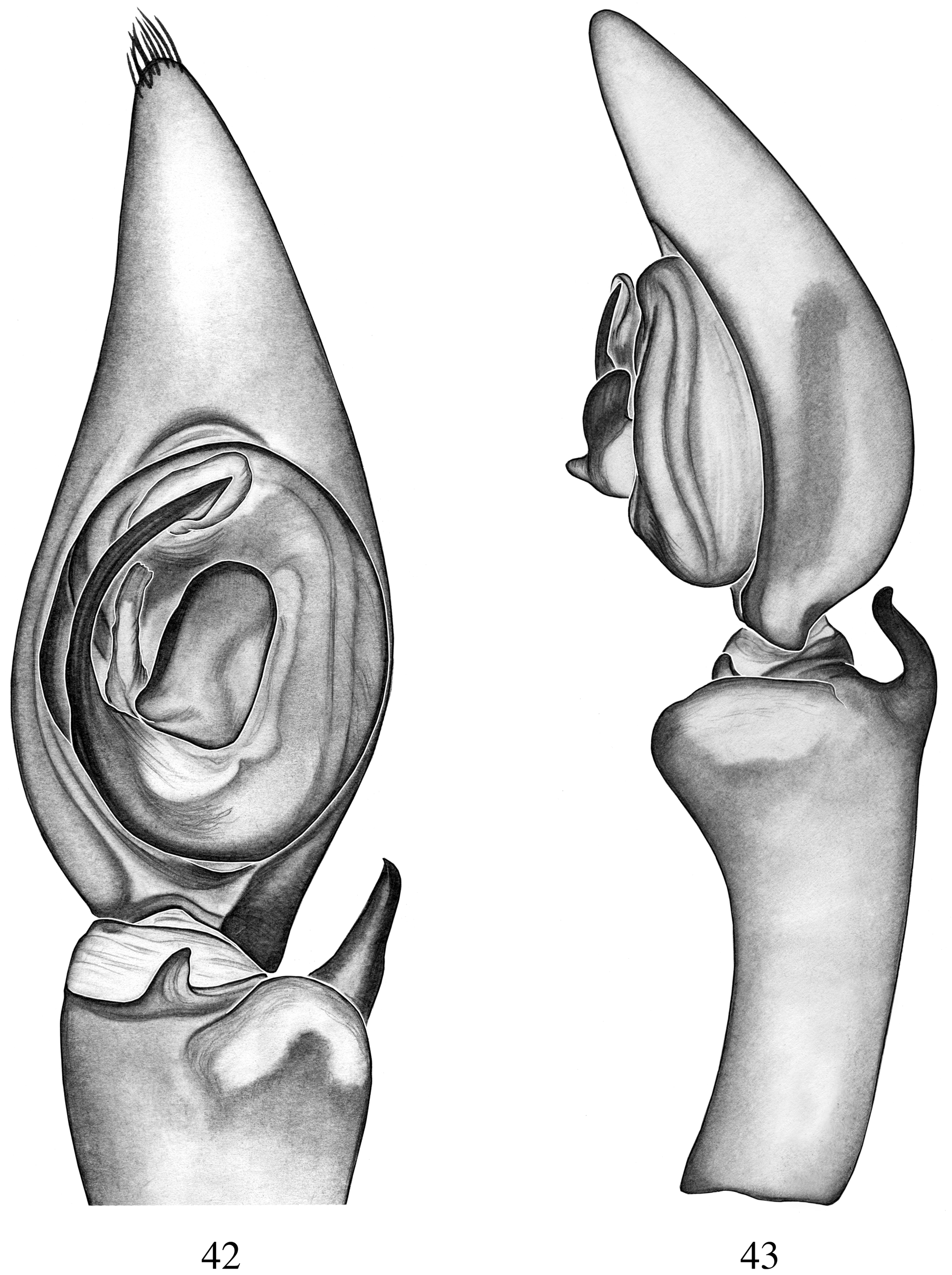
FIGURES 42, 43. Chococtenus piemontana new species. 42. Male palp, ventral view. 43. Male palp, retrolateral view.

FIGURES 18 – 19. Chococtenus waitti new species. Male. 18, 19. 18. Palp, ventral view. 19. Palp, retrolateral view.

FIGURES 38 – 41. Chococtenus kashakara new species. Male. 38, 39. Female. 40, 41. 39. Palp, ventral view. 39. Palp, retrolateral view. 40. Epigynum, ventral view. 41. Internal genitalia, dorsal view.
FIGURES 29 – 32. Chococtenus suffuscus new species. Male. 29, 30. Female. 31, 32. 29. Palp, ventral view. 30. Palp, retrolateral view. 31. Epigynum, ventral view. 32. Internal genitalia, dorsal view.
FIGURES 44, 45. Chococtenus luchoi new species. 44. Male palp, ventral view. 45. Male palp, retrolateral view.

FIGURES 46, 47. Chococtenus duendecito new species. 46. Male palp, ventral view. 47. Male palp, retrolateral view.
No known copyright restrictions apply. See Agosti, D., Egloff, W., 2009. Taxonomic information exchange and copyright: the Plazi approach. BMC Research Notes 2009, 2:53 for further explanation.
1 (by plazi, 2016-04-22 01:08:06)
2 (by ImsDioSync, 2017-01-20 16:21:16)
3 (by ImsDioSync, 2017-01-20 16:23:25)
4 (by ImsDioSync, 2017-06-22 01:58:06)
5 (by ExternalLinkService, 2019-09-26 10:20:34)
6 (by ExternalLinkService, 2022-01-30 01:54:21)
7 (by ExternalLinkService, 2022-02-15 23:53:14)
8 (by plazi, 2023-10-29 21:37:31)