
Diadiplosis smithi Felt
|
publication ID |
https://doi.org/10.11646/zootaxa.4847.1.1 |
|
publication LSID |
lsid:zoobank.org:pub:1F8E3DED-6EA9-4D8A-8DA9-CD8C0CC9147F |
|
DOI |
https://doi.org/10.5281/zenodo.4476850 |
|
persistent identifier |
https://treatment.plazi.org/id/A32D87D4-1C51-536D-55DE-F91E21FEE341 |
|
treatment provided by |
Plazi |
|
scientific name |
Diadiplosis smithi Felt |
| status |
|
Diadiplosis smithi Felt View in CoL
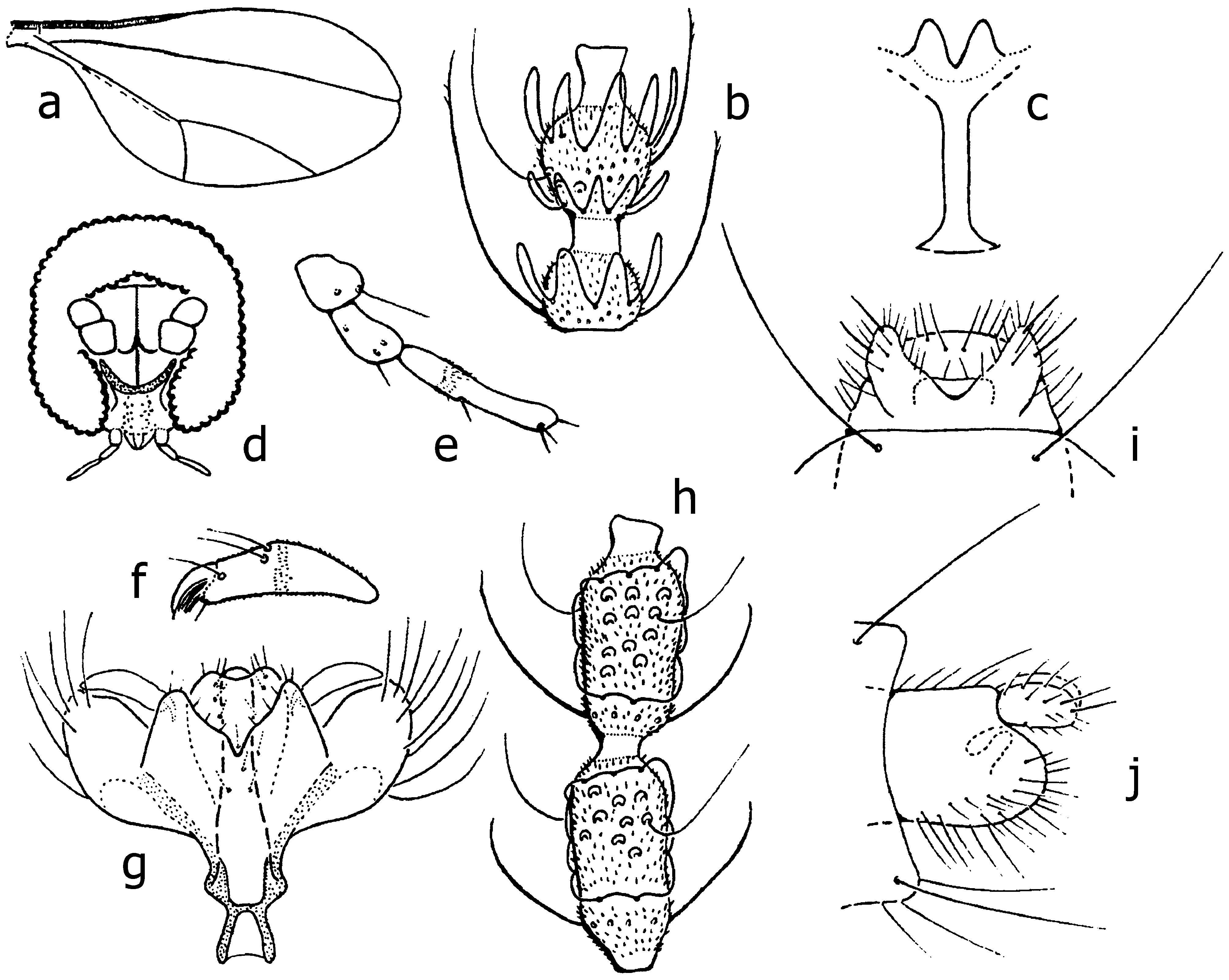
[ Figs 13 View FIGURES 13 a–j]
Diadiplosis smithi Felt, 1915: 178 View in CoL .
Coccodiplosis smithi (Felt) View in CoL : Harris (1968: 430), new combination, redescription.
Coccodiplosis pseudococci Meijere, 1917: 239 View in CoL ; Harris (1968: 430), junior synonym.
Types of names in this taxon. D. smithi View in CoL . Reared from larvae feeding on Planococcus View in CoL sp. ( Hemiptera View in CoL : Coccidae View in CoL ) [ Pulvinaria View in CoL sp. in Felt’s original description, corrected to Pseudococcus View in CoL sp by Harris (1968) based on slide label] on citrus in Manila, the Philippines, on or before 1915, Felt #a2495a. Not seen, presently on loan to Netta Dorchin, Tel Aviv University, Israel. We examined Felt’s topotypes, a male and a female (Felt #a2496), reared from larvae feeding on Planococcus View in CoL (as Pseudococcus View in CoL ) citri Risso View in CoL ( Hemiptera View in CoL : Pseudococcidae View in CoL ) presumably on citrus in Manila, the Philippines, 25-ii-1914. The specimens are mounted on separate slides as whole insects; the male is cleared, with distal flagellomeres missing, terminalia slightly misaligned; the female is uncleared, with all body parts partially shriveled. The morphology of these topotypes fits Harris’s (1968) description of Felt’s types except the cerci being strictly as long as opposed to slightly shorter than aedeagus [ Fig. 13g View FIGURES 13 ]. We assume the slight difference in the relative length of cerci is caused by the angle of mounting, a common artifact of Cecidomyiidae View in CoL slide preparation.
Coccodiplosis pseudococci View in CoL . Found feeding on the coffee mealybug Planococcus lilacinus View in CoL (as Pseudococcus crotonis View in CoL ) in Salatiga, Java ( Meijere 1917). We have not seen Meijere’s types (assumedly deposited in Zoological Museum, Amsterdam, now merged into Naturalis Biodiversity Center, Leiden The Netherlands). Meijere’s description fits Harris’s except that the male cerci are slightly longer ( Meijere 1917: Fig. 1d View FIGURE 1 ) as opposed to slightly shorter [ Fig. 13g View FIGURES 13 ] than the aedeagus. As stated above, the relative length of the cerci is often subject to the position of the terminalia on the slide mount.
Description. Length: male 0.8 mm, female 2 mm ( Felt 1915). Wing with R 5 joining C at wing apex [ Fig. 13a View FIGURES 13 ]. Palpus 3-segmented. Occipital protuberance absent. Flagellomeres 12. Male third flagellomere [ Fig. 13b View FIGURES 13 ]: basal node slightly longer than wide, internode half-length basal node, distal node round, slightly longer than wide, neck halflength distal node, circumfilar loops of basal node reaching node end, basal loops of distal node reaching node basal third, distal loops reaching neck midlength. Female flagellomeres [ Fig. 13h View FIGURES 13 ]: nodes twice as long as wide, necks short, circumfila consisting of two longitudinal and two longitudinal, interconnected bands. Tarsal claws toothed on fore and simple on mid and hindlegs. Male terminalia [ Figs 13f, g View FIGURES 13 ]: aedeagus as long as cerci and slightly longer than hypoproct, gonostylus widest at midlength, gonocoxal apodemes longer than distance between them. Female terminalia [ Figs 13i, j View FIGURES 13 ] with cerci tapered in dorsal view, rounded in lateral view, 1.5x longer than wide at base.
Pupa unknown.
Larva. Harris (1968) tentatively (due to the presence of another species of Cecidomyiidae at collection site) described the larva as bearing on the terminal segment two pairs of dorsal and one ventral papillae, and a sternal spatula with two triangular lobes divided by deep V-shaped incision. Harris’s (1968) spatula of D. smithi [ Fig. 13c View FIGURES 13 ] is similar to that depicted for this species by Meijere (1917) ( Fig. 1f View FIGURE 1 ) who describes the larva as orange-red, 1.5 mm in length.
Remarks. We also examined slides of a male and a female reared by L. Kikuchi from larvae feeding on Nipaecoccus viridis (as vastator) (Newstead), likely to have been infesting the commercially used tree Leucaena leucocephala (Lam.) de Wit [see Guam Agricultural Station (1979)] at Yigo, Guam, vii-1981. The specimens are mounted on separate slides as whole, cleared insects. These specimens fit Harris’s (1968) description of Felt’s types except the hypoproct is strictly blunt as opposed to slightly concave [ Fig. 13g View FIGURES 13 ]. We consider this slight difference part of the variability of the species.
Biology. Diadiplosis smithi is a predator of polyphagous plant-feeding mealybugs Planococcus citri and P. lilacinus ( Hemiptera : Pseudococcidae ).
Geographical distribution. The Philippines ( Felt 1915; Harris 1968), Papua New Guinea ( New Britain) ( Harris 1968) and Indonesia where it was found feeding on P. lilacinus in Java at Salatiga in or before 1917 ( Meijere 1917) and at Buitenzorg 2& 12.iv.1937 ( Harris 1968).
No known copyright restrictions apply. See Agosti, D., Egloff, W., 2009. Taxonomic information exchange and copyright: the Plazi approach. BMC Research Notes 2009, 2:53 for further explanation.
|
Kingdom |
|
|
Phylum |
|
|
Class |
|
|
Order |
|
|
Family |
|
|
Genus |
Diadiplosis smithi Felt
| Kolesik, Peter & Gagné, Raymond J. 2020 |
Coccodiplosis smithi (Felt)
| Harris, K. M. 1968: 430 |
Coccodiplosis pseudococci
| Harris, K. M. 1968: 430 |
| de Meijere, J. C. H. 1917: 239 |
Diadiplosis smithi
| Felt, E. P. 1915: 178 |