
Schizomyia villebrunneae Felt
|
publication ID |
https://doi.org/10.11646/zootaxa.4847.1.1 |
|
publication LSID |
lsid:zoobank.org:pub:1F8E3DED-6EA9-4D8A-8DA9-CD8C0CC9147F |
|
DOI |
https://doi.org/10.5281/zenodo.4407550 |
|
persistent identifier |
https://treatment.plazi.org/id/A32D87D4-1C0B-5330-55DE-FDB823EDE422 |
|
treatment provided by |
Plazi |
|
scientific name |
Schizomyia villebrunneae Felt |
| status |
|
Schizomyia villebrunneae Felt View in CoL
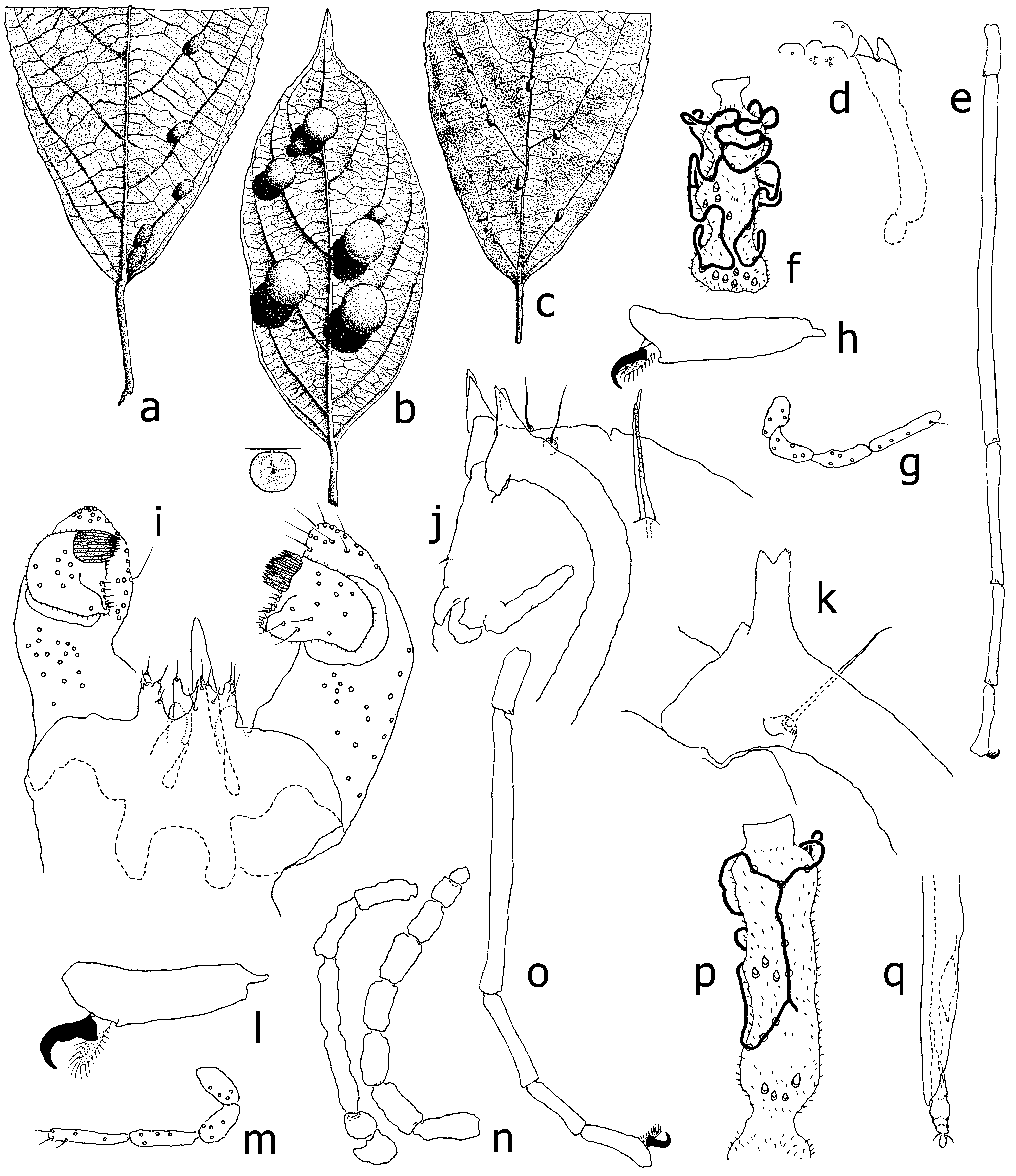
[ Figs 38a View FIGURES 38 , d–q, Figs 38 View FIGURES 38 b–c (references)]
Schizomyia villebrunneae Felt, 1921b: 146 View in CoL .
Material examined. Lectotype male, here designated from a syntypic series of two males, two pupae and a larva taken from a medium-sized, oval, thickly haired leaf gall on Oreocnide rubescens (Blume) Miq. (as Villebrunnea rubescens Bl. [note misspelling of Villebrunea ]) ( Urticaceae ) [ Figs. 38 View FIGURES 38 a–c], collected at Mt Ungaran, Java, alt. 1000 m, 13-iv-1914 by DvL, Felt #a3092. The types are mounted whole, uncleared, on two slides. The first slide contains the two males, with all body parts observable except for shrunken wings in both specimens, and only nine and three flagellomeres present in the lectotype. The second slide holds two pupae with all characters preserved, one containing a male, the other undifferentiated matter, and a larva mounted in lateral position with only spatula and posterior part of the head recognizable. Galls were illustrated in DvLR 1911, gall No. 247; DvLR & DvL (1926), gall No. 331, Fig. 237, [ Fig. 38a View FIGURES 38 ]). Felt (1921b) suggested that this species was also responsible for two other leaf galls on the same host (see under remarks), for one of which he described a series of two females (#a3091). Felt did not label them as types and only tentatively assigned them to this species. We show that the females do not appear to belong to this species.
Description. Male. Palpus 4-segmented, first segment as long as second, third and fourth progressively longer [ Fig. 38g View FIGURES 38 ]. Tarsus long, narrow, first tarsomere 1.3x, second 5x, third 2x, fourth 1.3x length last tarsomere [ Fig. 38e View FIGURES 38 ]. Tarsal claws slender, evenly narrowing towards tip, slightly bent at distal third, longer than empodia [ Fig. 38h View FIGURES 38 ]. Flagellomeres vaguely trinodal, circumifila irregular, anastomosing, strongly bowed [ Fig. 38f View FIGURES 38 ]. Terminalia [ Fig. 38i View FIGURES 38 ]: gonocoxite with large triangular ventroapical lobe; mesobasal lobe ovoid, small, 2x longer than wide; cerci irregularly incised on caudal margin; hypoproct as long as cerci, shallowly incised at apex, resulting lobes with single seta; aedeagus longer than hypoproct, tapered and pointed; gonostylus in dorsal view as long as wide, distal end covered about two-fifths width with spinose tooth, the remainder blunt, short-setose.
Female unknown.
Pupa [ Figs 38j, k View FIGURES 38 ]. Antennal horns elongate, bifid at apex, resulting lobes of equal length and minutely serrate apically. Prothoracic spiracle tapering, about 10x longer than basal width, trachea not quite reaching apex. Face with pair of setae and no horns. Abdominal segments uniformly covered with spiculae except for rows of spines on anterior third of terga.
Larva. Sternal spatula bilobed, lobes triangular, incision as long as lobes, with long shaft.
Remarks. To distinguish between this species and S. laporteae and S. nodosa , the two other species of the genus from Indonesia, see under the headings of Schizomyia and S. laporteae . The two females that Felt (1921b; specimens #a3091) only provisionally assigned to S. villebrunneae do not appear to fit this species because they have shorter tarsomeres and more robust and more strongly curved tarsal claws [ Fig. 38p View FIGURES 38 ]. For the record we show a palpus, a flagellomere, the end of the ovipositor and a tarsus [ Figs 38 View FIGURES 38 m–q] of these females. Another reason we consider the females a separate Schizomyia species from S. villebrunneae is that they came from a different leaf gall [ Fig. 38b View FIGURES 38 ].
Felt (1921b) originally received from DvL three series of insects from three distinct kinds of gall on Oreocnide rubescens (Blume) Miq. (as Villebrunnea rubescens Bl. [note misspelling of Villebrunea ]) [ Figs 38 View FIGURES 38 a–c]. The first kind, from which the syntype series #a3092 was reared, is an oval, thickly haired gall [ Fig. 38a View FIGURES 38 ], 7–9 mm long, 4–5 mm broad and about 4 mm high, attached by a short pedicel to veins on both sides of the leaf. The second kind [ Fig. 38b View FIGURES 38 ] is a large, spherical, thinly haired gall, 5–12 mm in diameter, attached by a very short pedicel to the leaf surface, and covered with thin, white, transparent, often pink hairs (DvLR & DvL 1926, gall No. 330, Fig. 236). The third kind is a small, oval, glabrous gall, 4 mm long and 2.5 mm broad, situated on veins of the upper side of the leaf (DvLR & DvL 1926, gall No. 332, Fig. 238). Felt (1921b) considered all three kinds to be made by the same species, yet he noticed a difference between males of the first kind and females from the second, causing him to only provisionally assign the females to S. villebrunneae . DvL (1921) was not convinced that the three distinct galls were caused by the same gall midge, but DvLR & DvL (1926) listed all three kinds of galls as made by S. villebrunneae . We consider only the first kind of gall [ Fig. 38a View FIGURES 38 ] to be made by S. villebrunneae .
Biology. As noted above, galls of S. villebrunneae [ Fig. 38a View FIGURES 38 ] develop on the veins of both sides of the leaf. Galls are oval, the surface is whitish, yellowish, but mostly dark red and covered with white hairlets. The single larval chamber is spherical and centered at the middle of each gall (DvLR & DvL 1926).
Geographical distribution. The known occurrence of all three gall types is confined to Java ( Felt 1921b; DvLR & DvL 1926). We expect that each of the three galls will one day be proven to be caused by a different gall midge, so we list here their geographical distribution separately. The first kind from which the syntypes came [ Fig. 38a View FIGURES 38 ] was found at the following localities. Mt Ungaran , alt. 1000 m, xii-1909; 13-iv-1914 ; Sekecer, near Weliri , alt. 100 m, x-1910 ; Cikadongdong, near Bandung , alt. 45 m, v-1915 ; Cisokan, near Cibeber , alt. 900 m, xii-1917 ; Mt Cibodas, Ciampea, near Bogor , alt. 300 m, iv-1919 ; Cianten, near Buitenzorg , alt. 400 m, viii-1924 . Additionally, this gall was found by Purnama Hidayat and Mahindra Dewi Nur Aisyah ( IPB University (pers. comm.) at Naringgul , near Cianjur ( 7°21’6.15”S 107°7’8.87”E), alt. 252 m, 12-iv-2018 GoogleMaps . The second kind [ Fig. 38b View FIGURES 38 ] was found at the following localities. Mt Ungaran , alt. 600 m, xii-1909 & v-1910 ; alt. 1000 m, 13-iv-1914; Mt Andong , alt. 1200 m, x-1911 ; Mt Kelud , alt. 600 m, v-1912 ; Pateungteung, near Garut , alt. 1500 m, xi-1918 ; Mt Gede, Cibodas , alt. 1300 m, xii-1914; ii-1916; ix-1918; i-1919 ; Pangalengan, near Bandung , alt. 1400 m, vi-1921 . Additionally, this gall was found at Mt Gede Pangrango National Park, Cianjur ( 6°44‘52“S, 106°59‘31“E), alt. 1591 m, 17-iv-2018 ( Hidayat & Nur Aisyah, pers. comm.) GoogleMaps . The third kind [ Fig. 38c View FIGURES 38 ] was found at the following localities. Mt Ungaran , alt. 1000 m, ix-1910 ; alt. 1400 m, 14-iv-1914; Cadas, Malang, near Cibeber , alt. 1200 m, ix-1917 ; Mt Salak , alt. 1200 m, vii-1920 .
| IPB |
Institut fuer Palaeontologie |
No known copyright restrictions apply. See Agosti, D., Egloff, W., 2009. Taxonomic information exchange and copyright: the Plazi approach. BMC Research Notes 2009, 2:53 for further explanation.
|
Kingdom |
|
|
Phylum |
|
|
Class |
|
|
Order |
|
|
Family |
|
|
Genus |
Schizomyia villebrunneae Felt
| Kolesik, Peter & Gagné, Raymond J. 2020 |
Schizomyia villebrunneae
| Felt, E. P. 1921: 146 |