
Holocynips illinoiensis Melika & Nicholls, 2023
|
publication ID |
https://doi.org/ 10.11646/zootaxa.5301.4.5 |
|
publication LSID |
lsid:zoobank.org:pub:198A6F76-D8B4-41B8-BDBD-7589FB0D433F |
|
DOI |
https://doi.org/10.5281/zenodo.8036065 |
|
persistent identifier |
https://treatment.plazi.org/id/0D8D00B9-CB7A-41C2-BD1A-D9DD20F1C51B |
|
taxon LSID |
lsid:zoobank.org:act:0D8D00B9-CB7A-41C2-BD1A-D9DD20F1C51B |
|
treatment provided by |
Plazi |
|
scientific name |
Holocynips illinoiensis Melika & Nicholls |
| status |
sp. nov. |
Holocynips illinoiensis Melika & Nicholls , sp. nov.
Figs 1–16 View FIGURES 1–6 View FIGURES 7–10 View FIGURES 11–12 View FIGURES 13–16
urn:lsid:zoobank.org:act:0D8D00B9-CB7A-41C2-BD1A-D9DD20F1C51B
Type material. Holotype female “ USA, Illinois, Lake Co., 17 December 2022, 42.203566, -87.838975. Quercus macrocarpa . Coll. N.E. Furlan ”, red label “Holotype female Holocynips illinoiensis ”. GoogleMaps One paratype female with the same labels as the holotype GoogleMaps . Holotype deposited at the USNM, one paratype female at the PHDNRL .
Etymology. Named after the USA state of Illinois, where the species was collected.
Diagnosis. Two Holocynips species are known from the eastern half of the USA, also from Illinois in particular (see Burks 1979), H. badia and H. maxima , but their galls differ from those of H. illinoiensis , sp. nov.. In H. badia and H. maxima the galls are woody and rounded, just below the soil surface, large and polythalamous in H. maxima , small and monothalamous in H. badia ; while in H. illinoiensis the galls are monothalamous, just above the soil surface and structurally complex with multiple spongy protrusions surrounding the larval cell. In H. badia the antennae have 11 flagellomeres, the propleuron uniformly reddish brown, mesoscutellar foveae ovate, broader than high, with broad elevated sculptured area between them, the central propodeal area with dense setae only along anterior half of lateral propodeal carinae, the prominent part of ventral spine of hypopygium 6.0× as long as broad, 2nd metasomal tergum with deep punctures. In H. maxima the antenna has 13 flagellomeres, the anterior parallel line extends to a maximum 1/3 of the mesoscutum length, the central propodeal area coriaceous with strong irregular rugae on posterior half, the prominent part of the ventral spine of the hypopygium nearly 10.0× as long as broad in ventral view. In contrast, in H. illinoiensis sp. nov. the antenna has 12 flagellomeres, the propleuron is reddish brown only in its mediocentral part but black along edges, the anterior parallel line extends to 1/2 of the mesoscutum length, mesoscutellar foveae in the form of an anteriorly transverse, smooth, glabrous impressed area, the central propodeal area smooth, glabrous, with dense setae on anterior half, the prominent part of the ventral spine of the hypopygium nearly 3.5–4.0× as long as broad in ventral view.
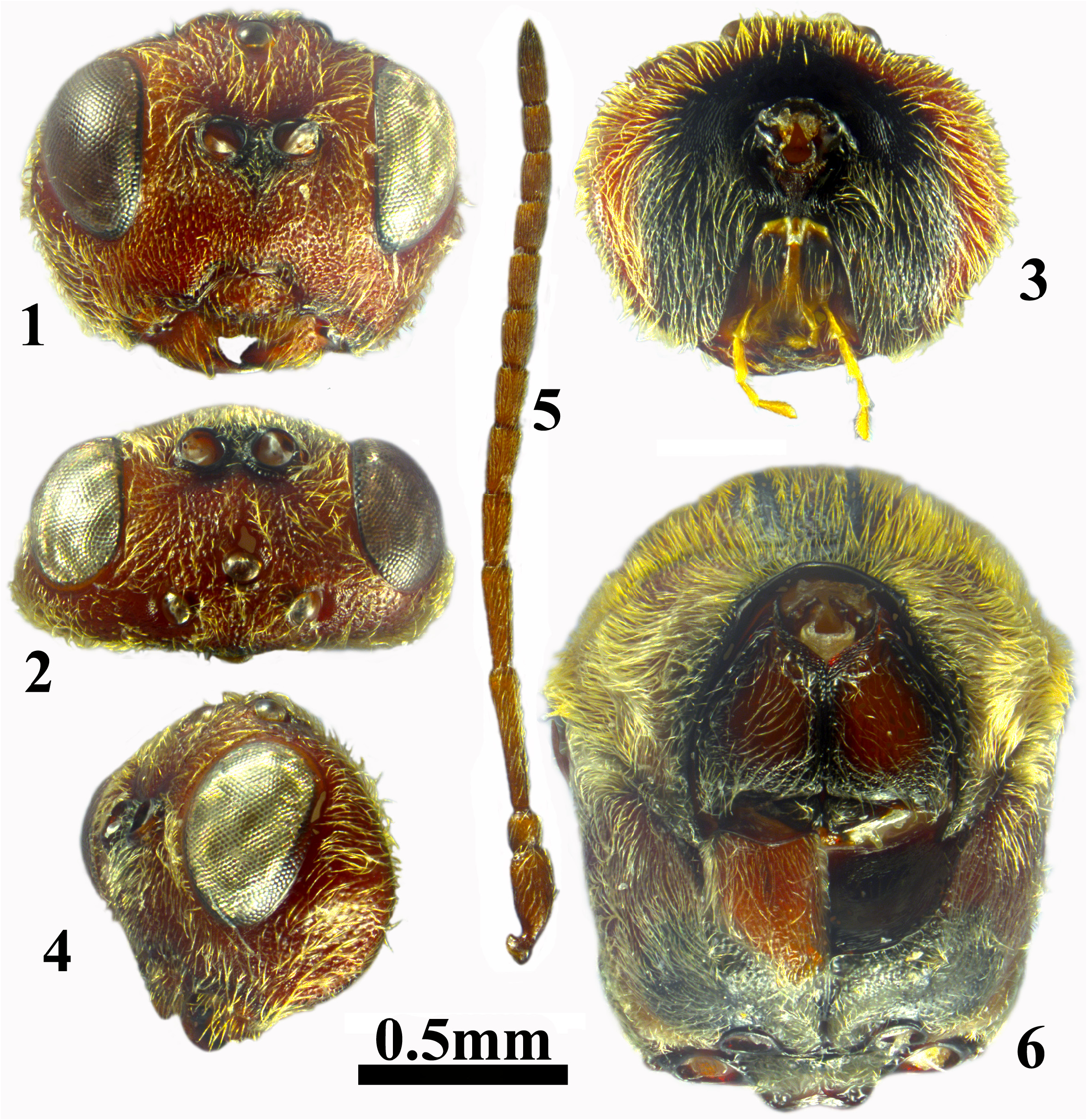
Description. Asexual female ( Figs 1–13 View FIGURES 1–6 View FIGURES 7–10 View FIGURES 11–12 View FIGURES 13–16 ). Head dark reddish brown anteriorly, black posteriorly, mouthparts yellow, antenna slightly lighter than mesosoma; mesosoma predominantly dark reddish brown, with black stripes along anterior parallel and parapsidal lines; propodeum black; legs uniformly dark reddish brown, metasoma dark reddish brown with almost black posterior terga. Head and mesoscutum with dense yellowish-white setae.
Head ( Figs 1–4 View FIGURES 1–6 ) with dense yellowish white setae, 1.2× as broad as high and as broad as mesosoma in frontal view; 1.8× as broad as long in dorsal view. Gena with deep punctures, broadened behind eye in frontal view, narrower than transverse diameter of eye in lateral view. Malar space punctured, without sulcus; eye 2.2× as high as length of malar space. Inner margins of eyes parallel. POL 1.5× as long as OOL, OOL 1.6× as long as diameter of lateral ocellus and 1.5× as long as LOL, all ocelli ovate, of same size. Transfacial distance 1.2× as long as height of eye; toruli located in the upper half of head and frons shorter than lower face, diameter of antennal torulus nearly 1.8× as long as distance between them, distance between torulus and eye slightly longer than diameter of torulus; lower face and slightly elevated median area with deep rounded punctures, with sparse white setae. Clypeus trapezoid, broader than high, with deep punctures, delicately coriaceous, with a few long setae scattered all over; ventrally rounded, emarginate, without median incision; anterior tentorial pit small, rounded, indistinct, epistomal sulcus distinct, clypeo-pleurostomal line well impressed. Frons, interocellar area, vertex uniformly with deep punctures and setae; area under central ocellus impressed, shining; occiput and postocciput smooth, shining, area beside occipital foramen reticulate, postgena punctured, with dense setae; posterior tentorial pit large, elongated, area below impressed; occipital foramen as high as height of postgenal bridge; hypostomal carina emarginate, continuing into distinct postgenal sulci which diverge strongly toward occipital foramen, postgenal bridge anteriorly slightly broader than occipital foramen. Antenna longer than head+mesosoma, with 12 flagellomeres (suture between F12 and F11 indistinct); pedicel slightly longer than broad; F1 3.7× as long as pedicel and 1.3× as long as F2; F2 1.4× as long as F3; F3=F4, F5 until F7 equal in length, shorter than F4; F8 until F11 shorter and all equal in length, F12 as long as F11; placodeal sensilla on F3–F12 ( Fig. 5 View FIGURES 1–6 ).
Mesosoma longer than high, with dense setae all over ( Fig. 7 View FIGURES 7–10 ). Pronotum with punctures, foveolate along propleuron, laterally with punctures; propleuron reddish brown, smooth, glabrous in mediocentral part, black and with punctures along sides ( Fig. 6 View FIGURES 1–6 ). Mesoscutum longer than broad (greatest width measured across mesoscutum level with base of tegulae), uniformly punctured, with dense setae ( Fig. 8 View FIGURES 7–10 ). Notaulus complete, deep, posteriorly converging and broader than anteriorly, bottom smooth, glabrous; at posterior end the distance between notauli shorter than distance between notaulus and side of mesoscutum; anterior parallel line black, distinct, elevated above mesoscutum, smooth, glabrous, extending to 1/2 length of mesoscutum; parapsidal line distinct, black, smooth; median mesoscutal line absent; circumscutellar carina broad, reaching slightly above level of tegulae. Mesoscutellum trapezoid, longer than broad, with subparallel sides, broader at posterior end; disc of mesoscutellum punctured, with setae, overhanging metanotum; circumscutellar carina indistinct ( Fig. 9–10 View FIGURES 7–10 ). Mesoscutellar foveae in the form of an anteriorly transverse, smooth, glabrous impressed area, black, well-delimited anteriorly and smoothly continuing into disc of mesoscutellum. Mesopleuron entirely uniformly punctured, with dense setae; mesopleural triangle rugose, with sparse short white setae; dorsal and lateral axillar areas punctured, with dense setae; axillula with delicate parallel longitudinal striae; subaxillular bar smooth, glabrous, triangular, posteriorly as high as height of metanotal trough; metapleural sulcus reaching mesopleuron at half of its height, lower part of sulcus delimiting broad triangular punctured area, with dense setae; upper part of sulcus also distinct, separating smooth, glabrous area with dense setae ( Fig. 7 View FIGURES 7–10 ). Metascutellum granulose, 2.0× as high as height of smooth, glabrous ventral impressed area; metanotal trough smooth, with dense short setae; central propodeal area with parallel sides, smooth, glabrous, with short irregular rugae and dense setae in anterior half; lateral propodeal carinae parallel, bent slightly outwards in posterior 1/3; lateral propodeal area smooth, glabrous, with long dense white setae. Nucha with irregular rugae dorsally and laterally ( Fig. 10 View FIGURES 7–10 ). Tarsal claws simple, without basal lobe.
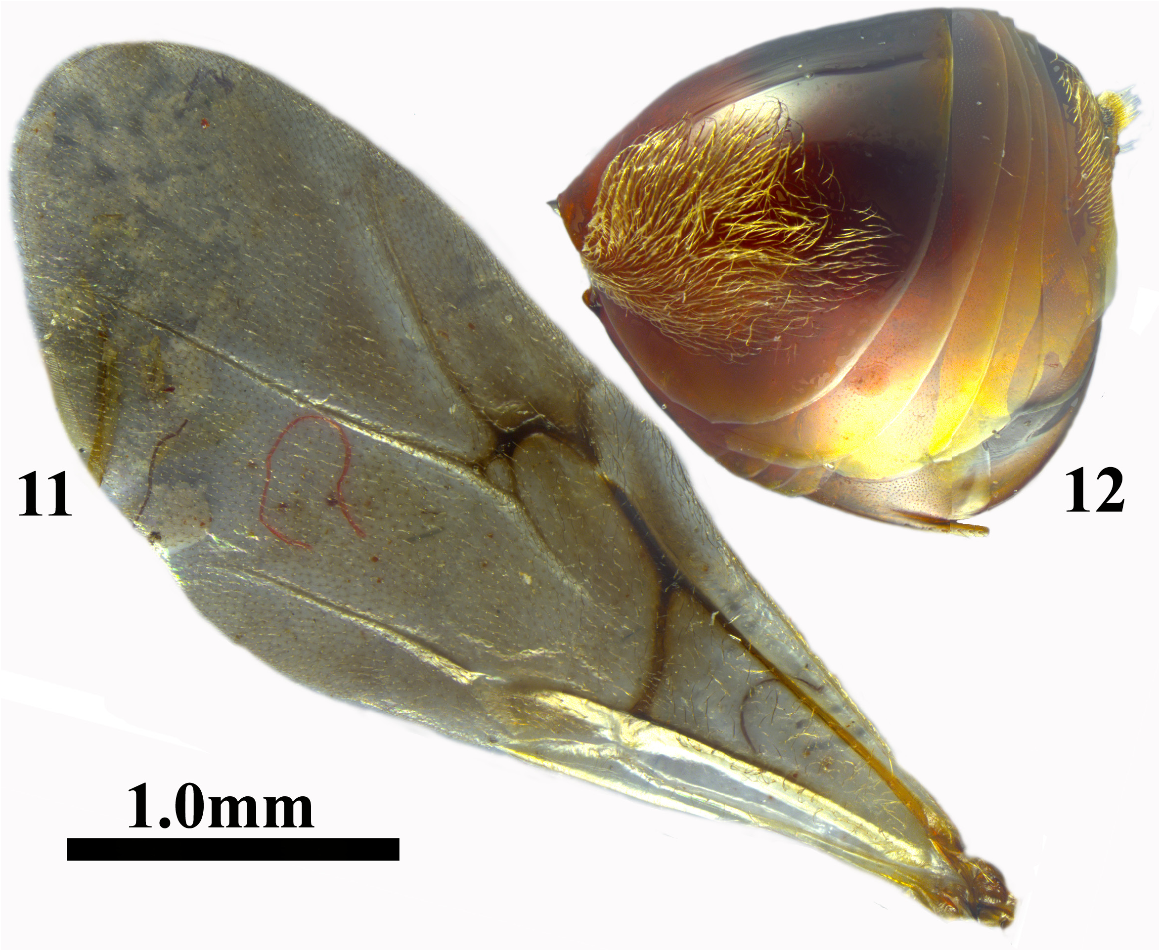
Fore wing longer than body, slightly infuscated, with short cilia on margin, veins light brown, radial cell open, 2.7× as long as broad; Rs and R1 nearly reaching wing margin; areolet triangular, closed by distinct veins. Rs+M indistinct, visible on 2/3 of distance between areolet and basalis, its projection reaching basalis at half of its height ( Fig. 11 View FIGURES 11–12 ).
Metasoma as long as head+mesosoma, slightly longer than high in lateral view; second metasomal tergum smooth, extending to 2/3 length of metasoma in dorsal view, with patch of dense white setae anterolaterally, without micropunctures; all subsequent terga and hypopygium with minute micropunctures; prominent part of ventral spine of hypopygium 3.5–4.0× as long as broad in ventral view, with a few short white setae ventrally ( Fig. 12 View FIGURES 11–12 ). Body length 4.7– 4.9 mm (n = 2).
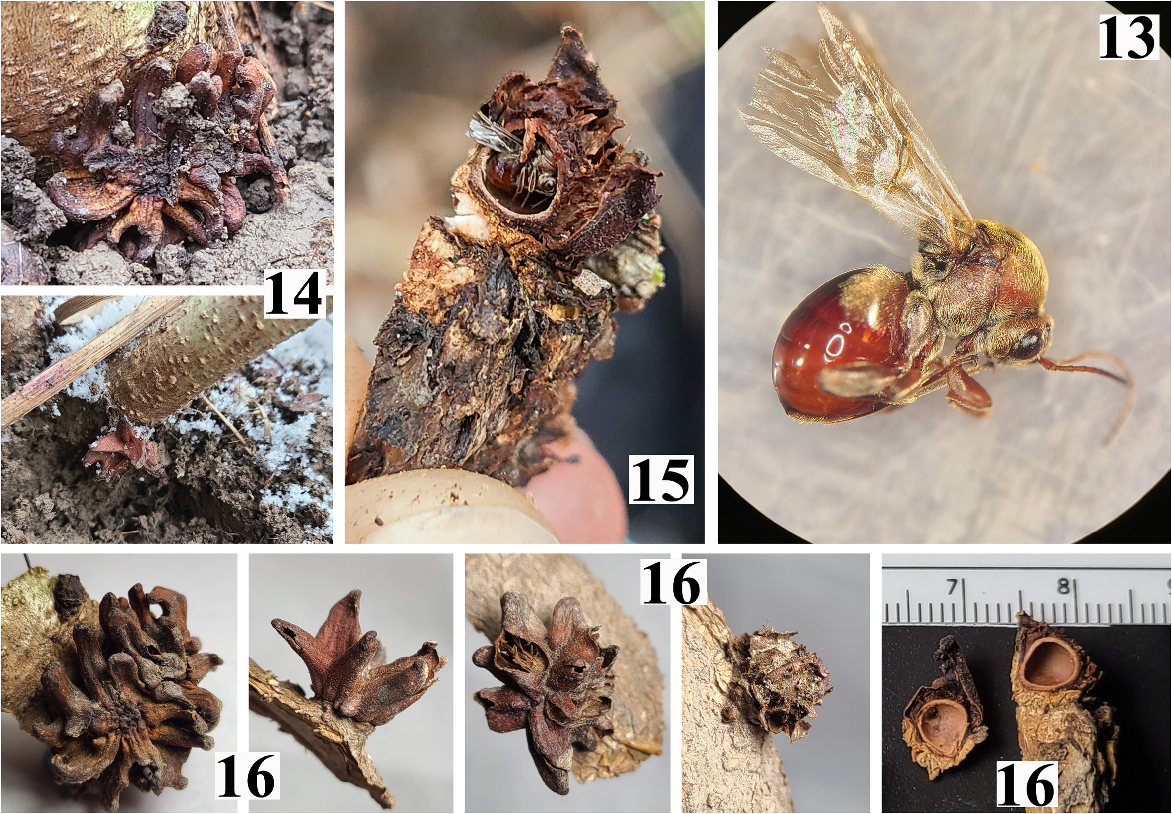
Gall ( Figs 14–16 View FIGURES 13–16 ). Occurring singly or in closely appressed clusters of 3–6 at the base of small stems (diameter at breast height ≤ 3 cm) or shoot regrowth from cut stumps, most at ground level and either hidden by leaf litter and vegetation or partially buried in the soil. Occasional galls have been observed higher on the stem, up to 75 cm above the ground.
Galls are dull red to reddish-brown, darkening to purple-brown or black with age and elemental exposure. Surface texture is finely granulate and dull. Each individual gall consists of a single larval cell surrounded by 6–10 thin-walled parenchyma-filled protrusions in a radial or whorled configuration. Protrusions are septate, becoming hollow and brittle with age and eventually disintegrating. Overall gall morphology ranges from irregularly globular (as when protrusions are small or absent) to more elaborate shapes reminiscent of flowers or dried star anise. Individual galls range in size from 11–20 mm wide and 8–10 mm in height. Gall detachment from the stem surface requires some force and leaves a conically depressed scar 4–6 mm in diameter, paler than the surrounding tissue and enclosed by an elevated margin 1–3 mm high.
The larval cell is round or ovate to teardrop-shaped, 4–5 mm wide and 4–5 mm tall. Cell walls are woody, 0.5–0.6 mm thick and lighter in color than the surrounding protrusions.Adult emergence holes observed on old galls are ovate, approximately 2 mm wide by 3mm tall.
Biology. Only the asexual generation is known, which induces galls on Q. macrocarpa Michx. (subgenus Quercus , section Quercus ). Galls mature in November–December; adults were cut out in December and January under laboratory conditions.
Molecular taxonomy. Cytb sequences were compared between a specimen of the new species H. illinoiensis and a specimen of H. badia , a morphologically similar species that also induces unilocular galls at the base of Q. macrocarpa stems. The two species differed by 2.3% at this locus (10 bases different, including one base substitution resulting in an amino acid change), consistent with expected variation between distinct species, especially given that the two specimens were collected only 14.5km apart and hence represent essentially sympatic populations of their respective species. ITS2 sequences from these two species differed by two base substitutions (0.4%) and two indels, which are levels of variation at the lower end of that expected for two close congeners. Variation between H. illinoiensis and H. hartmani was much higher, with 50 bases differing beween their respective cytb sequences (11.5%). All sequences are deposited on GenBank (accessions OQ716584–OQ716586 for cytb, OQ716467– OQ716468 for ITS2).

| USNM |
Smithsonian Institution, National Museum of Natural History |
No known copyright restrictions apply. See Agosti, D., Egloff, W., 2009. Taxonomic information exchange and copyright: the Plazi approach. BMC Research Notes 2009, 2:53 for further explanation.
|
Kingdom |
|
|
Phylum |
|
|
Class |
|
|
Order |
|
|
Family |
|
|
Genus |