
Molinaranea mitnickii, Saupe & Selden & Penney, 2010
|
publication ID |
https://doi.org/10.1111/j.1096-3642.2009.00581.x |
|
DOI |
https://doi.org/10.5281/zenodo.10545531 |
|
persistent identifier |
https://treatment.plazi.org/id/A01B87BB-E372-9D6F-AB1A-A1943C17DF3F |
|
treatment provided by |
Valdenar |
|
scientific name |
Molinaranea mitnickii |
| status |
sp. nov. |
MOLINARANEA MITNICKII SP. NOV. ( FIGS 1–3 View Figure 1 View Figure 2 View Figure 3 )
Material examined: Holotype and only known specimen: Amber Fossil Collection, University of Kansas Natural History Museum KU-NHM-ENT, DR-018, adult male, Dominican amber, La Toca mines, northern Dominican Republic; coll. TerraTreasures.
Diagnosis: Molinaranea mitnickii can be distinguished from all other species by the median apophysis with long, thin/spindly, subequal prongs, resembling a lop-sided wishbone, with a proximal lobe/elbow. The ventral femora of legs 1 and 2 possess a row of strong macrosetae.
Etymology: The specific epithet is after Justin Mitnick, nephew of Keith Luzzi, the owner of Terra- Treasures who found and donated the specimen for study.
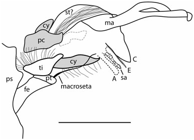
Description: Body length 6.95. Carapace 2.82 long, 2.0 wide, ± 1.88 tall; pars cephalica only slightly elevated (approximately 0.42). Eyes small; ALE appear to be on small tubercles; numerous macrosetae in the ocular region. Details of chelicerae and fangs obscured; small. Sternum 1.41 long, 1.04 wide; relatively short and rounded; lateral margins project between coxae. Endites 0.38 long, longer than wide, suboval, tooth present. Labium 0.38, as wide as long, suboval to subcircular. Petiole attached 0.95 from anterior of opisthosoma immediately above book lungs; not sclerotized. Opisthosoma 4.13 long including spinnerets ( Fig. 2D View Figure 2 ), 3.6 long without spinnerets, 2.23 at widest point, height uncertain because of flattened nature of specimen; likely to have been elongate and suboval in life; dorsal surface bears abundant, long, scattered setae; concentrated setae on two anterior tubercles ( Fig. 1B View Figure 1 ). PLS longer than MS and AS, PLS defined in two segments ( Fig. 2E View Figure 2 ); AS 0.43 and PLS 0.51; colulus present, tongue-shaped with nine setae. Spiracle situated 0.03 anterior to co and 0.13 to base of AS; anal tubercle 0.34.
Leg formula 2143; leg 1 cx 0.52, tr 0.30, fe 3.16, pt 1.07, ti 4.13, mt 2.17, ts 0.63, total 11.98; leg 2 cx 0.50, tr 0.28, fe 3.44, pt 0.90, ti 4.13, mt 2.36, ts 0.64, total 12.25; leg 3 cx 0.42, tr 0.28, fe 2.36, pt 0.82, ti 1.25, mt 1.26, ts 0.48, total 6.87; leg 4 cx 0.45, tr 0.14, fe 2.43, pt 0.98, ti 2.10, mt 2.03, ts 0.46, total 8.59. Legs long; all legs possess strong macrosetae ( Figs 1 View Figure 1 , 2A, B View Figure 2 ); macrosetae originate from strong cuticular bases; variable in length, longer macrosetae 0.7–0.8, shorter macrosetae 0.4–0.6; longer macrosetae appear to be concentrated on the lateral margins of tibiae 1 and 2 and ventral surfaces of most leg segments, although this is variable; row of macrosetae on prolateral to ventral margin of tibia 1 and 2; row of seven to ten macrosetae on inferior surface of femora of legs 1 and 2; row of three to four macrosetae on superior surface of femora of legs 1 and 2; femora of leg 1 with lateral row of seven to eight macrosetae; scattered macrosetae, semialigned, on ventral femora of legs 3 and 4; tibia and femur of legs 1 and 2 thicker and more robust. Hook on distal margin of the first coxa; fourth coxa with at least one macroseta. Paired tarsal claws with teeth, unpaired claw simple.
Palps large ( Figs 1 View Figure 1 , 2F View Figure 2 ); length of palpal bulb without median apophysis ± 1.09, width 0.79; median apophysis with bifurcation into long, thin spindly prongs ( Figs 2A, F View Figure 2 , 3 View Figure 3 ), resembling a wishbone; median apophysis 1.41 long; prongs on median apophysis equal, with recurved, semipointed tip; median apophysis with proximal lobe or elbow; embolus distally filiform and situated between conductor and terminal apophysis ( Fig. 3 View Figure 3 ); conductor broader than terminal apophysis and attached in middle of bulb with a semipointed tip; subterminal apophysis present as a narrow band between embolus and terminal apophysis ( Fig. 3 View Figure 3 ); terminal apophysis lobate to truncate and narrow, larger than subterminal apophysis ( Fig. 3 View Figure 3 ); one macroseta on patella.
Female: Unknown.
Distribution and age: Dominican Republic amber; probably middle Miocene (16–19 Mya) (see Iturralde- Vinent, 2001).
Remarks: The species can be distinguished from Molinaranea vildav Levi, 2001 by the presence of a proximal lobe or elbow below the radix of the median apophysis ( Fig. 3 View Figure 3 ) instead of above it, by the curved tip on the lower prong of the median apophysis, and by the prongs, which appear more separated (like a wishbone) in M. mitnickii than in M. vildav . Further, M. mitnickii possesses a row of macrosetae on the ventral surfaces of femora 1 and 2, unlike in M. vildav . The length of the median apophysis prongs distinguishes M. mitnickii from M. vildav , Molinaranea mammifera Tullgren, 1902 , and Molinaranea clymene Nicolet, 1849 (significantly shorter in M. vildav , M. mammifera , and M. clymene ). Molinaranea mitnickii lacks the short, wide median apophysis characteristic of M. mammifera and the tufts of setae on the abdomen that are present in M. clymene ( Levi, 2001: figs 27, 30). Unfortunately, much of Levi’s description and diagnostic characters are based on colour pattern, which is usually not discernible in amber specimens.


No known copyright restrictions apply. See Agosti, D., Egloff, W., 2009. Taxonomic information exchange and copyright: the Plazi approach. BMC Research Notes 2009, 2:53 for further explanation.
|
Kingdom |
|
|
Phylum |
|
|
Class |
|
|
Order |
|
|
Family |
|
|
Genus |