
Pilobatella kovaci, Ermilov & Starý, 2020
|
publication ID |
https://doi.org/ 10.24349/acarologia/20204384 |
|
DOI |
https://doi.org/10.5281/zenodo.4527425 |
|
persistent identifier |
https://treatment.plazi.org/id/9F7787C3-FFC6-FF9D-FE46-F8CEDE4BB8E6 |
|
treatment provided by |
Felipe |
|
scientific name |
Pilobatella kovaci |
| status |
sp. nov. |
Pilobatella kovaci View in CoL n. sp.
Zoobank: A9C8AE64-9D65-4F32-98CE-704D77A4E4D0
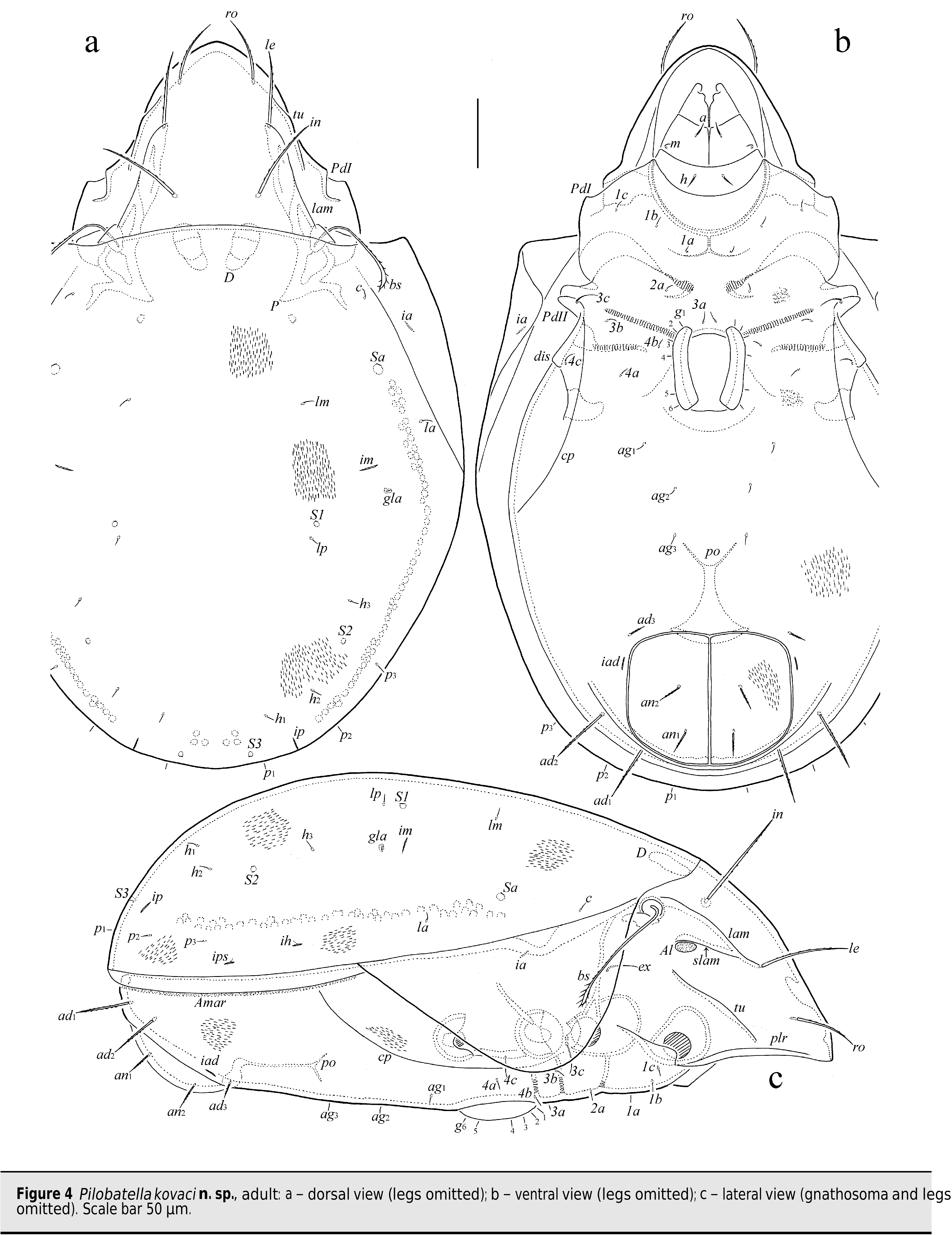
( Figure 4 View Figure 4 )
Diagnosis — Body size 531–581 × 249–332. Rostral, lamellar and interlamellar setae long, setiform, barbed, ro shortest, in longest. Bothridial seta long, gradually thickening distally, beginning at mid-length, ciliate. Tutorium long. All notogastral setae short, setiform, smooth Epimeral, six pairs of genital and aggenital setae short, setiform, roughened. Medial end of apodeme 2 well removed from midline. Anal and adanal setae setiform, erect, densely barbed, ad 1 and ad 2 long. All legs monodactylous. Trochanter IV rounded anterodorsally.
.
Description — Measurements – Body length 564 (holotype), 531–581 (three paratypes); body width 282 (holotype), 249–332 (three paratypes). Females larger than males: 564–581 ×
282–332 versus 531–547 × 249–265.
Integument – Body light brown. Surface of body and all legs microporose (visible under high magnification, ×1000). Notogaster , anogenital region and anal plate lineolate, with short depressed, distinct lines. Epimeral region and genital plate microlineolate. Lateral part of body microgranulate. Antiaxial side of femora I, II and paraxial side of femora III and IV striate.
Prodorsum ( Figs 4a, 4c View Figure 4 ) – Rostrum rounded. Lamella about 1/2 length of prodorsum, strong. Prolamella absent. Sublamella about 1/2 length of lamella. Sublamellar porose area (14–16 × 8–10) oval, located ventral to sublamella. Tutorium long, similar in length to lamella, ridgelike, almost reaching rostral margin. Prodorsal lateral ridge distinct. Rostral (53–61), lamellar (61–69) and interlamellar (73–82) setae setiform, slightly barbed; le located on lamellar end. Bothridial seta (86–90) gradually thickening distally, beginning at mid-length, shortly ciliate bilaterally. Exobothridial seta (28–41) setiform, thin, slightly barbed. Sejugal porose area not observed.
Notogaster ( Figs 4a, 4c View Figure 4 ) – Anterior notogastral margin slightly convex medially. Pteromorph triangular, rounded laterally, with slightly developed hinge. Ten pairs of notogastral setae (6–8) setiform, thin, smooth. Four pairs of sacculi pyriform. Notogastral lyrifissure, opisthonotal gland opening, circumgastric scissure and circumgastric sigillar band distinct.
Gnathosoma – Generally, similar to those of Pilobatella mikoi n. sp. Subcapitulum size 131–135 × 77–86. Subcapitular setae (a, 16–20; m, 8–10; h, 20–24) setiform, roughened, m thinnest. Adoral seta (12) setiform, densely barbed. Palp (73–82) with typical formula. Postpalpal seta (4) spiniform, smooth. Chelicera (131–135) with two setiform, barbed setae (cha, 45–49; chb, 24–28).
Epimeral and lateral podosomal regions ( Figs 4b, 4c View Figure 4 ) – Epimeral setal formula 3–1–3–3. All setae setiform, thin, roughened, 3c (28–41) longer than 4c (16–20) and others (10–14). Medial end of apodeme 2 well removed from midline. Pedotectum II bifurcate apically in ventral view. Circumpedal carina long, directed to pedotectum II. Discidium triangular.
Anogenital region ( Figs 4b, 4c View Figure 4 ) – Six pairs of genital (6–8) and three pairs of aggenital (8–12) setae setiform, thin, roughened. Two pairs of anal (20–24) and three pairs of adanal ad (1, ad 2, 41–49; ad 3, 12–16) setae setiform, erect, barbed. Adanal setae ad 1 inserted on arcuate ridge, ad 2 near this ridge. Adanal lyrifissure distinct. Marginal porose area complete, narrowly band-like. Preanal organ goblet-like.
Legs – Generally, similar to those of Pilobatella mikoi n. sp. Monodactylous. Claw of all tarsi strong, slightly barbed on dorsal side, with tubercle ventrobasally. Claw of tarsi I and II thicker than that of tarsi III and IV. Tibiae I and II with tubercle ventrobasally. Femur II with broadly rounded ledge distoventrally. Trochanter IV rounded distodorsally. Dorsoparaxial porose area on all femora and on trochanters III, IV, and proximoventral porose area on all tarsi and distoventral porose area on all tibiae well visible. Formulas of leg setation and solenidia: I (1–5–3–4–20) [1–2–2], II (1–5–3–4–15) [1–1–2], III (2–3–1–3–15) [1–1–0], IV (1–2–2–3–12) [0–1–0]; homology of setae and solenidia indicated in Table 1. Famulus of tarsus I short, erect, slightly dilated distally, inserted between solenidion ωand seta ft ″. Solenidion ω on tarsus I, 2 1 ω and ω on tarsus II and σ on genu III bacilliform, other solenidia setiform. Seta pl ′ on tarsus 1 2 I located dorsally on segment, posterior to seta ft ″. Seta l ″ on genu I inserted on tubercle.
Material examined — Holotype (female) and three paratypes (two males and one female): Madagascar, Andasibe-Mantadia National Park , evergreen rain forest18, °49 ′ 36 ″ S, 48°26 ′ 52 ″ E, 550 m a.s.l., Winkler apparatus extraction of sifted leaf litter, 28.I.2014 (sample MAG-290 collected by R. Ravebolun and L. Rabotenoson).
Type deposition — The holotype is deposited in the collection of the Senckenberg Institute, Görlitz, Germany. Three paratypes are deposited in the collection of the Tyumen State University Museum of Zoology, Tyumen, Russia. Specimens are preserved in ethanol with a drop of glycerol.
Etymology — The species name is dedicated to our friend and colleague, the well-known soil ecologist Prof. Dr. Ľubomír Kováč ( Slovakia, Košice), for his extensive contribution to our knowledge of taxonomy and ecology of soil and troglophilous species of springtails (Collembola) and other groups of cave mesofauna.
Remarks — In having monodactylous legs, long interlamellar setae and adanal setae ad 1 and ad 2, Pilobatella kovaci n. sp. is morphologically most similar to P. mikoi n. sp. from Madagascar, but differs from the latter by the presence of its lineolate notogaster and anogenital region (versus lineolate markings absent), long tutoria (versus tutoria of medium length), rounded trochanters anterodorsally (versus pointed) and clearly distanced medial ends of apodemes 2 (versus nearly touching at midline).
| R |
Departamento de Geologia, Universidad de Chile |
No known copyright restrictions apply. See Agosti, D., Egloff, W., 2009. Taxonomic information exchange and copyright: the Plazi approach. BMC Research Notes 2009, 2:53 for further explanation.