
Chimarra vitiensis, Johanson, Kjell Arne & Oláh, János, 2012
|
publication ID |
https://doi.org/10.5281/zenodo.210736 |
|
DOI |
https://doi.org/10.5281/zenodo.5664482 |
|
persistent identifier |
https://treatment.plazi.org/id/9F3E87DD-5638-FFCE-E89A-FA72FE50FDCE |
|
treatment provided by |
Plazi |
|
scientific name |
Chimarra vitiensis |
| status |
sp. nov. |
Chimarra vitiensis , new species
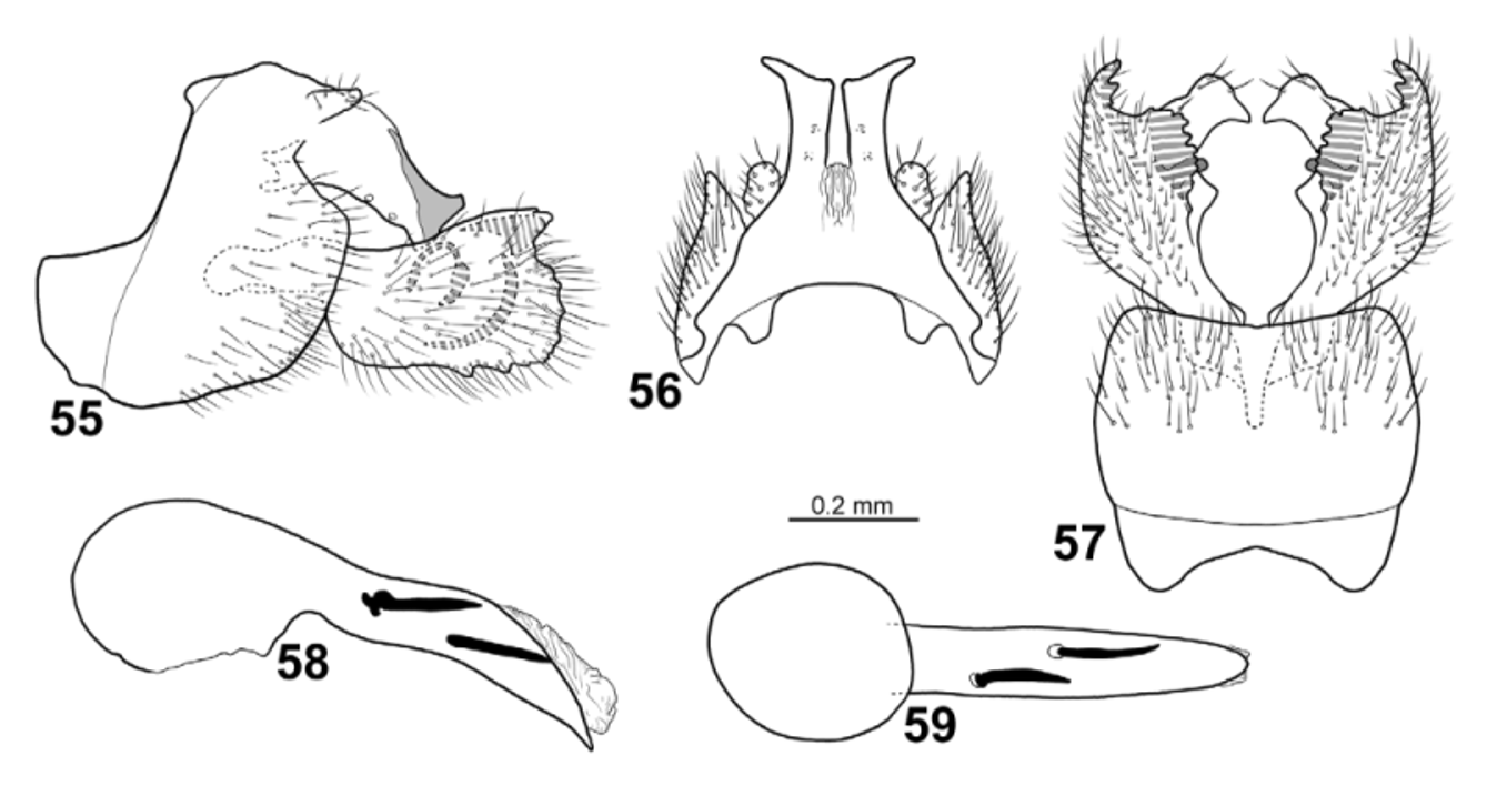
Figs. 12 View FIGURES 6 – 13 , 65–69 View FIGURES 65 – 69
Chimarra vitiensis resembles C. braueri , C. nathani , C. schlingeri , and C. signata in having wings with a large, pale, hyaline area located centrally. Chimarra vitiensis is distinguished from the other species by the genitalia of the male, particularly in that the broad gonopods are strongly and darkly sclerotized along their dorsal margin; and also in having a dorsal branch of each gonopod that appears short in lateral view, but is seen to be produced into a short, slender, finger-like process in dorsal view.
Male. Body yellowish-brown, dorsal part of meso- and metathorax brown. Large dark area between lateral and anterior ocelli. Foreleg anterior claw as long as foreleg spur.
Wings ( Fig. 12 View FIGURES 6 – 13 ). Forewings 4.6 mm (n=1), brown; relatively broad, ratio of length to breadth 3.4; R1 slightly undulating before crossvein r; radial sector slightly produced anterad immediately before discoidal cell; discoidal cell originating at mid-length of wing, 3x longer than wide; median cell nearly as long as discoidal cell; crossvein r situated at base of fork I; fork I originating before crossvein s at distance equal to 1/2 length of crossvein s; nygma located near base of fork II; fork III 1 /6th as long as wing; fork V as long as fork I, shorter than fork II; Cu2 nearly touching from A at wing margin. Hind wings 3.8 mm (n=1), brown; broad, ratio of length to breadth 3.1; margin slightly incurved at arculus, where Cu1 and Cu2 fused with margin; fork III as long as discoidal cell and 1/9 as long as wing; fork I weakly developed, originate from anterodistal corner of discoidal cell, about as long as fork V; 1A+2A 3x longer than 1A.
Male genitalia ( Figs. 56–59 View FIGURES 55 – 59 ). Segment IX as long as high; anterior plates narrowing anteriorly, rounded apically; posterior 1/2 of segment broad, expanded dorsally, parallelogram shaped, slightly produced anterad and laterad ( Fig. 66 View FIGURES 65 – 69 ); each anterodorsal margin widely and deeply concave in lateral view; each ventral margin stepwise convex, with incision at vertical apodeme; posterior margins nearly straight, curved anterad well below cercus. In dorsal view segment IX with short, wide anterior lobes; in dorsal view anterodorsal margin forming deep, narrow, hyperbolic incision without anterad-orienting processes on posteromesal margins. In ventral view segment IX nearly rectangular, except slightly convex lateral margins and slightly incised at transverse apodeme; anterior margin deeply concave; posterior margin slightly concave; posterior margin without central projection. Tergum X undivided in lateral view; with weakly concave ventral margin; dorsal margin with large, rounded lobe near basis; apex slightly produced dorsad into short finger-like processes. In dorsal view tergum X divided into 2 laterally diverging branches, apically curved laterad. Sensillae on tergum X located ventrolaterally at 3/4 its length. Cerci located near dorsal margin of segment IX and tergum X; directed dorsad in lateral view, club-shaped; forming setose wart-like structures in dorsal view; covered by long setae. Gonopods broad, nearly quadrate, except narrow at basis, slightly shorter than segment IX, 2-branched. Each dorsal branch reaching as far posteriorly as tergum X in lateral view; weakly produced dorsad; bending mesad into short, rounded plate with finger-like mesal process; megasetae absent. Ventral branch of each gonopod rectangular in lateral view, with small undulations along posterior margin and undulating ventral margin; each ventral branch with mesal margin strongly irregular in ventral view, with strongly sclerotized wide, short teeth. Mesal branches absent. Phallic apparatus slightly longer than rest of genitalia, nearly straight; phallotheca, in lateral view, with anterior part 1.5x thicker than posterior part; in ventral view anterior part nearly 2x wider than posterior part; apicoventral spine absent on phallotheca; phallotremal sclerite not observed; 2 very long, brownish endothecal spines present.
Female. Unknown.
Holotype male: VITI LEVU: Vuda Prov., Koroyanitu N.M.P., Savuione Trail, Malaise trap, 12–19.x.2002, 17°40’S, 177°33’E, 450 m, M. Irwin, E. Schlinger & M. Tokota’a [loc#02] [ FNIC].
Paratypes: S ame data as holotype [loc#02] — 3 males [ NHRS]. Vuda Prov., Koroyanitu Pk., 1 km E Abaca Vlg., Savuione Trail, Malaise trap, 11–19.iii.2003, 17.667°S, 177.55°E, 800 m, M. Irwin, E. Schlinger & M. Tokota’a [loc#03] — 1 male [ BPBM]. Same data, except: 7–12.x.2002 [loc#03] — 1 male [ BPBM]. Same data, except: 19–26.x.2002 [loc#03] — 2 males [ BPBM]. Naitasiri Prov., Nakobalevu Mt., 22.ix–9.x.2002, 18°03’S, 178°25’E [ 18.0500°S, 178.4167°E], 340 m, M. Irwin, E. Schlinger & M. Tokota’a [loc#10] — 1 male [ BPBM]. Pabitra Wabu Baseline Survey, Malaise trap, 17–20.xi.2003, 17.5833°S, 178.0833°E, 1034 m, leg. Delena Veikovi [loc#13] — 1 male [ NHRS, DNA voucher IN2].
Etymology: Vitiensis , named after Viti Levu, the type locality of the species.
Distribution: Viti Levu.


No known copyright restrictions apply. See Agosti, D., Egloff, W., 2009. Taxonomic information exchange and copyright: the Plazi approach. BMC Research Notes 2009, 2:53 for further explanation.
|
Kingdom |
|
|
Phylum |
|
|
Class |
|
|
Order |
|
|
Family |
|
|
Genus |