
Chimarra helomyzida, Johanson, Kjell Arne & Oláh, János, 2012
|
publication ID |
https://doi.org/10.5281/zenodo.210736 |
|
DOI |
https://doi.org/10.5281/zenodo.5664512 |
|
persistent identifier |
https://treatment.plazi.org/id/9F3E87DD-5600-FFE6-E89A-FA67FDE7FD86 |
|
treatment provided by |
Plazi |
|
scientific name |
Chimarra helomyzida |
| status |
sp. nov. |
Chimarra helomyzida , new species
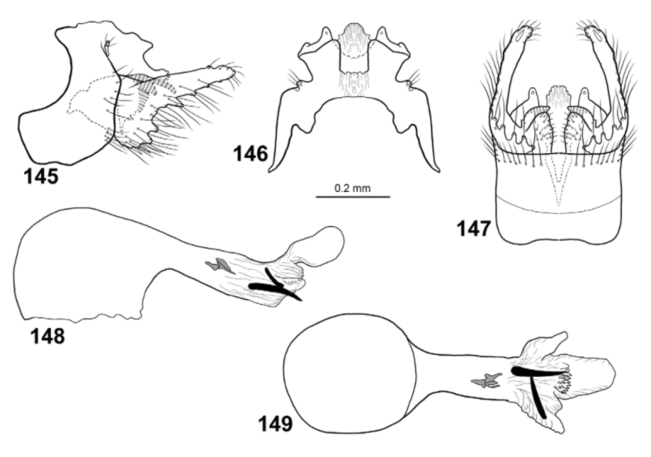
Figs. 27 View FIGURES 22 – 29 , 145–149 View FIGURES 145 – 149
The male genitalia of C. helomyzida resemble those of C. kimminsi , C. cakaudrovensis , C. cartwrighti , C. manni , C. lavensis , C. devoensis and C. vanuensis , particularly in the shape of the gonopods in lateral view. Chimarra helomyzida is distinguished from C. kimminsi by the presence of a dorsal process on tergum X in lateral view; from C. lavensis , C. devoensis , C. cakaudrovensis and C. manni by the presence of only 2 endothecal spines in the phallic apparatus; from C. cartwrighti by the narrower anterior plates of segment IX in lateral view; and from C. vanuensis by the absence of a slender, dorsad oriented process mid-way on tergum X in lateral view.
Male. Head and thorax brown, dorsal part of meso- and metathorax dark brown. Area between ocelli nearly black. Foreleg anterior claw as long as foreleg spur.
Wings ( Fig. 27 View FIGURES 22 – 29 ). Forewings 4.2 mm (n=1), greyish-brown. Forewings broad, ratio of length to breadth 3.1; R1 weakly curved before crossvein r; radial sector not produced anterad immediately before discoidal cell; discoidal cell originating at mid-length of wing, nearly 3x longer than wide; median cell as long as discoidal cell; crossvein r originating from basis of fork I; fork I originating before crossvein s at distance equal to length of crossvein s; nygma located near base of fork II; fork III 1 /5th as long as wing; fork V about as long as fork II; Cu2 ending in wing margin well separated from A. Hind wings 3.4 mm (n=1), brown; ratio of length to breadth 2.9; margin weakly incurved at arculus, where Cu1 and Cu2 fused with margin; fork III slightly longer than discoidal cell and 1/6th as long as wing; fork V shorter than fork I; 1A+2A about 4x as long as 1A.
Male genitalia ( Figs. 145–149 View FIGURES 145 – 149 ). Segment IX clearly taller than long; anterodorsal margins with deep rounded concavity in lateral view; ventral margins irregularly convex, incised at vertical apodeme; each posterior margin shallowly concave; ventral side of posterior margin of segment IX with few setae in marginal row ( Fig. 147 View FIGURES 145 – 149 ). In dorsal view with short, rounded anterior lobes; anterodorsal margin with wide, U-shaped incision in dorsal view, mesal margins with triangular process corresponding to anterad oriented anterodorsal process seen in lateral view. In ventral view segment IX with parallel lateral margins, shallowly concave anterior margin and nearly straight posterior margin; posterior margin without central projection. Tergum X short, nearly straight and oriented posteroventrad; divided into 2 lateral branches at basis; in lateral view each lateral branch divided into short, broad dorsal branch and slightly longer, more slender, tube-shaped ventral branch with 2 apical sensillae. In dorsal view dorsal branches of tergum separated by rectangular gap, processes forming posteriorly broadening plates with laterally narrow corners and posterior rounded corners; ventral branches of tergum X separated by ellipsoid gap, processes similar in shape as dorsal processes. Cerci very short, wart-liked in dorsal view, forming narrow ridge in lateral view; located at mid-height of basis of tergum X in lateral view; covered by long setae. Gonopods slightly longer than segment IX,unbranched in lateral view, but each with well-developed dorsomesal lobe and ventromesal lobe of mesal process. Dorsal margin nearly straight, smooth; basal 1/2 clearly broader than distal 1/2, posteroventral margin slightly convex, with prominent erect setal bases; gonopods about 2x longer than tergum X. In ventral view, gonopods narrow, bent posterad at basis, straight and slightly converging after basis; each with ventromesal and dorsomesal lobe; ventromesal lobe triangular, covered by minute setae; dorsomesal lobe smooth, hook-shaped, directed mesad. Phallic apparatus about 1.5x longer than rest of genitalia, straight along its length; phallotheca, in lateral and ventral view about 3x thicker than posterior part; apicoventral spine absent; phallotremal sclerite small, composed of 4 minute sclerites; 2 nearly black, long, posterad directed and laterad directed, sub-equally large endothecal spines present, slightly longer than diameter of narrowest part of phallotheca; group of ventrad oriented spicules present distally of spine pair.
Female. Unknown.
Holotype male: VITI LEVU: Naitasiri Prov., 3.3 km N Veisari, logging rd. to Waivudava, Malaise trap, 8–31.iii.2003, 18.0592°S, 178.367°E, 300 m, leg. M. Tokota’a [loc#20] [ FNIC]. Paratypes: TAVEUNI: Cakaudrove Prov., Devo Forest Reserve, Malaise trap, 10–16.i.2003, 16°50’S, 179°59’E [ 16.8333°S, 179.9833°E], 800 m, leg. M. Irwin, E. Schlinger & M. Tokota’a [loc#37] — [ 2 males in BPBM, 1 male in NHRS].
Etymology: Helomyzida , named after the Diptera family Helomyzidae (sun flies) into which the genus Trichoptera Lioy was described.
Distribution: Viti Levu and Taveuni.

No known copyright restrictions apply. See Agosti, D., Egloff, W., 2009. Taxonomic information exchange and copyright: the Plazi approach. BMC Research Notes 2009, 2:53 for further explanation.
|
Kingdom |
|
|
Phylum |
|
|
Class |
|
|
Order |
|
|
Family |
|
|
Genus |