
Tegoribates Ewing, 1917
|
publication ID |
https://doi.org/10.11646/zootaxa.4337.2.1 |
|
publication LSID |
lsid:zoobank.org:pub:A712D90D-FCD1-49C9-928F-21C95814815B |
|
persistent identifier |
https://treatment.plazi.org/id/9D02C578-FF93-FFAC-FF12-C89FFA01F8A0 |
|
treatment provided by |
Plazi |
|
scientific name |
Tegoribates Ewing, 1917 |
| status |
|
Tegoribates Ewing, 1917 View in CoL Type species: Tegoribates subniger Ewing, 1917
Synonym: Lepidoribates Sellnick, 1920 . Type species: Zetes latirostris C. L. Koch, 1844
Included extant species:
Tegoribates subniger Ewing, 1917 View in CoL ; known from Indiana, USA.
Tegoribates americanus Hammer, 1958 View in CoL ; known from the western Nearctic , Russian Far East, Argentina and Vietnam. Tegoribates bryophilus Woolley, 1965 View in CoL ; known from Colorado, USA.
Tegoribates latirostris ( Koch, 1844) ( Zetes) View in CoL (= Oribata alzanii Coggi, 1900 ); known from the Palaearctic. Tegoribates montana Engelbrecht, 1986 ; known from South Africa
Tegoribates natalensis Engelbrecht, 1986 ; known from South Africa
Tegoribates nuda Engelbrecht, 1986 ; known from South Africa
Revised diagnosis: Adults: With character states of Tegoribatidae ( Grandjean (1953, 1954), Woolley (1965) Fredes & Martínez (2016), and see below). Prodorsum with lamellae fused medially. Genal tooth absent; open taenidium present in usual position of genal incision, extending to acetabulum I. Humerosejugal organs Am, Ah present as porose area; Al absent. Dorsophragmata fused medially, elongated. Pedotectum I large, extending to base of bothridium. Bothridium cup-shaped, positioned laterally, closely adjacent to lamella, with anterior pointed scale. Notogaster with continuous posterior tectum; pteromorphs with hinge; lenticulus present or absent. Notogastral setation 10 pairs; setae apobasic. Octotaxic system developed as porose areas or saccules or tubules. Genital setation 6 pairs. Postanal porose area present. Subcapitular mentum with complete tectum. Axillary saccule of subcapitulum present. On palptarsus eupathidium acm running parallel to solenidion (Acm S of Grandjean 1954), but bases slightly separated. Femur I long, with medial, distinct bend. Tibia IV with solenidion. Tibia II and tarsus II without anterodorsal carina. Monodactylous or heterotridactylous.
Immatures: Plicate, eupheredermous, line of dehiscense complete, without hysterosomal macrosclerites or excentrosclerites, at least partially tuberculate; cerotegument as tightly packed platelets, 1–2 in diameter, covering all of body and leg segments. Prodorsal porose regions present. Gastronotal setation unideficient; larva with 12 pairs, nymphs with 15 pairs of setae ( f1 absent). Gastronotal setae dimorphic with those of c, d and p series similar, setose; some or all setae of l and h series brush-like. Seta d present on tibiae I to IV and genua I to III.
Description. Adult. Integument. Cerotegument present laterally on podosoma, finely and densely granulate. Integument microtuberculate throughout; U-shaped shallow groove extending from coxisternal region to posterior of anal plates ( Figs. 10 View FIGURES 10 – 11 , 21B View FIGURE 21 ).
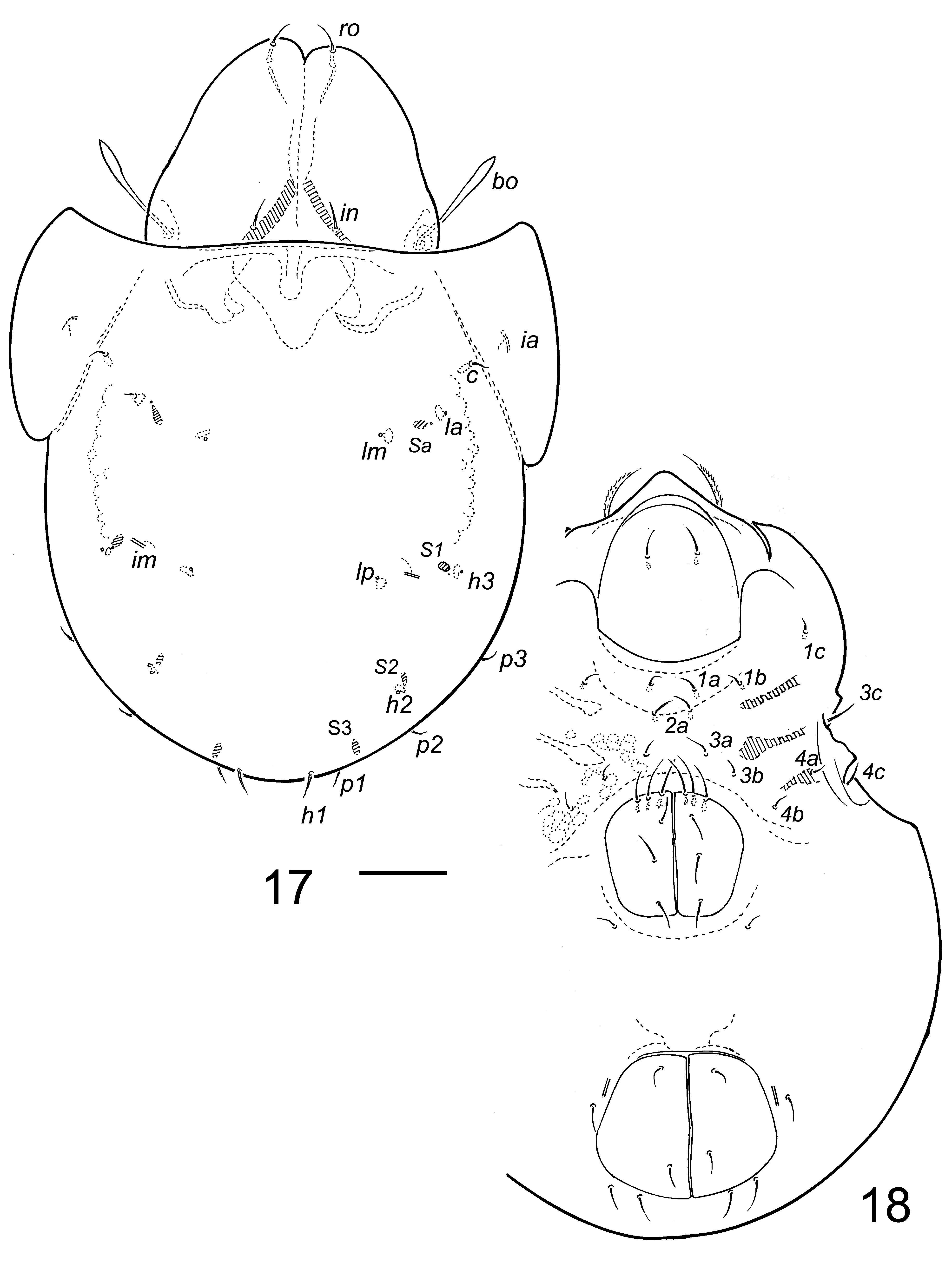
Prodorsum. Rostrum with smooth margin, without medial incision; weak medial keel extending posteriorly to underneath lamella ( Fig. 21C, D View FIGURE 21 ). Rostral setae heavily barbed laterally, directed anteromedially ( Fig. 11 View FIGURES 10 – 11 ). Lamella long, pair fused, covering all of prodorsum in dorsal aspect ( Fig. 9 View FIGURE 9 ), with faint longitudinal line medially (visible with light microscopy), with almost parallel areas accommodating nerve canal for seta le ( Figs. 8F View FIGURE 8 , 22A View FIGURE 22 ). Lamella with short, V or U-shaped indentation anteriorly; concave posterolaterally, accommodating bothridium ( Figs. 8E View FIGURE 8 , 13B View FIGURE 13 , 21D View FIGURE 21 ). Seta le arising dorsally on lamella ( Figs. 9 View FIGURE 9 , 17 View FIGURES 17 – 18 , 20 View FIGURE 20 ). Interlamellar setae subequal in thickness to lamellar setae. Exobothridial seta present, or absent; alveolus usually present. Bothridial wall expanded anterodorsally with pointed scale, otherwise cup-shaped, without lateral indentation ( Figs. 13B View FIGURE 13 , 22A View FIGURE 22 ). Porose area Ad present. Dorsophragmata fused ( Figs. 9 View FIGURE 9 , 17 View FIGURES 17 – 18 , 20 View FIGURE 20 ).
Notogaster. Longer than wide; with hinged pteromorphs having smooth margins without dens anteroventrally ( Figs. 19B View FIGURE 19 , 21D View FIGURE 21 , 22B View FIGURE 22 ). Anterior margin of notogaster straight, transverse, without ridges in region lateral to bothridium ( Fig. 9 View FIGURE 9 ). Anterior of notogaster without hexagonal pattern in transmitted light. Octotaxic system expressed as 4 pairs of either porose areas, saccules or tubules ( Figs. 8D View FIGURE 8 , 9 View FIGURE 9 , 17 View FIGURES 17 – 18 , 20 View FIGURE 20 ); without sexual dimorphism. With 10 pairs of short setae.
Lateral Region of Podosoma. Genal incision and genal tooth absent; open taenidium present in usual position of genal incision, extending to acetabulum I ( Figs. 8B View FIGURE 8 , 13A View FIGURE 13 , 21E View FIGURE 21 ). Tutorium narrow, lamelliform, cusp triangular, lying parallel to dorsal contour of prodorsum in lateral aspect, extending anterior of insertion of rostral seta ( Figs. 11 View FIGURES 10 – 11 , 13A View FIGURE 13 ). Pedotectum I flattened or rounded dorsally, pointed distally, without ventral depression ( Fig. 11 View FIGURES 10 – 11 ). Pedotectum II present, without tubercle close to body wall. Custodium present as very short extension to circumpedal carina ( Figs. 8F View FIGURE 8 , 11 View FIGURES 10 – 11 , 19E View FIGURE 19 , 22C View FIGURE 22 ). Discidium strongly curved lamina ( Fig. 8F View FIGURE 8 ). Porose area Al absent. Humerosejugal porose organs Am and Ah present, expressed as porose areas ( Figs. 8E View FIGURE 8 , 11 View FIGURES 10 – 11 ).
Ventral. Epimere I without necklace of small tubercles anteriorly. Epimeral setal formula 3-1-3-3. Genital plates with 6 setae, 3 setae on anterior margin; 1 pair aggenital setae; 3 pairs adanal setae and 2 pairs anal setae ( Fig. 10 View FIGURES 10 – 11 ). Lyrifissure iad at anterolateral edge of anal plate ( Fig. 11 View FIGURES 10 – 11 ). Postanal porose area present ( Fig. 13E View FIGURE 13 ). Band of darker integument absent between genital and anal plates. Preanal organ large cup-shaped ( Fig. 8C View FIGURE 8 ).
Gnathosoma . Axillary saccule present at base of palp. Chelicera chelate-dentate ( Fig. 13C View FIGURE 13 ). Mentum with tectum covering camerostome when elevated ( Figs. 10 View FIGURES 10 – 11 , 21C, E View FIGURE 21 ). Palp setal formula 0-2-1-3-9(1); eupathidium acm subequal to solenidion, forming double horn with solenidion along length, bases slightly separate ( Fig. 8G View FIGURE 8 ).
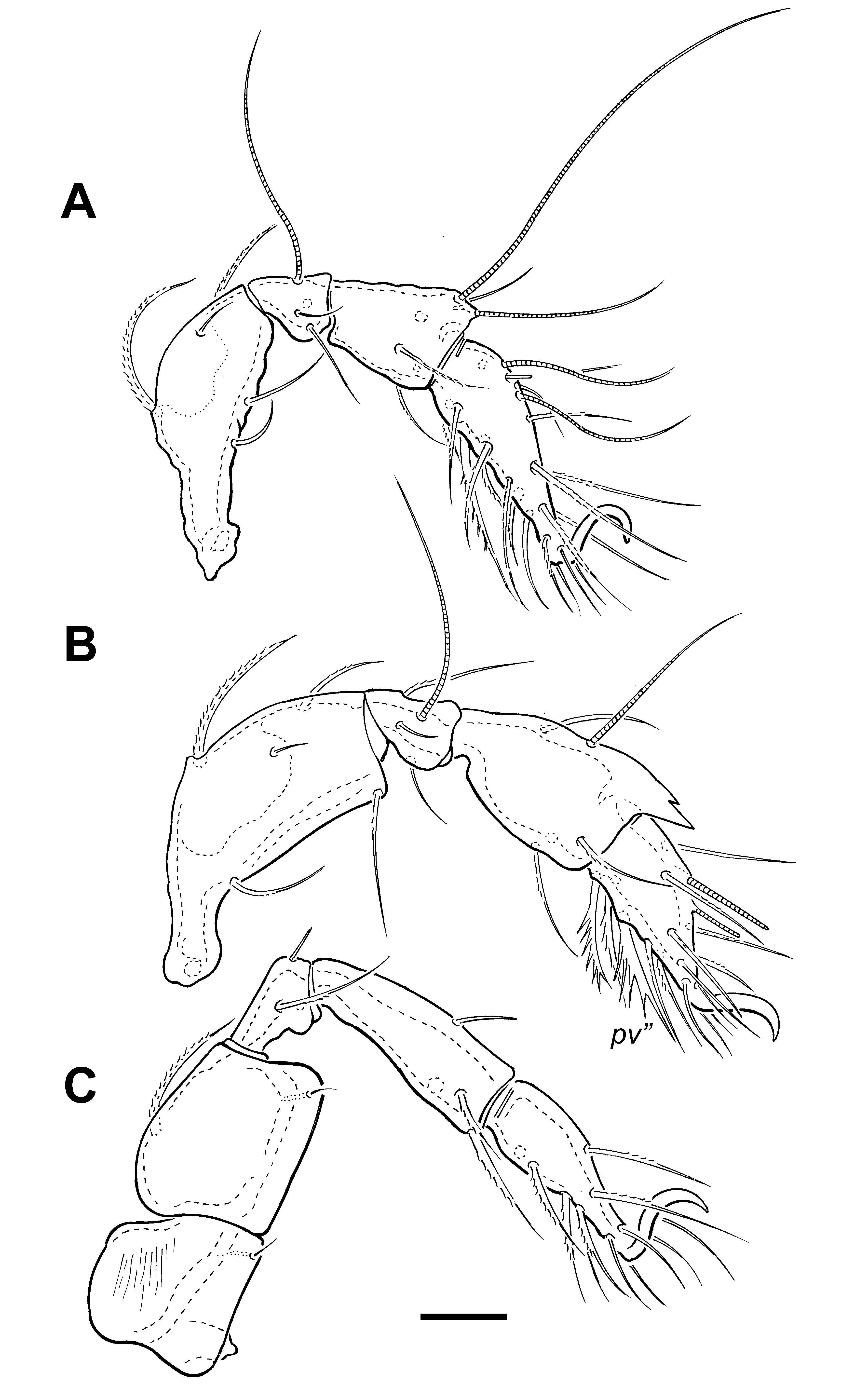
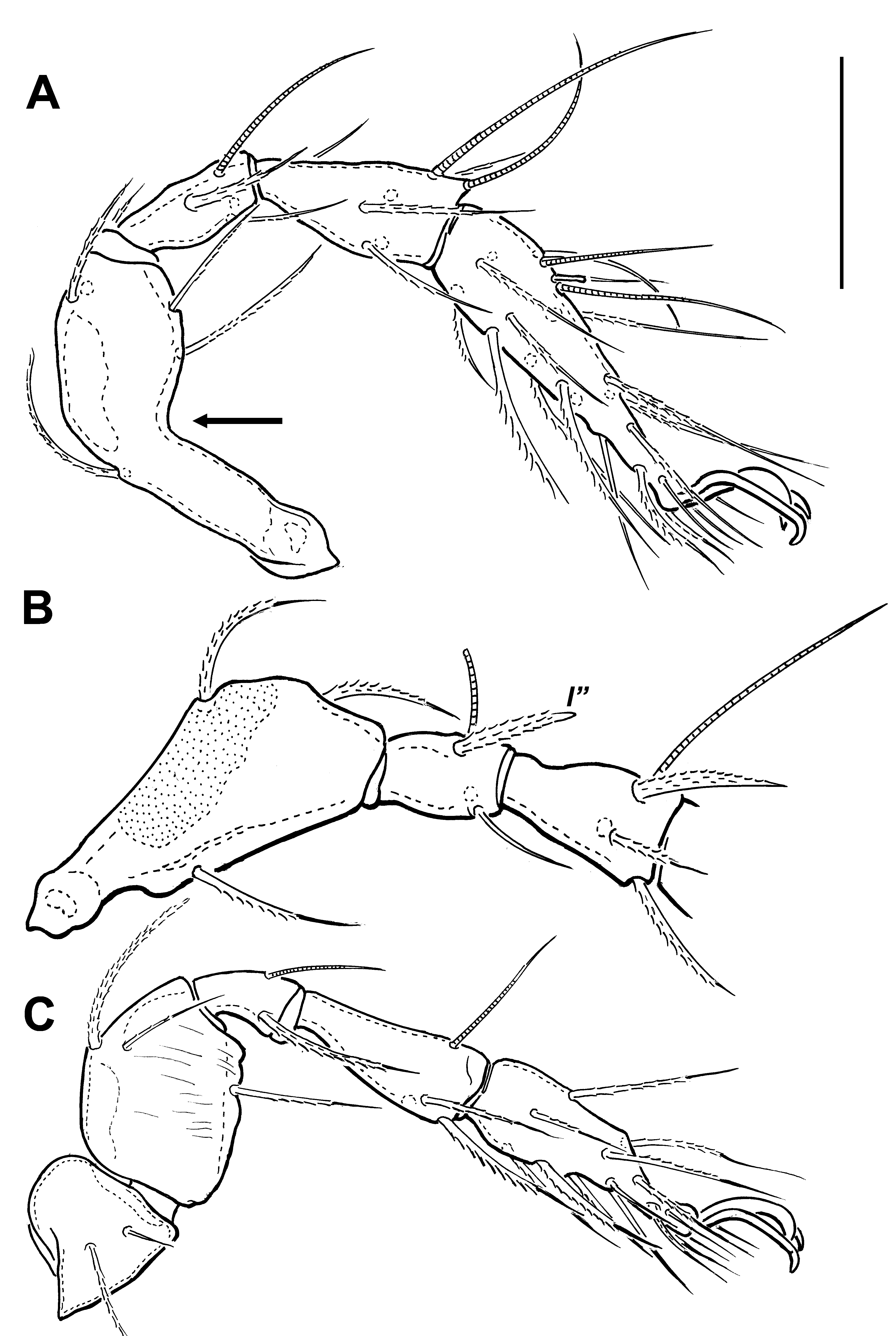
Legs. Monodactylous or heterotridactylous. Leg setal formulas I–IV, solenidia in parentheses: I: 1-5-3(1)-4(2)- 20(2); II: 1-5-3(1)-4(1)-15(2); III: 2-3-1(1)-3(1)-15; IV: 1-2-2-3(1)-12. Genua I and II without tooth ventrally ( Figs 12A, B View FIGURE 12 ). Porose areas on femora I to IV and trochanters III and IV. Femur I with obtuse bend medially ventral to seta d ( Fig. 12A View FIGURE 12 , Fig. 22E View FIGURE 22 ). Tibia II without anterodorsal dentate carina. Seta s on tarsus I eupathidial, positioned distal of setae ( a). Solenidia on tarsus II shorter than setae on segment; ω1 not most proximal setiform structure on segment;. Genua I and II with distinctive and spinous seta l” ( Figs. 12B View FIGURE 12 , 19F View FIGURE 19 , insert, 21C). Femur III with seta l’ present; seta v' of genua I and II present. On tibiae I and II seta l” slightly more spinous than l’. Anterodorsal knobs or spines absent from tibia I.
Remarks. 1. Sellnick (1920) proposed Lepidoribates to accommodate Zetes latirostris Koch. Jacot (1929) considered it a junior synonym of Tegoribates and this was recognized by Sellnick (1955) and subsequent authors.
2. Other than a brief comment on larva and nymphs of T. americanus ( Behan-Pelletier 2001) , immatures of Tegoribates have been known only for T. latirostris (larva and nymph; Tuxen 1943, Grandjean 1954, Norton and Ermilov 2014). Tuxen (1943 Figs 12 View FIGURE 12 , 13 View FIGURE 13 ) described a “spinous knob” on the dorsal of the hysterosoma of the larva; this is the hysterosomal region bearing setae dp, as found in other species of Tegoribates , described below.
3. Grandjean (1954) diagnosed Tegoribates as lacking a genal notch. This is the case in all described species. However, a taenidium is present in the same region in all members of this genus, running from close to the border of the rostrum posteriorly to acetabulum I. Following the terminology of Grandjean (1968; see also Travé 1986), this appears to be a “respiratory taenidium”, which he defined as a furrow, almost closed or not, banded or smooth, hollowed into the integument, and communicating with the stigma of trachea I or with those of tracheae I, sejugal and III. The taenidium of Tegoribates extends to acetabulum I, and possibly communicates with trachea I. A taenidium, with a similar morphology, is found in the same region in the licneremaeoid genus Phylleremus, which is a unique expression in the superfamily ( Behan-Pelletier & Walter 2007). A ‘taenidial gutter’ is also found on the prodorsum of the oppioid Hypogeoppia belgicae Wauthy & Ducarme, 2006 . Travé (1986) noted how easy it is to overlook this structure. When viewed with incident illumination of flattened, slide-mounted specimens the taenidium appears as in Figs. 8B View FIGURE 8 , 13A View FIGURE 13 , and 19A View FIGURE 19 ; on heavily sclerotized whole-mounts, it has the appearance indicated in Figs 21 D, E View FIGURE 21 . This is the lateral structure outlined by Engelbrecht (1986) on the prodorsum of Tegoribates natalensis (his Fig. 65). Hypozetes also lacks a genal notch, and while the rostral ridge has a narrow tectum there is no evidence of a taenidium ( Behan-Pelletier 2001, her Fig. 3 View FIGURES 2 – 3 ).
4. The octotaxic system in Tegoribates is developed in various forms. Grandjean (1953, 1954) noted that the octotaxic system is represented by 4 pairs of small “trachea”, called ‘tubules’ by Norton et al. (1997; their Fig. 5l View FIGURE 5 ) and herein. This is the expression of the octotaxic system in T. latirostris and T. subniger (see below), and these tubules are closely associated with setae. I follow Norton et al. (1997) and use the notation Ta, T1-T3 for these structures, corresponding to the notation for porose areas ( Aa, A1–A3) and saccules ( Sa, S1–S3). Norton et al. (1997) also noted the presence of tubules in Plakoribates . Fine tubules also are found in the ceratozetoid genera Lamellobates, Paralamellobates and Sacculozetes , but these are not closely associated with setae. Woolley (1965) expanded the diagnosis of Tegoribates to include T. bryophilu s with the octotaxic system of saccules, an expression also found in T. americanus . The expression of the octotaxic system is further expanded in T. walteri sp. nov. (described below) to include porose areas. The close association of the octotaxic system with setae is most evident in T. latirostris and T. subniger . Other genera of poronotic Brachypylina have the octotaxic system as either porose areas or saccules, according to species (e.g., Ceratozetidae : Trichoribates , Achipteriidae : Anachipteria , Tegoribatidae : Tectoribates ); only Tegoribates includes species with either porose areas, saccules or tubules.
5. A distinct apomorphy of all known species of Tegoribates is femur I distinctly bent to an obtuse angle near the level of seta d. Femur I is also relatively longer than in other genera of Tegoribatidae where legs have been studied; e.g., compare Figs 12A View FIGURE 12 , 22E View FIGURE 22 with Fig. 4A View FIGURE 4 herein, and Fig. 7A View FIGURE 7 ( Behan-Pelletier & Walter 2013), Fig. 21 View FIGURE 21 ( Ermilov & Anichkin 2013), Fig. 3A View FIGURES 2 – 3 ( Ermilov & Minor 2015) and Fig. 2A View FIGURES 2 – 3 ( Fredes & Martínez 2016). This elongated and bent femur I could possibly be a modification to accommodate the depth of the prosoma of Tegoribates , and the associated length of pedotectum I ( Fig. 22E View FIGURE 22 ).
6. The preanal organ of Tegoribates ( Fig. 8C View FIGURE 8 ) is most similar to that of Podacarus auberti Grandjean (1955) (Podacaridae) with a large, wide cup, the anterior face of which serves for attachment of muscles for the genital aperture, and protuberance ob. It differs in that the struts for P. auberti are parallel; those of Tegoribates are directed laterally at an obtuse angle. The preanal organ has not been studied in much detail in poronotic Brachypylina other than by Grandjean (especially Grandjean 1969). This character seems to be highly variable in Tegoribatidae , with that of Protectoribates ( Figs. 22F, G View FIGURE 22 ) different from any described to date. In contrast, that of Tectoribates ( Fig. 22I View FIGURE 22 ) is similar to that of described Ceratozetoidea (e.g., Euzetes ; Grandjean 1969). The preanal organ of Hypozetes is similar to that of Tectoribates , except that the cup is very wide and the struts are thick, strongly sclerotized and parallel ( Fig. 22H View FIGURE 22 ),
7. Ermilov and Minor (2015) disagreed with the transfer of Tegoribates montana Engelbrecht, 1986 and T. nuda Engelbrecht, 1986 (both from South Africa) to Neophysobates by Subías (2004, 2017) and these species are retained in Tegoribates herein. Similarly, Fredes and Martínez (2016) questioned the change in classification of Tegoribates natalensis Engelbrecht, 1986 to Paraphysobates by Subías (2004), and this species is also retained in Tegoribates herein.
8. A tutorium is present, but difficult to see, in Tegoribates species as it is overlain by the lamella dorsally and the pedotectum laterally. This possibly explains the absence of this character from previous descriptions of species.


No known copyright restrictions apply. See Agosti, D., Egloff, W., 2009. Taxonomic information exchange and copyright: the Plazi approach. BMC Research Notes 2009, 2:53 for further explanation.
|
Kingdom |
|
|
Phylum |
|
|
Class |
|
|
Order |
|
|
SubOrder |
Oribatida |
|
Family |
Tegoribates Ewing, 1917
| Behan-Pelletier, Valerie M. 2017 |
Tegoribates montana
| Engelbrecht 1986 |
Tegoribates natalensis
| Engelbrecht 1986 |
Tegoribates nuda
| Engelbrecht 1986 |
Tegoribates bryophilus
| Woolley 1965 |
Tegoribates americanus Hammer, 1958
| Hammer. Setae (Roman 1958 |
Tegoribates subniger
| Ewing 1917 |
Oribata alzanii
| Coggi 1900 |
Tegoribates latirostris (
| Koch 1844 |