
Hyoidea Reuter, 1876
|
publication ID |
https://doi.org/10.5281/zenodo.5740129 |
|
publication LSID |
lsid:zoobank.org:pub:A5DF029E-037F-418D-BD77-BBC7C951592E |
|
persistent identifier |
https://treatment.plazi.org/id/9B7B87D3-FFED-CF18-FDBC-E189FD74FDEC |
|
treatment provided by |
Marcus |
|
scientific name |
Hyoidea Reuter, 1876 |
| status |
|
Hyoidea Reuter, 1876 View in CoL
Hyoidea Reuter, 1876: 34 View in CoL (original description)
Hyoidea: CARVALHO (1958: 7) View in CoL (catalogue); HOBERLANDT (1963: 261) (redescription, key to species); KERZHNER (1964: 971) (redescription); WAGNER (1974: 138) (redescription); SCHUH (1995: 123) (catalogue); KERZHNER & JOSIFOV (1999: 249) (catalogue); KNYSHOV & KONSTANTINOV (2012) (habitus illustration, figures of male and female genitalia, measurements, discussion).
Type species. Hyoidea notaticeps Reuter, 1876 View in CoL (by monotypy).
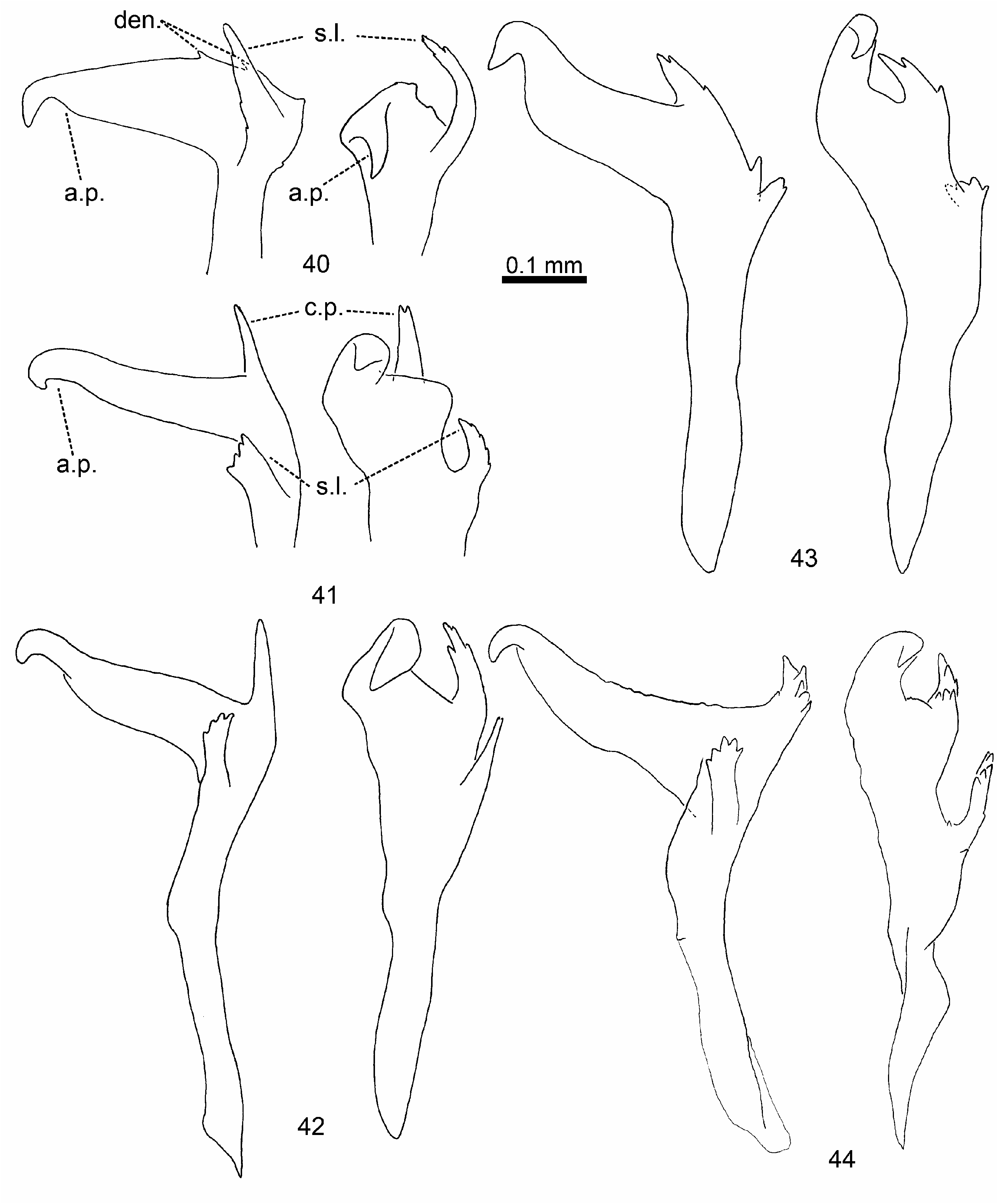
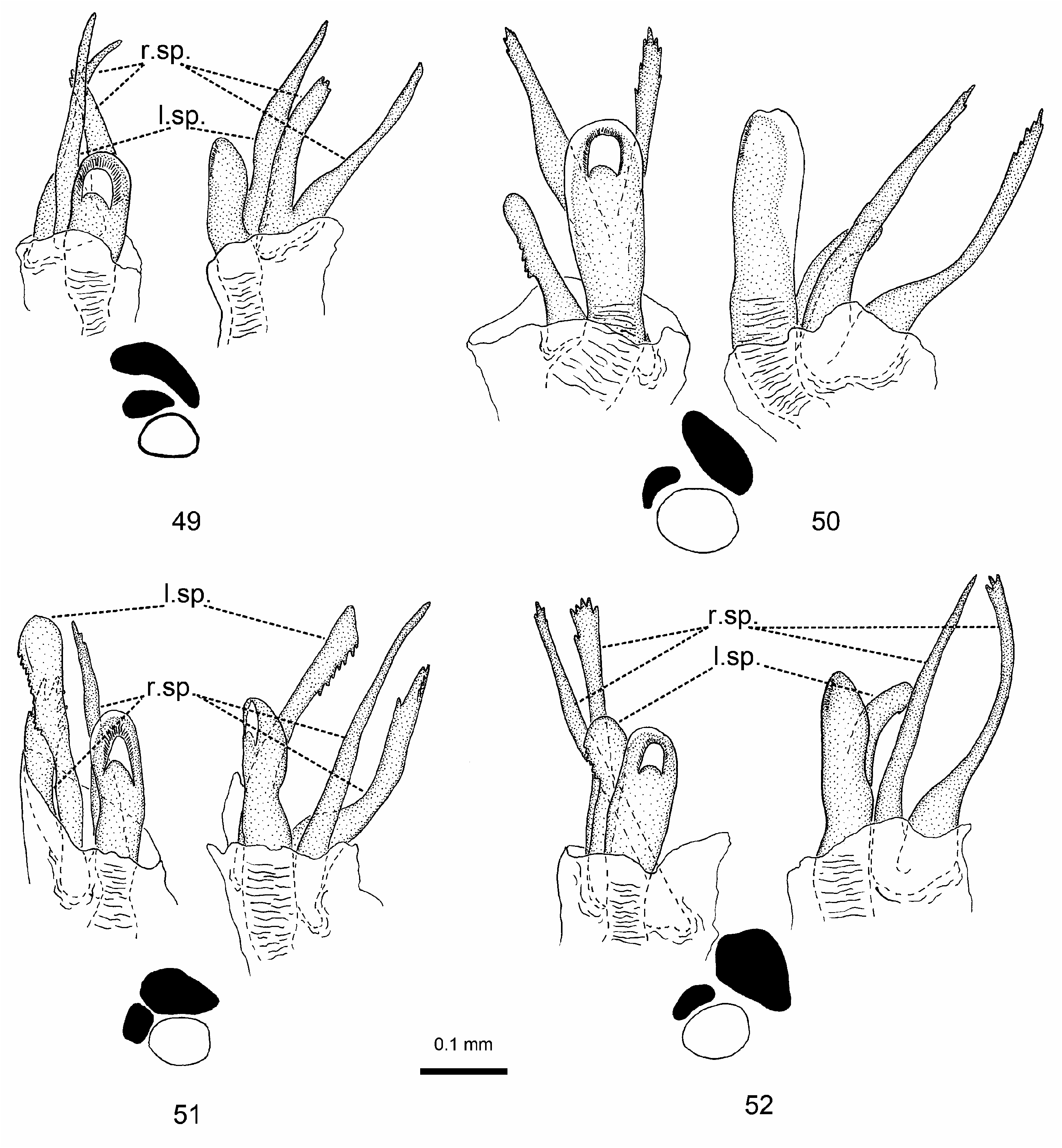
Redescription. Male: Macropterous, nearly parallel-sided, small to middle-sized, 3.7–5.6. COLOURATION ( Figs 1–17 View Figs 1–9 View Figs 10–17 ): Dirty yellow to brown, dorsum in many species covered with minute dark spots. Head: Clypeus typically with dark medial spot at base and two dark longitudinal stripes at sides extending from base to middle or even to apex of clypeus; sometimes spots on clypeus large and confluent or clypeus entirely dark; frons with a series of large dark stripes radiating from midline and with small lateral dark spots; vertex with two large rounded spots near eyes, in H. hannothiauxi with large transverse dark stripe; antenna dark brown to black with uniformly coloured segments, sometimes segment II with somewhat darkened apical half; labium brown to dark brown with darkened apex of segment IV. Thorax: Pronotal collar pale, with darkened middle part or entirely darkened; pronotal disc dirty yellow to brown, usually with dense, minute, more or less round dark spots; calli pale to brown with black confluent spots or entirely black; two pits between calli always dark; exposed part of mesonotum pale to dark brown, sometimes with orange tinge; scutellum pale to dark brown, usually with more or less expressed pale midline; thoracic pleurites pale brown. Hemelytron: Dirty yellow to brown, usually with dense minute, round dark spots; hemelytron uniformly coloured in all species except H. flavolimbata with contrastingly yellow exocorium and H. hannothiauxi with reddish brown cuneus; membrane uniformly pale brown to brown, with somewhat darker veins. Legs: Femora pale brown, with dark spots on dorsal and ventral surfaces, uniformly brown in dark specimens; tibiae pale to dark brown, without spots at bases of tibial spines; tarsi uniformly pale to dark brown. Abdomen: Uniformly pale brown or dorsally darker than ventrally. SURFACE AND VESTITURE: Dorsum dull, finely rugose, scutellum and basal part of pronotum frequently with fine transverse wrinkles; pronotum always with two minute pits between calli; pronotum and clavus in some species with dense, shallow and darkened punctation; clothed with mixture of contrastingly short semiadpressed simple setae ranging in color from pale to black and small, oval, silvery scales ( Figs 22–24, 26 View Figs 20–29 ), in H. horvathi replaced with longer and moderately flattened, silvery scalelike setae; each anterior angle of pronotum with long dark stiff seta; tibial spines brown and short, located on inner surface of tibia, almost equal in length to width of tibia, absent at base, scarce and usually arranged in two rows at middle and dense at apex of tibia; thoracic and abdominal venter with pale simple setae. STRUCTURE: Head: Eyes ovate in lateral view ( Fig. 21 View Figs 20–29 ), facets flat ( Fig. 25 View Figs 20–29 ); frons convex, vertex with two dark, round, distinctly sculptured spots ( Figs 21–22 View Figs 20–29 ); head posteriorly with more or less raised transverse carina ( Figs 21, 23 View Figs 20–29 ); labium reaching middle coxa. Thorax: Pronotum trapeziform, with straight or slightly concave lateral and posterior margins; anterolateral angles distinctly carinate ( Figs 20–21, 23–24 View Figs 20–29 ); pronotal collar thin, but distinct ( Figs 20–21, 22–24 View Figs 20–29 ); calli convex ( Figs 21, 23 View Figs 20–29 ); scutellum slightly convex, with weakly pointed apex; metepisternal scent gland evaporatory area relatively large, triangular, with abundant mushroom bodies, peritreme covered with microtrichia ( Fig. 27 View Figs 20–29 ). Hemelytron: Cuneus twice as long as broad at base; membrane of forewing relatively long, far extending beyond apex of abdomen. Legs: Femora somewhat flattened, tibiae cylindrical; third tarsal segment equal in length to first and second segments combined; pretarsus with long, smoothly curved claws and lamellate, apically convergent parempodia, pulvilli absent ( Figs 28–29 View Figs 20–29 ). GENITALIA: Genital capsule with distinct large tooth on left side of genital opening ( Figs 30–32, 34–36 View Figs 30–36 ) in all species except H. hannothiauxi ( Fig. 33 View Figs 30–36 ); genital opening located at apex of genital capsule; cuplike sclerite noticeably protruding posteriorly beyond margin of genital capsule; left paramere L-shaped, often with well-developed sensory lobe and additional caudal process ( Figs 40–48 View Figs 40–44 View Figs ); apical process of left paramere strongly curved at apex, hook-like; right paramere club-shaped, with curved and serrate apex (Figs 37–39); aedeagus with strongly sclerotized phallotheca and two spicules, with right spicule usually larger than left one and divided into two branches at extreme base ( Figs 49 View Figs 49–52 –56).
Female: Macropterous, small to middle-sized, 3.0–5.5. COLOURATION, SURFACE AND VESTITURE: Similar to male. STRUCTURE: Usually larger in body size than male, with larger interocular distance, less developed anterolateral angles of pronotum, shorter hemelytron and particularly cuneus, and relatively large, broader abdomen usually noticeably extending beyond apex of membrane. GENITALIA: Posterior wall with dorsally projecting interramal lobes and weakly sclerotized central area; interramal lobes varying in shape from weakly cleft to smoothly rounded (Figs 59–60); dorsal labiate plate membranous or partly sclerotized, with clearly ovate sclerotized rings (Figs 57–58); vestibular sclerites relatively simple, only slightly asymmetrical (Fig. 61); first gonapophysis apically widened ventrally (Figs 63, 65, 67); second gonapophysis apically arrow-shaped (Figs 62, 64, 66).
Differential diagnosis. Distinguished from other Palaearctic genera of Orthotylus group by the following combination of characters: lateroapical pronotal angles distinctly carinate and protruding ( Figs 20–21, 23–24 View Figs 20–29 ); pronotal collar thin, but distinct ( Figs 20–21, 23–24 View Figs 20–29 ); vertex with transverse posterior carina ( Figs 21, 23 View Figs 20–29 ); hemelytron in male relatively long, with base of cuneus far surpassing abdominal apex; vestiture represented by simple setae and oval, short scalelike setae ( Figs 24, 26 View Figs 20–29 ); vertex always with two dark rounded spots, sometimes merging into single dark stripe; left paramere L-shaped, with well-developed additional processes and serrations on sensory lobe and body of paramere ( Figs 40–48 View Figs 40–44 View Figs ); right paramere club-shaped, with curved and serrate apex (Figs 37–39); aedeagus with two large spicules, right spicule bifurcated at extreme base ( Figs 49 View Figs 49–52 –56); sclerites encircling vulva asymmetrical (Fig. 61); first gonapophysis slightly widened apically (Figs 63, 65, 67); second gonapophysis apically arrow-shaped (Figs 62, 64, 66).
Host associations. Hosts are known for seven of the nine species currently recognized within this genus. Based on current evidence, Hyoidea is one of the two Palaearctic mirid genera that are exclusively associated with Ephedra spp. (Ephedraceae) (for review see KMENT & BRYJA 2007).
Distribution. Hyoidea is a Palaearctic genus, principally distributed in the Mediterranean region (Figs 18–19), with two species, H. notaticeps and H. kerzhneri , ranging Eastern Europe to Central Asia, and northern China. Distributional data on Hyoidea species are scarce, with only two species, H. notaticeps and H. kerzhneri sufficiently sampled. Two species, Hyoidea horvathi and H. lindbergi , were recorded from a few localities that are distant from localities of the remaining species ( H. flavolimbata , H. hannothiauxi , H. hermione , H. lopezcoloni , and H. stehliki ), which are known from the type localities only.
Discussion. Hyoidea belongs to the Orthotylus group of genera ( sensu Schuh 1974, see KNYSHOV & KONSTANTINOV 2012 for details). The combination of several peculiar diagnostic features separates Hyoidea from all other orthotyline genera. It seems to be most similar to Angulonotus , and is putatively its sister-genus. Both genera share distinctly carinate anterolateral angles of the pronotum, a thin but distinct pronotal collar, and several large dark spots on the frons and vertex. All Hyoidea species can be clearly distinguished from Angulonotus by the distinct mushroom bodies of the evaporatory area of the metathoracic scent gland, the short and broad scale-like setae on the dorsum, flat eye ommatidia, and the shape of the
Figs 18–19. 18 – Distribution map for Hyoidea hermione Linnavuori, 1989 , H. notaticeps Reuter, 1876 , and H. kerzhneri Hoberlandt, 1963 . 19 – Distribution map for H. flavolimbata Ribes & Ribes, 2000 , H. hannothiauxi Carapezza, 1997 , H. horvathi Montandon, 1890 , H. lindbergi Hoberlandt, 1963 , H. stehliki Baena & Günther, 2001 , and H. lopezcoloni Baena & Günther, 2001 . Literature data are excerpted from the following sources: H. notaticeps ( KIRITSHENKO 1918, 1951; LINNAVUORI & MODARRES 1999; PUTSHKOV & PUTSHKOV 1989; QI et al. 1995); H. kerzhneri ( GIDAYATOV 1971, LINNAVUORI 1989, QI et al. 1995); H. flavolimbata ( RIBES & RIBES 2000) ; H. lindbergi ( CARAPEZZA 1997) .
male and female genitalic structures (also see KNYSHOV & KONSTANTINOV 2012 for further discussion).
The nine species of Hyoidea currently are easily separated from each other by the structure of the male genitalia and, to a lesser extent, by the colouration, ratios and measurements. The genus can also be divided into several geographically defined groups of species. The first group includes H. notaticeps and H. kerzhneri , which are distributed in southeastern Europe and Asia, and H. hermione , which is restricted to Israel. The second group comprises those species restricted to the Iberian Peninsula, namely H. flavolimbata , H. lopezcoloni , and H. stehliki . Hyoidea hannothiauxi , H. horvathi and H. lindbergi comprise a third group, and are known from proximate localities on the northwestern coast of Africa. Species in each group can be easily distinguished from each other, except H. horvathi and H. lindbergi , which are discussed in relevant sections.
Female genitalic structures of Hyoidea are not distinctive between species, and in some cases accurate identifications of females requires their association with conspecific males. Sclerotized rings of the dorsal labiate plate, interramal lobes as well as first and second gonapophysis vary only slightly in shape within Hyoidea and also do not differ from those of many other orthotyline taxa. Dissections of many female specimens of the largely sympatric H. notaticeps and H. kerzhneri revealed only slight intraspecific variability in genitalic structures, despite their strikingly different habitus.


No known copyright restrictions apply. See Agosti, D., Egloff, W., 2009. Taxonomic information exchange and copyright: the Plazi approach. BMC Research Notes 2009, 2:53 for further explanation.
|
Kingdom |
|
|
Phylum |
|
|
Class |
|
|
Order |
|
|
Family |
Hyoidea Reuter, 1876
| Knyshov, Alexander & Konstantinov, Fedor V. 2013 |
Hyoidea : CARVALHO (1958: 7)
| KERZHNER I. M. & JOSIFOV M. 1999: 249 |
| SCHUH R. T. 1995: 123 |
| WAGNER E. 1974: 138 |
| KERZHNER I. M. 1964: 971 |
| HOBERLANDT L. 1963: 261 |
| CARVALHO J. C. M. 1958: ) |
Hyoidea
| REUTER O. M. 1876: 34 |