
Rhyacophila palmeni McLachlan 1879
|
publication ID |
https://doi.org/ 10.11646/zootaxa.4514.1.8 |
|
publication LSID |
lsid:zoobank.org:pub:D8F6FE7E-7EDE-476A-B1B1-79F206801BAC |
|
DOI |
https://doi.org/10.5281/zenodo.6492336 |
|
persistent identifier |
https://treatment.plazi.org/id/995487A7-FF94-FFD6-FF6B-FC0F2480FE2C |
|
treatment provided by |
Plazi |
|
scientific name |
Rhyacophila palmeni McLachlan 1879 |
| status |
|
Description of the fifth instar larva of Rhyacophila palmeni McLachlan 1879 View in CoL
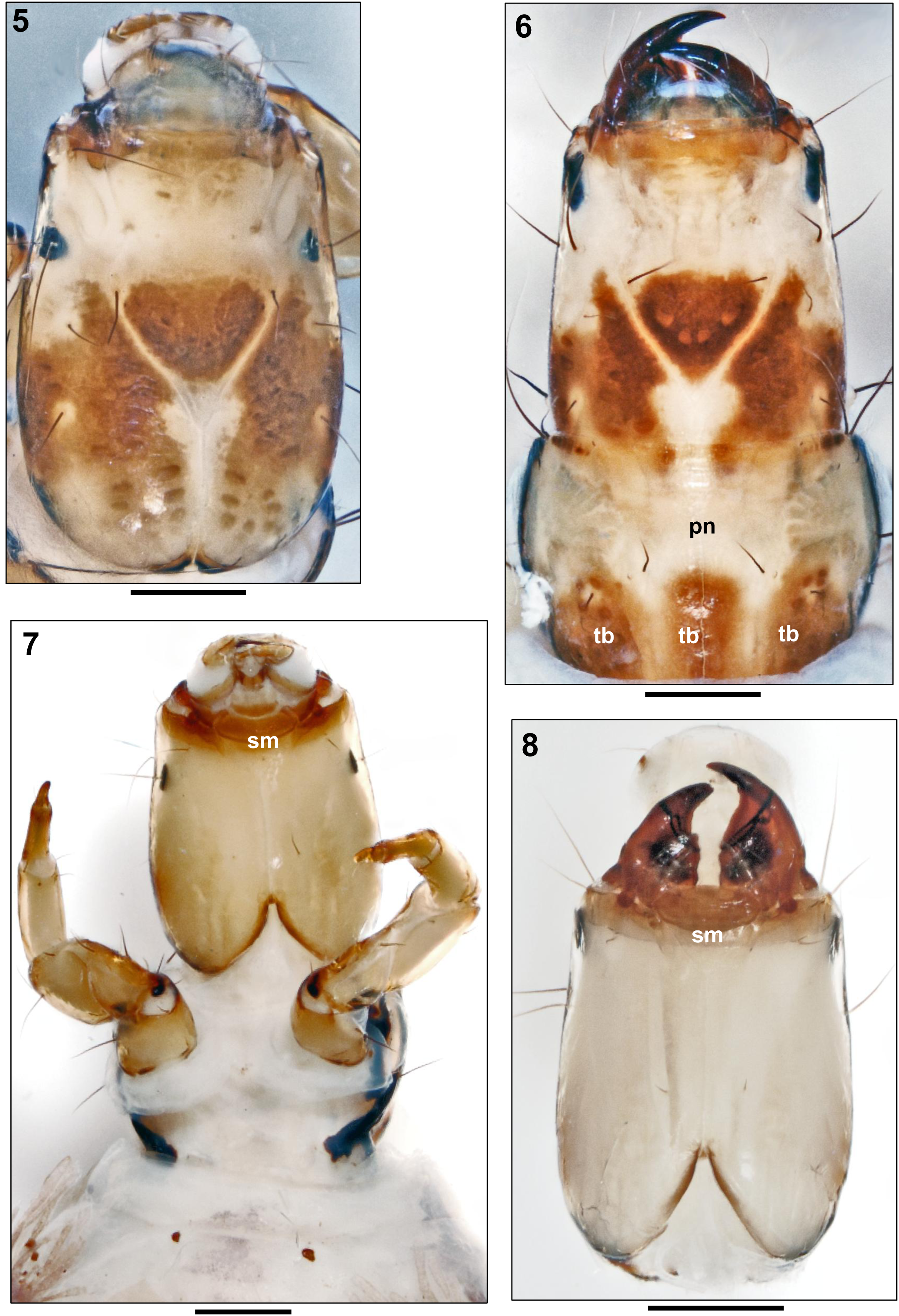
Biometry. Body length ranging from 16.4 to 17.5 mm, head width from 1.08 to 1.39 mm (n = 13). Soft body parts (mesonotum, metanotum, and abdomen) of live specimens green dorsally, pale green ventrally. Specimens in alcohol, purplish-green or purplish brown dorsally; lightly coloured ventrally. Sclerotised areas (head, pronotum, legs, abdominal dorsum IX and anal prolegs) light brown or yellowish with dark brown or black markings ( Figures 1–17 View FIGURES 1–4 View FIGURES 5–8 View FIGURES 9–14 View FIGURES 15–18 ).
Head. Head capsule elongated, yellowish brown, with lateral margins almost parallel, slightly convergent anteriorly, with slight constriction at eyes ( Figs. 1–8 View FIGURES 1–4 View FIGURES 5–8 ). Broad, transverse brown or light brown band across posterior 1/3 of frontoclypeus and adjacent sides of head interrupted by thin pale lines along frontoclypeal sutures, broad pale areas along coronal suture, and pale areas surrounding primary setae, especially setae 15 and 16 ( Figs. 1–6 View FIGURES 1–4 View FIGURES 5–8 , 9 View FIGURES 9–14 ). Conspicuous brown muscle attachment spots at dorsal and lateral sides of parietalia, and along coronal suture ( Figs. 1–6 View FIGURES 1–4 View FIGURES 5–8 , 9 View FIGURES 9–14 ). Dark brown marking at aboral end of frontoclypeus forming spade- or heart-shaped with crescentic row of four characteristic light muscle attachment spots ( Figs. 1–6 View FIGURES 1–4 View FIGURES 5–8 , often inconspicuous). Anterior edge of frontoclypeus (anteclypeus) almost straight, margined by light brown. Ventral view pale yellow except pale brown anterior and posterior margins, without any spots ( Figs. 7–8 View FIGURES 5–8 ). Ventral apotome triangular, light brown or yellowish, merged with rectangular submentum ( Figs. 7–8 View FIGURES 5–8 sm). Mouthparts ( Figs 4 m View FIGURES 1–4 , 8 View FIGURES 5–8 ) and labrum ( Figs 1–3 View FIGURES 1–4 lb) typical of the genus.
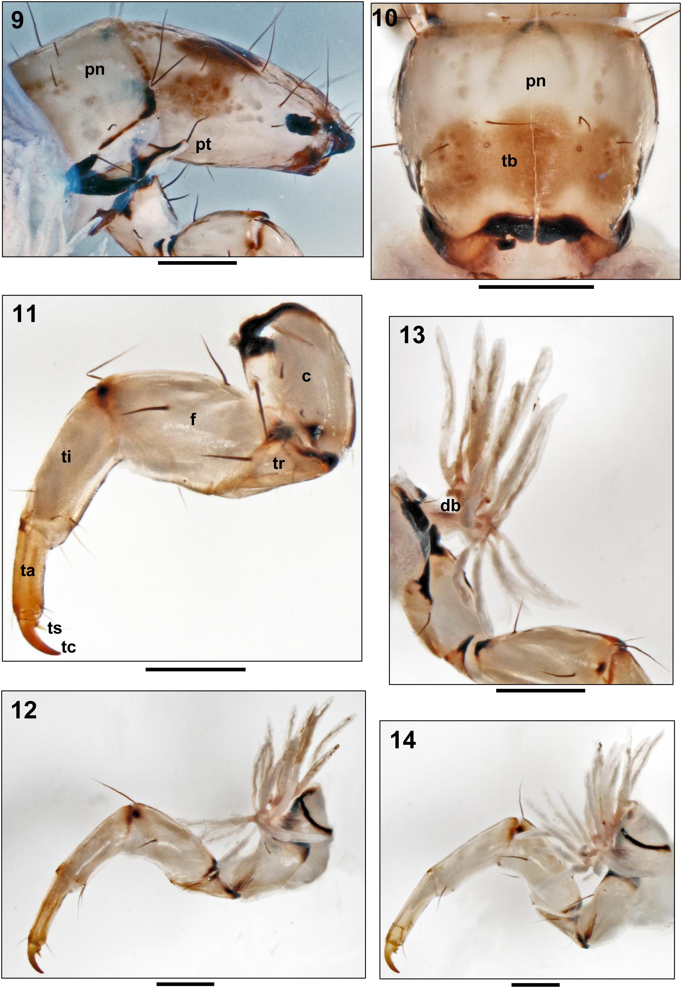
Thorax. Pronotum trapezoidal, covered by two large sclerites tapering posteriorly, wider than head capsule ( Fig. 10 View FIGURES 9–14 ). Yellowish and usually with few darker attachment spots in anterior half; posterior half mostly covered by transverse brown band bearing group of dark attachment spots, not always visible ( Figs. 6 View FIGURES 5–8 , 10 View FIGURES 9–14 tb). Posterior and lateral margins thickened and darkly striped, including anterolateral corners ( Figs. 9, 10 View FIGURES 9–14 ). Legs pale brown, with numerous setae on coxae, trochanters, and femora; tibiae and tarsi brownish with few smaller and thinner setae ( Figs. 11–14 View FIGURES 9–14 ). Forelegs shorter and stronger than mid- and hind legs. Forefemora distinctly wider than mid- and hind femora. Tarsal claws long and slender, curved, pointed, each with basal spur ( Figs. 11, 12, 14 View FIGURES 9–14 ). Thoracic gills with slender, long filaments arising laterally from two basal branches above each coxa, characteristic for the Rhyacophila s. str. Group ( Figs. 12–14 View FIGURES 9–14 ).
Abdomen. Abdomen slightly flattened dorsoventrally ( Fig. 16 View FIGURES 15–18 ); most specimens in alcohol pale brown to yellowish-green ventrally and darker purplish-brown to purplish-green dorsally. Tufted gill filaments present on each side of segments I to VIII each arising from common base ( Figs. 15, 16 View FIGURES 15–18 ). As in all species of Rhyacophila s. str., tracheal gills each consisting of short, thick ventral branch (vb) with numerous filaments near apex, and longer, more-slender dorsal branch (db) with numerous filaments on its dorsal side ( Figs. 15, 16 View FIGURES 15–18 ). Dorsal sclerite on segment IX ( Fig. 17 View FIGURES 15–18 ) trapezoidal and light brown in colour, with a darker posterior margin. Setae at posterior end long and black ( Fig. 17 View FIGURES 15–18 ). Anal appendages each with proximal sclerite ( Fig. 18 b View FIGURES 15–18 ) with ventral basal hook ( Fig. 18 h View FIGURES 15–18 ) and with long lateral sword process ( Figs 17, 18 View FIGURES 15–18 sp), extending beyond suture line but shorter than anal claw; anal claws each with three ventral teeth ( Fig. 18 View FIGURES 15–18 ac).


No known copyright restrictions apply. See Agosti, D., Egloff, W., 2009. Taxonomic information exchange and copyright: the Plazi approach. BMC Research Notes 2009, 2:53 for further explanation.