
Pleurodeles
|
publication ID |
https://doi.org/ 10.1093/zoolinnean/zlac063 |
|
DOI |
https://doi.org/10.5281/zenodo.7696798 |
|
persistent identifier |
https://treatment.plazi.org/id/9750C307-FFB6-4C01-FEB3-F029FA15F975 |
|
treatment provided by |
Plazi |
|
scientific name |
Pleurodeles |
| status |
|
PLEURODELES MICHAHELLES, 1830 View in CoL View at ENA
Species: Pleurodeles waltl * Michahelles, 1830 .
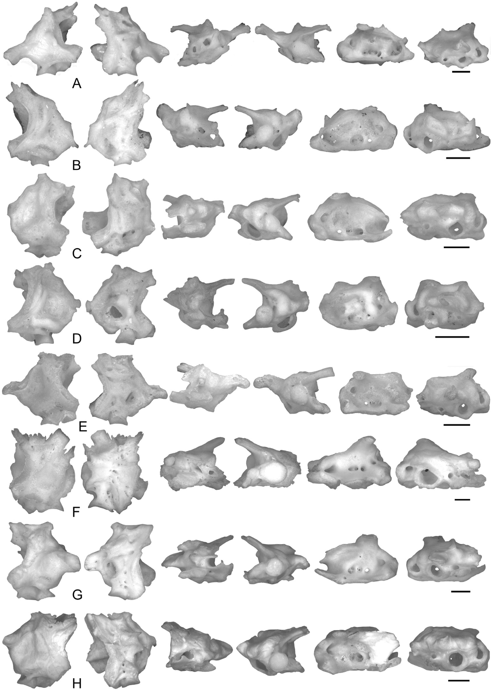
Otic–occipitum complex ( Fig. 5F View Figure 5 )
The otic–occipitum complex is generally sharp, because the prominentiae semicircularis anterioris and posterioris are usually covered by numerous crests (except for MNCM 16165, in which the crests are low). The prominentia semicircularis lateralis is more rounded and clearly recognizable. The middle depression is not visible between the prominentiae semicircularis. The circular fenestra ovalis is visible in posterior and ventral views. Its dorsal edge is also visible in dorsal view. Its anterior edge is located at the mid-length of the complex, and it is connected to the cotyles with a horizontal lamina. The parietal crest is low and limited to parts of the tectum synoticum (e.g. in HNHM 2004.90.1 and MNHN 2013-27) or, alternatively, it starts from the mid-length of the distal edge of the tectum synoticum, and it reaches the mid-length of the prominentia semicircularis anterioris (e.g. in MNHN 2013-26 and HNHM 2004.91.1). The parotic crest and process generally are not present. Instead, anterior and lateral to the anterior half of the prominentia semicircularis lateralis and parallel to the otic process, there are several small, anteriorly pointing crests or a single crest that ends in a sharp and anteriorly directed process. The anterior edge of the tectum synoticum is posterior to the mid-length of the complex. The tectum is developed medially as the prefacial and hypochordal commissures, and no concavity is visible between the prefacial commissure and the tectum. Posteriorly, the cotyle surrounds the circular or kidney-shaped postoticum foramen. The cylindrical otic process is much more anteriorly projecting than the processus basalis and bears a subcircular articular surface and sometimes an anterodorsal spine. The articular surface of the processus basalis is kidney-shaped, with the foramen faciale posterodorsal to it. A deep sulcus petrosus separates the otic and basalis processes. The foramen prooticum is entirely surrounded by bone. The auditory cavity is not deep. The basicapsular commissure bears an anteromedian ventral spine, also visible in dorsal view. The crista retrosellaris between the hypochordal and basicapsular commissures covers the whole fenestra basicranialis, which, therefore, is absent. The middle of the ventral side of the complex is depressed and strongly perforated, especially next to the ventral spine, and no medial crest is visible.
Remarks: In MNCM 16167, the parietal crest contacts a low parotic crest in the mid-length of the prominentia semicircularis anterioris. In MNCM 16165, the prooticum foramen is not entirely surrounded by bone.
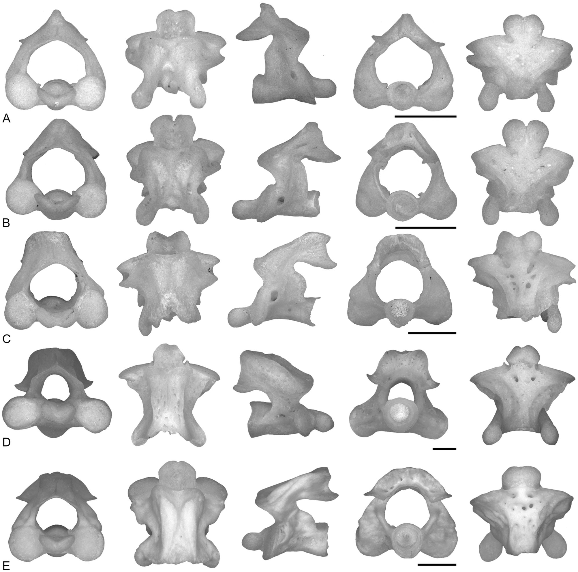
Atlas ( Fig. 8D View Figure 8 )
The neural canal is circular in anterior view and is as high as each occipital joint or slightly higher. In the same view, the dorsal edge of the neural canal is V-shaped. In posterior view, the neural canal is as wide as the circular cotyle. The occipital joints are circular or elliptical, with the major axis being horizontal. The articular facets of the odontoid process are separated by a narrow groove visible in ventral view. In this view, the base of the odontoid process is as wide as or narrower than each occipital joint. The low neural crest starts posterior to the anterior edge of the neural arch, and it is posteriorly broadened or forked (in this case, forming an incisura dorsalis visible in dorsal view and rarely in posterior view). The secondary crests are as inclined as the neural crest or ventrally concave. They reach the posterior edge of the neural arch. The neural spine is absent or low. The lateral surface of the atlas bears several foramina per side. The incisura vertebralis cranialis is small and V-shaped. In lateral view, the dorsal edge of the neural arch is strongly inclined. The deep incisura caudalis reaches the atlantal centrum, and its maximum concavity lies in the horizontal plane containing the maximum concavity of the incisura cranialis. The low lateral crests generally reach the postzygapophyses. The inferior crests are low, from the ventral edge of the occipital joints, forming a deep lateral concavity, and extending straight until the end of the centrum. In lateral view, the postzygapophyses extend posteriorly beyond the cotyle for more than half of their length. The posterodorsal edge of the atlas is flat in lateral view, and its posterior surface shows several small foramina. In dorsal view, the neural arch is concave anteriorly (U- or V-shaped concavity), and the cotyle is not visible posteriorly. The ventral surface bears two or more foramina.
Remarks: In MDHC 253, the neural crest is equally developed throughout its length. The anterior edge of the lateral walls of the neural arch is not convex in lateral view in MNHN 2013-27, hence the incisura vertebralis cranialis is only slightly visible. The lateral crests are not visible dorsal to the first spinal nerve foramen in MDHC 253. In HNHM 2004.90.1 and in the right side of MDHC 253, a third crest is present between the lateral and inferior crests of the atlas. In MNCN 161165, the incisura cranialis is roundly concave, and the incisura caudalis is not deep (neural arch slightly concave), whereas the anterior edge of the neural arch is flat in dorsal view. The incisura dorsalis is not visible in this specimen, in which the neural crest is clearly thicker posteriorly, forming a perforated bony triangle.
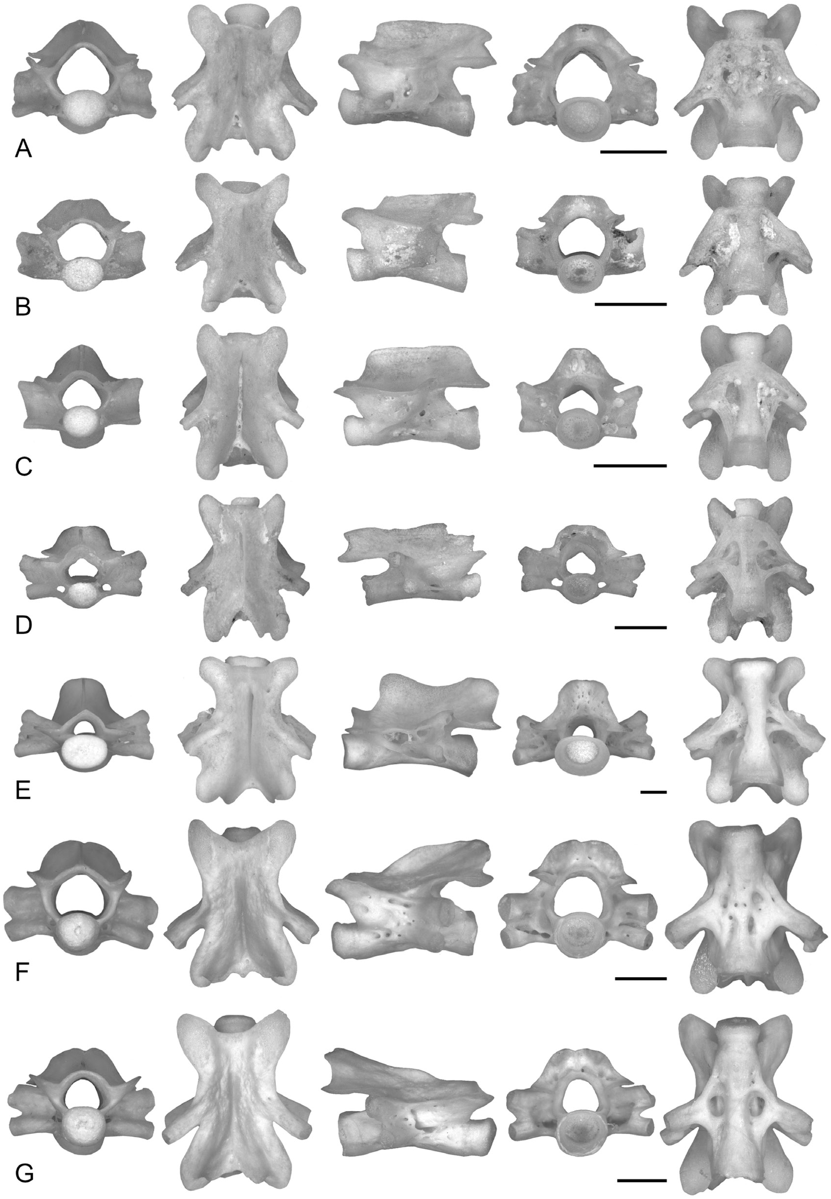
Precaudal vertebrae ( Fig. 11E View Figure 11 )
The precaudal vertebrae are opisthocoelous. The neural canal is triangular and, in general, lower than the condyle. The condyle is elliptical, with a horizontal major axis. In lateral view, the neural arch between the condyle and the elliptical prezygapophyses is weakly inclined. Diapophyses and parapophyses are distinguishable, connected by a smooth lamina that reaches their distal ends and is distally indented. The lamina is smooth anteriorly and bears some foramina posteriorly. The transverse processes are subvertical or with a slightly posterior orientation, covering the incisura caudalis only marginally in lateral view or, in some cases, not covering it. In lateral view, the neural arch dorsal to the prezygapophyses is visible and vertical or anteriorly concave. Half of the height of the vertebra is formed by the neural crest and neural arch dorsal to the postzygapophyses. The neural crest is blade-like and high, starting posterior to the line connecting the posterior edge of the prezygapophyses. The neural crest gently broadens posteriorly, being high in lateral view, and is pierced by several small foramina. The neural spine is absent. The anterior zygapophyseal crests contact the dorsal part of the parapophyses or the lamina connecting the diapophyses and parapophyses (the anterior zygapophyseal crest never contacts the dorsal part of the diapophyses; see also Stoetzel et al., 2011). The posterior zygapophyseal crests are well developed and horizontal. The small anterior ventral crest starts from the anteroventral edge of the parapophyses and contacts the prezygapophyses in their ventral part, thus being dorsally concave. The posterior ventral crests are absent or small. The ventral lamina is trapezoidal. Generally, the lateral surface of the vertebrae is perforated. In lateral view, the dorsal edge of the neural arch is anterodorsally concave. The incisura vertebralis caudalis is deep and generally reaches the centrum. In posterior view, the dorsal edge of the neural arch is horizontal or dorsally convex (inverted U-shaped). The neural arch dorsal to the postzygapophyses is sigmoid in lateral view. In lateral view, the postzygapophyses extend posteriorly beyond the cotyle for more than half of their length. In dorsal view, the neural arch is flat or anteriorly concave (U-shaped). Part of the condyle is visible in dorsal view, whereas the cotyle is not visible. The ventral surface is generally perforated.
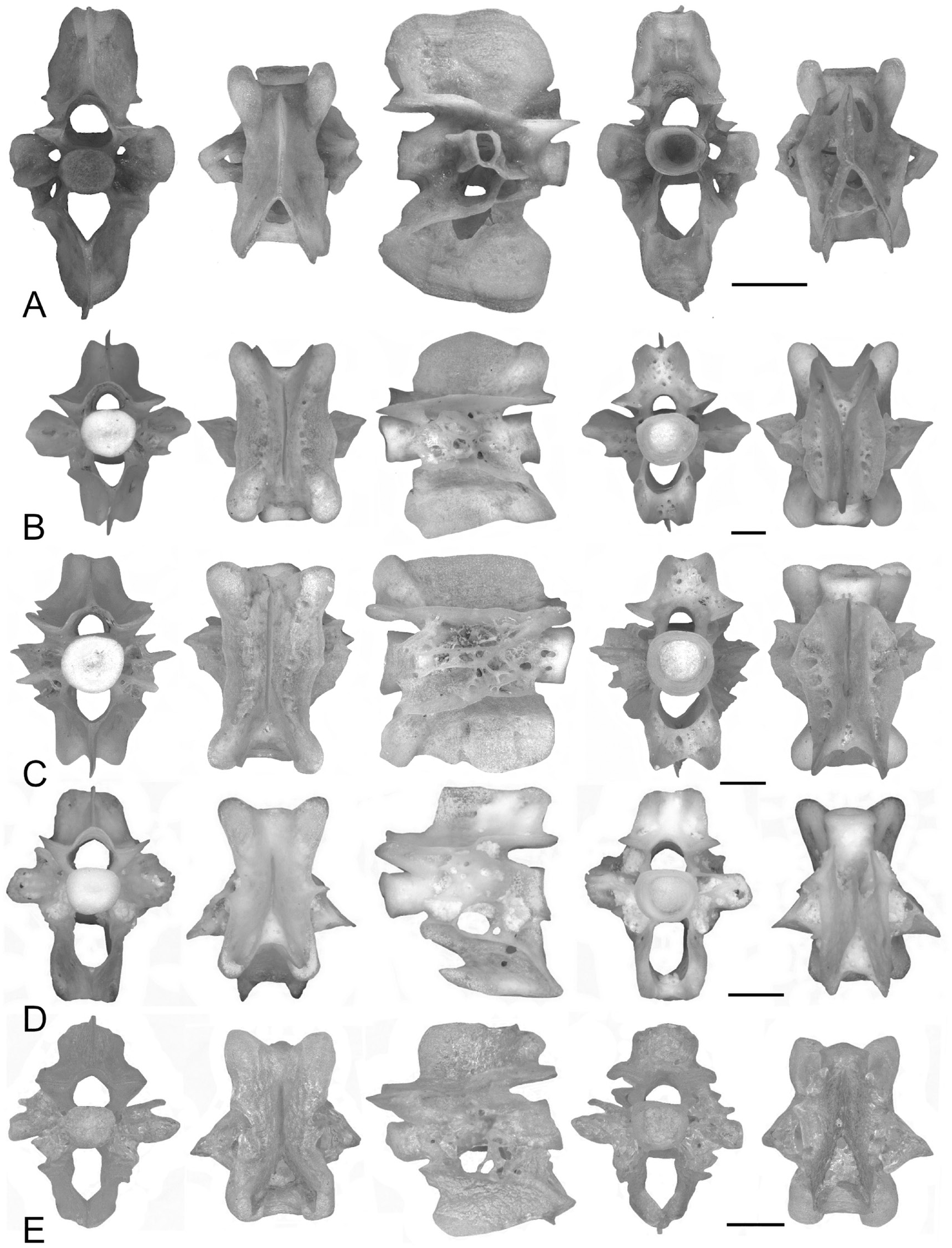
Caudal vertebrae ( Fig. 14B, C View Figure 14 )
The caudal vertebrae are not particularly high (height/ length ratio <1.25). The neural canal is pentagonal or circular, and the haemal canal is elliptical or U-shaped. The neural canal is narrower than the haemal canal and is as high as the haemal canal. The transverse processes are horizontal or rectangular (with the longest side vertical) in anterior view and sub-vertical in lateral view; in dorsal view, they are triangular. The neural and haemal crests are high and posteriorly enlarged or forked; if they are enlarged, the dorsal surface may be perforated. The lateral surface is not smooth, because several crests and small foramina are present. There is no large foramen on the haemal arch. The zygapophyseal crests are marked and horizontal. They are poorly connected with the transverse processes. The anterior ventral crests are marked and reach the anterior edge of the haemal arch. The posterior ventral crests start from the ventral edge of the haemal canal. In lateral view, the anterior and posterior ventral crests form an angle> 130°, almost reaching 180° in some cases. In lateral view, the anterior margin of the haemal arch is concave (or posteriorly inclined) between the centrum and the anterior ventral crest, and convex (or anteriorly inclined) ventral to it. The haemal crest projects anteriorly beyond the haemal arch, forming a sharp or rounded tip in lateral view. The haemal arch and crest form a sharp tip posteriorly.
Remarks: In MDHC 253, the transverse processes a r e V- s h a p e d, f o r m i n g a n a n t e r i o r c o n c av i t y. MNCN 16165 has procoelous caudal vertebrae and small anterior middle crests not in contact with either the prezygapophyses or the anterior edge of the cotyle.
No known copyright restrictions apply. See Agosti, D., Egloff, W., 2009. Taxonomic information exchange and copyright: the Plazi approach. BMC Research Notes 2009, 2:53 for further explanation.