
Spio blakei Maciolek, 1990
|
publication ID |
https://doi.org/10.11646/zootaxa.4019.1.15 |
|
publication LSID |
lsid:zoobank.org:pub:54E60C63-EC98-424A-B66E-A72CA79B65E8 |
|
DOI |
https://doi.org/10.5281/zenodo.5665408 |
|
persistent identifier |
https://treatment.plazi.org/id/971C0501-8924-FFBA-DFCD-951CFBD060D8 |
|
treatment provided by |
Plazi |
|
scientific name |
Spio blakei Maciolek, 1990 |
| status |
|
Spio blakei Maciolek, 1990 View in CoL
( Figs 5–7 View FIGURE 5 View FIGURE 7 )
Spio blakei Maciolek, 1990: 1110 View in CoL , Tables 1 View TABLE 1 –2. [Rename of homonym: identification of Spio pacifica Blake & Kudenov, 1978 View in CoL as homonym of Spio martinensis pacifica Berkeley, 1927 View in CoL ]
Spio pacifica Blake & Kudenov, 1978: 228 View in CoL –230, figs 28a–k.
Material examined. AM W.43926, MI QLD 2340 (2 af), formalin; AM W.43927, MI QLD 2340 (2 af), formalin; AM W.44119, MI QLD 2366 (2 mf), formalin; AM W.44371, MI QLD 2376 (>50), formalin; AM W.44478, MI QLD 2376, complete but fragmented, formalin; AM W.44381, MI QLD 2376 (6 af), formalin; AM W.44372, MI QLD 2376 (18 afs), 96% ethanol; AM W.44374, MI QLD 2378 (17 af), formalin; AM W.44565, MI QLD 2422 (3 af), formalin; AM W.44841, MI QLD 2429, af, formalin, with eggs; AM W.44860, MI QLD 2439, complete but fragmented, formalin; AM W.44863, MI QLD 2438, complete but fragmented, formalin; AM W.44476, MI QLD 2394, af, mf, formalin.
Diagnosis. Prostomium broadly rounded, posterior end short, extending to chaetiger 1, barely tapered. Nuchal organs with short median and long lateral ciliary bands, median bands extending to tcb of chaetiger 2 and recurved lateral bands up to chaetiger 3; metameric dorsal ciliated organs double-paired, usually present from chaetiger 3. Branchiae from chaetiger 1, continuous to almost end of body, length of first pair of branchiae about two thirds length of second pair; branchiae mostly free from notopodial lamellae. Ventral epidermal glands present from about chaetiger 3 to posterior middle body chaetigers; two pairs of glands per chaetiger. Postchaetal lamellae rounded, notopodial prechaetal lamellae present in anterior and middle chaetigers. From chaetiger 11 row of 4–5 tridentate hooded hooks replacing posterior row of capillaries in neuropodia, uppermost tooth very inconspicuous. Pygidium with two pairs of anal cirri, dorsal pair thinner and shorter.
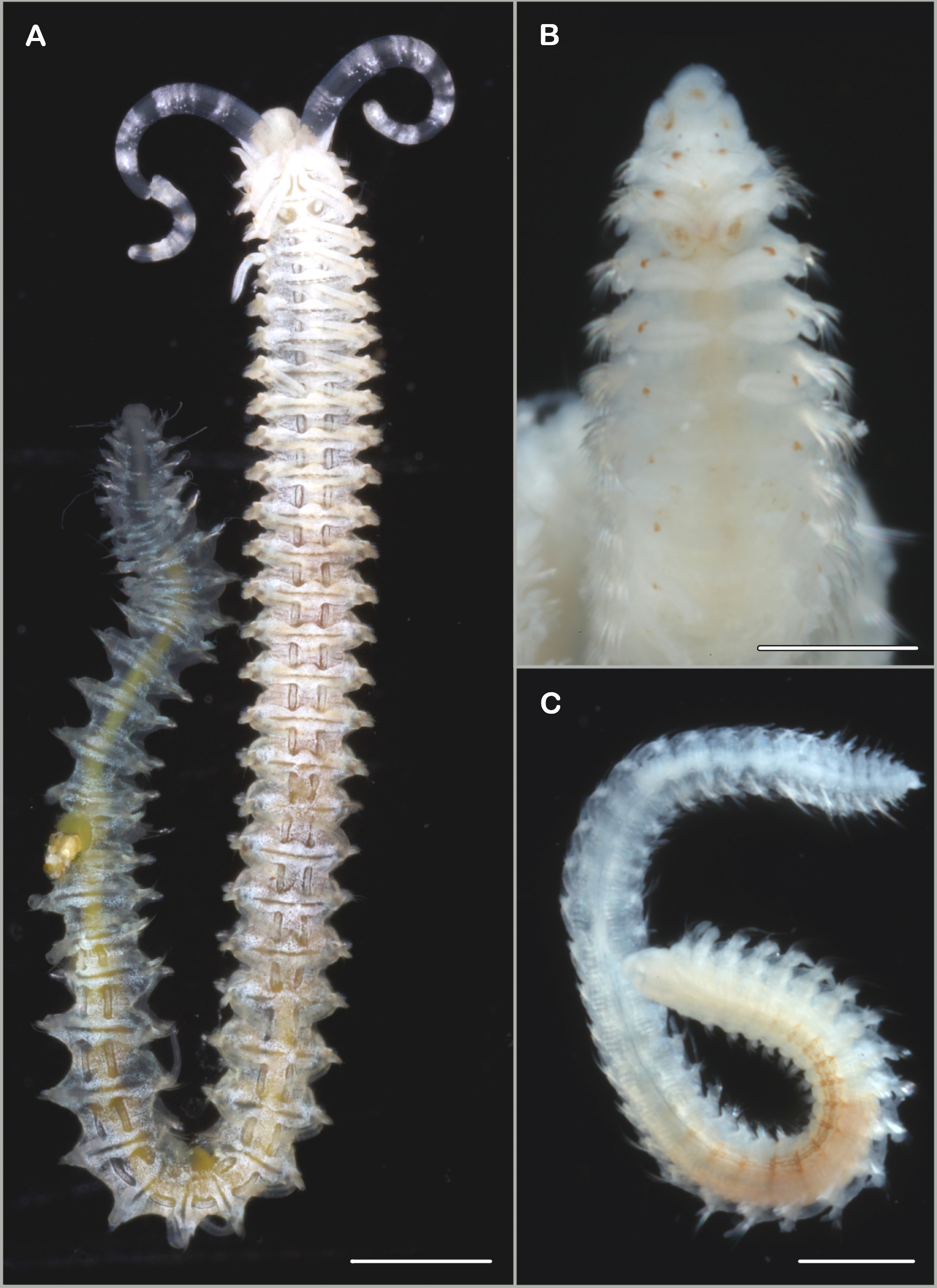
Description. (based on specimens examined in the course of the present study) Longest specimen 61 chaetigers, 0.75 mm wide and about 11 mm long. Prostomium broadly rounded, slightly expanded at anterolateral margin, with inconspicuous median furrow ( Figs 5 View FIGURE 5 A, B, 6A), anterior margin slightly projecting over peristomium; posterior end short, extending to chaetiger 1, barely tapered (Fig. 6A); usually with two pairs of black eyes arranged in trapezoid, anterior pair larger, almost crescent-shaped, further apart than posterior pair; prostomium separated from peristomium by a considerable furrow (Fig. 6A).
Nuchal organs and metameric dorsal ciliated organs distinct in well-preserved and living specimens; nuchal organs with short median and long lateral ciliary bands, median bands extended to tcb of chaetiger 2 and recurved lateral bands up to chaetiger 3 ( Figs 5 View FIGURE 5 A, B, 6A); metameric dorsal ciliated organs double-paired, usually present from chaetiger 3 ( Figs 5 View FIGURE 5 A, 6A), posteriorly extending to the end of the middle body region, slightly longer in further posterior segments (Fig. 6B); tcb´s discernable throughout the body (Fig. 6A–C).
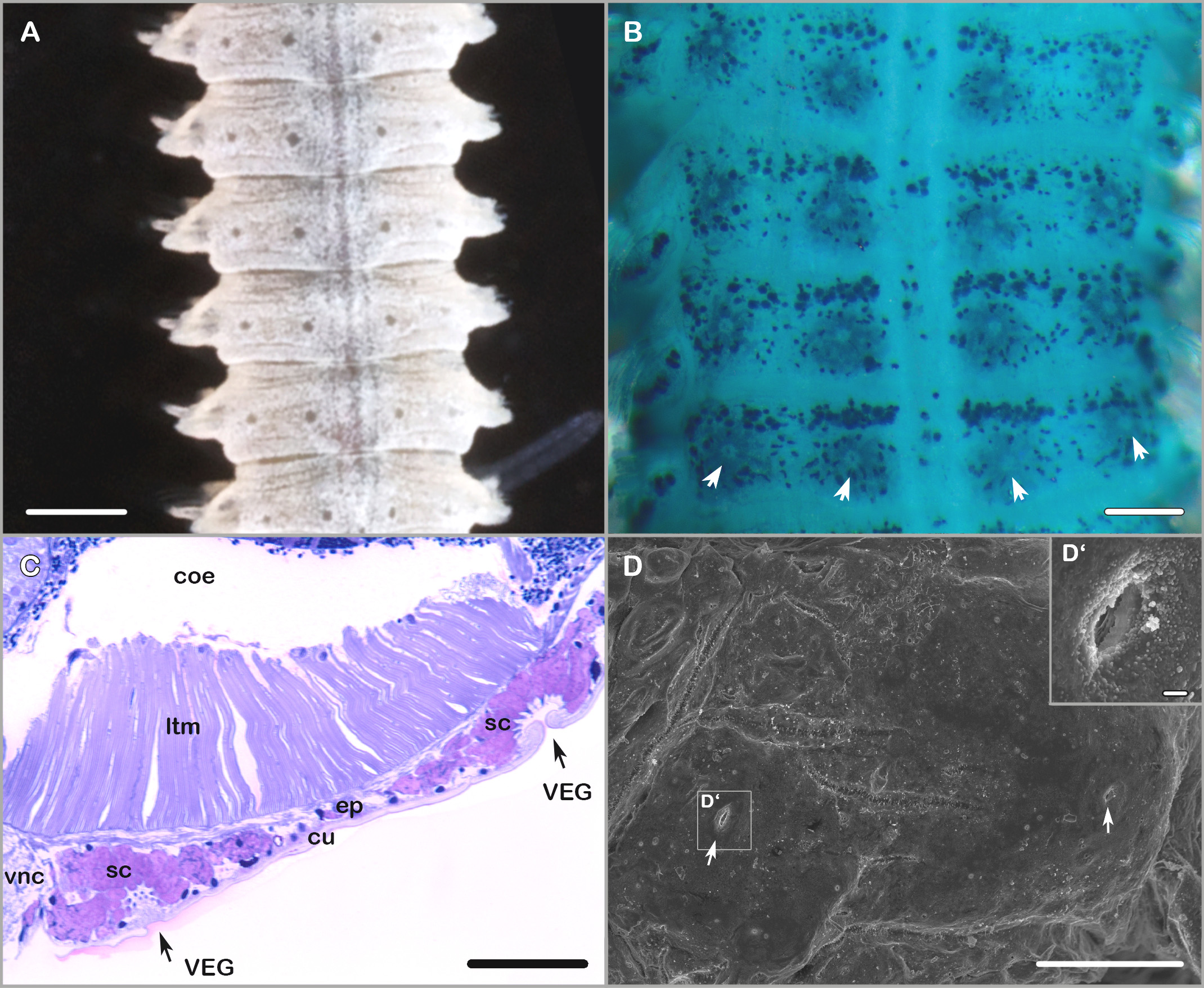
Ventral epidermal glands present from about chaetiger 3 to posterior middle body region; two pairs of glands per chaetiger: median pair slightly posteriorly to centerline on ventral side of the respective glandophorous chaetiger, second pair in lateral position directly at the centerline of the chaetiger ( Fig. 7 View FIGURE 7 A–D) (best observed with SEM or after methyl green staining, also observable in live specimens).
Branchiae from chaetiger 1, continuous to near end of body, with only about last three chaetigers abranchiate; length of first pair of branchiae about two thirds the length of second pair (Fig. 6A–C); longest branchiae on chaetigers 2–10, almost reaching dorsal midline (Fig. 6A), after about chaetiger 10 branchiae decreasing in length (Fig. 6B); branchiae mostly free from postchaetal notopodial lamellae. First notopodium not distinctly shifted dorsally. Notopodial postchaetal lamellae rounded, slightly tapered superiorly and longer than chaetal row. Neuropodial postchaetal lamellae also rounded, only slightly longer than chaetal row in anterior and middle chaetigers. Notopodial prechaetal lamellae present in anterior and middle chaetigers.
Notopodial chaetae all capillaries; in anterior chaetigers arranged in two rows, anterior capillaries slightly shorter than capillaries of posterior row (Fig. 6A), stout, heavily granulated, with distinct sheaths, capillaries of posterior row less stout, slightly granulated, with narrow sheath; additional superior fascicle of 2–3 long, thin capillaries without granulations present from chaetiger 1; capillaries of middle and posterior chaetigers not clearly arranged in rows, thin, non-granulated, of different length within a fascicle. Capillaries of anterior neuropodia arranged in two rows, capillaries of both rows of about same length, anterior capillaries stout, heavily granulated with distinct sheaths, capillaries of posterior row less stout, slightly granulated near tip, with sheaths; from chaetiger 11 posterior row of capillaries replaced by single row of 4–5 tridentate hooded hooks, uppermost tooth inconspicous, anterior row of thin, smooth and alimbate capillaries present in hook-bearing chaetigers, capillaries slightly longer than hooks (Fig. 6E); inferior fascicle of 3–4 long capillaries in position of sabre chaetae usually present from anteriormost chaetigers, hook-bearing chaetigers with 1–2 stout granulated sabre chaetae in inferiormost position, with narrow sheath or alimbate, appearance of sheath variable near the tip (Fig. 6D, also see Remarks).
Pygidium with two pairs of anal cirri: dorsal pair thinner and shorter, ventral pair in comparison very stout, almost cone-shaped and longer than dorsal pair (Fig. 6C).
Pigmentation. Live specimens of whitish colour and with orange-brown pigment on the anterior part of the prostomium, on the dorsal side of the peristomium next to the prostomium, on the dorsum in vicinity of the nuchal organs, and with pigment spots of the same colour anteriorly mid-way along the branchiae of about first eight chaetigers ( Fig. 5 View FIGURE 5 A); palps with white circular bands ( Fig. 5 View FIGURE 5 A). In formalin and ethanol fixed specimens orangebrownish pigment usually still observable following the same pattern as in live specimens ( Fig. 5 View FIGURE 5 B), but in some specimens pigmentation completely lost; several specimens with brownish pigment on the ventrum from about the chaetigers 7–20 with pigmentation being most intense on segmental margins ( Fig. 5 View FIGURE 5 C).
Methyl green staining pattern. Inconspicuous; prostomium, peristomium, branchiae and neuropodial prechaetal lobes most intensely stained; two longitudinal stripes become apparent on the ventrum in some specimens after methyl green staining. Position of ventral epidermal glands indicated by white dots in the centre of larger blue spots on the ventral surface of glandophorous chaetigers (most anterior and middle body chaetigers).
Remarks. In general, the specimens collected in the Lizard Island area are in good agreement with the description by Blake & Kudenov (1978). Minor deviations concern the prostomial shape. In the original description it is stated that a caruncle divided into two lobes surrounded laterally by paired, curved ciliated nuchal organs extending to middle of chaetiger 3 is present ( Blake & Kudenov 1978). This observation is not corroborated by our results: SEM studies revealed that the posterior end of the prostomium is rather blunt and short whereas the nuchal organs follow the pattern typical for this genus (with short median ciliated bands extending to first tcb on chaetiger 2 and long recurved lateral ciliary bands not extending tcb on chaetiger 3). Blake & Kudenov (1978) described distally falcate sabre chaetae with partial hoods formed by extension of sheath. By means of SEM an unusual though variable development of the chaetal sheaths of sabre chaetae near the tip was discovered in specimens from Lizard Island (Fig. 6D) which might refer to the observations by Blake & Kudenov (1978).
FIGURE 6. Spio blakei ( Blake & Kudenov, 1978) , AM W.44381, MI QLD 2376. A. Anterior end, dorsal view, palps removed; B. Middle body region, dorsal view; C. Posterior end, dorsal view; D. Sabre chaetae from 12th chaetiger; E. Neuropodium from 11th chaetiger, anterior view. Scale bars: A–C = 100 µm, D = 1 µm, E = 10 µm.
S. blakei can be distinguished from other Spio spp. with tridentate neuropodial hooks commencing on chaetiger 11 by the following combination of characters (compare Maciolek 1990, Table 2): small species, prostomium entire and rounded anteriorly, first branchiae about two-thirds the length of second pair of branchiae, 4–5 neuropodial hooks per fascicle in hook-bearing chaetigers, pygidium with four cirri with dorsal pair being shorter and more slender than ventral pair. Also the observation of nuchal organs, dorsal ciliated organs and the distribution of ventral epidermal glands can be used for species delimitation but unfortunately these characters are not sufficiently described for all currently known Spio species.
Habitat / Ecology. In the Lizard Island area occurring intertidally in sand and fine sand. Other records in Australia from along the Eastern coasts in Queensland, New South Wales and Victoria ( Blake & Kudenov 1978); also found in estuaries of New South Wales, in sandy mud at depths of 4–10 m in salinities of 29.8–35 ‰ ( Hutchings & Murray 1984). Outside Australia the species has been reported from the Golfo de Nicoya ( Costa Rica) from subtidal depths ( 20 m) and muddy sand ( Dean 2004), and at Baja California Sur ( Mexico) it was collected from 74 m water depth (de León-González 1998).
Distribution. Australia: Queensland, New South Wales, Victoria American Pacific coasts: Mexico: Baja California, Baja California Sur, Bahia San Quintin, Sinaloa peninsula (van der Heiden & Hendrickx 1982, de León- González 1998, Díaz-Castañeda et al. 2005); Costa Rica: Golfo de Nicoya ( Dean 2004).
No known copyright restrictions apply. See Agosti, D., Egloff, W., 2009. Taxonomic information exchange and copyright: the Plazi approach. BMC Research Notes 2009, 2:53 for further explanation.