
Hansreia Halffter & Martínez 1977
|
publication ID |
https://doi.org/ 10.11646/zootaxa.4027.2.2 |
|
publication LSID |
lsid:zoobank.org:pub:3381D7F2-F548-4A67-B690-8BB94223635D |
|
DOI |
https://doi.org/10.5281/zenodo.6103436 |
|
persistent identifier |
https://treatment.plazi.org/id/95638791-FFFA-A514-01AB-F8D5CC7AD694 |
|
treatment provided by |
Plazi |
|
scientific name |
Hansreia Halffter & Martínez 1977 |
| status |
|
Hansreia Halffter & Martínez 1977 View in CoL
Hansreia Halffter & Martínez 1977: 64 View in CoL , 65 (original description); Vaz-de-Mello 2000: 193 (checklist); Medina et al. 2001: 133 (checklist); Vaz-de-Mello et al. 2011: 11, 19, 26, 34, 41, fig. 96 (checklist and identification key to the New World genera and subgenera of Scarabaeinae View in CoL dung beetles).
Type. Ateuchus affinis Fabricius 1801 = Hansreia affinis ( Fabricius 1801) [designated by Halffter & Martínez 1977].
Habitat. Amazon Rainforest.
Diagnosis. Distinguished from the others genera of the Deltochilini tribe by the following combination of characters: pronotum with green, red or yellow metallic sheen. Elytra dark brown and opaque. Sides of pronotum flattened and expanded; margin between anterior and lateral angles oblique, with smooth indentation at the halflength of margin, forming a small tooth directed back ( Fig. 6 View FIGURES 1 – 9 ). Lateral pronotal angles with acute tooth most visible ventrally. Elytral lateral carina strong and complete.
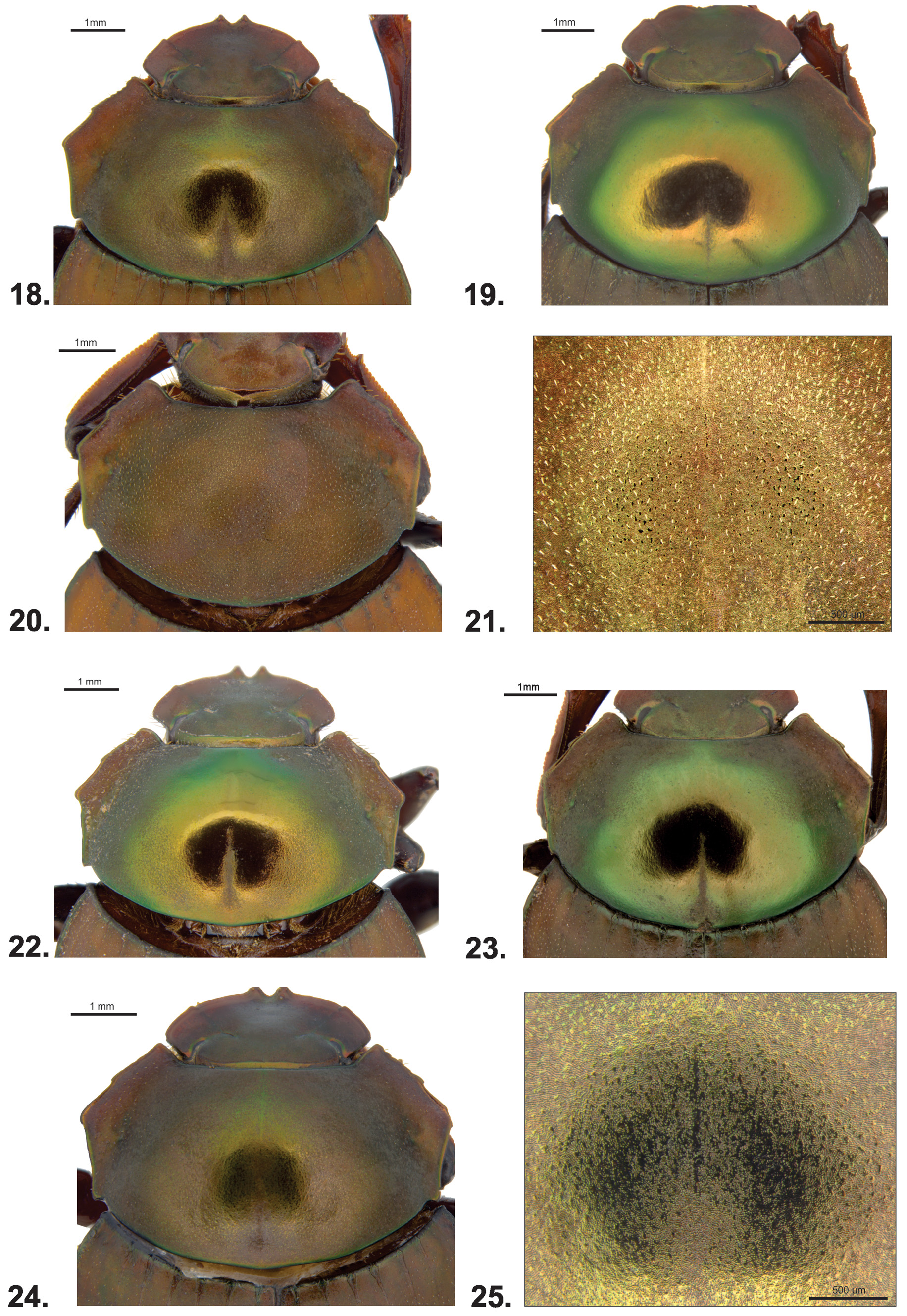
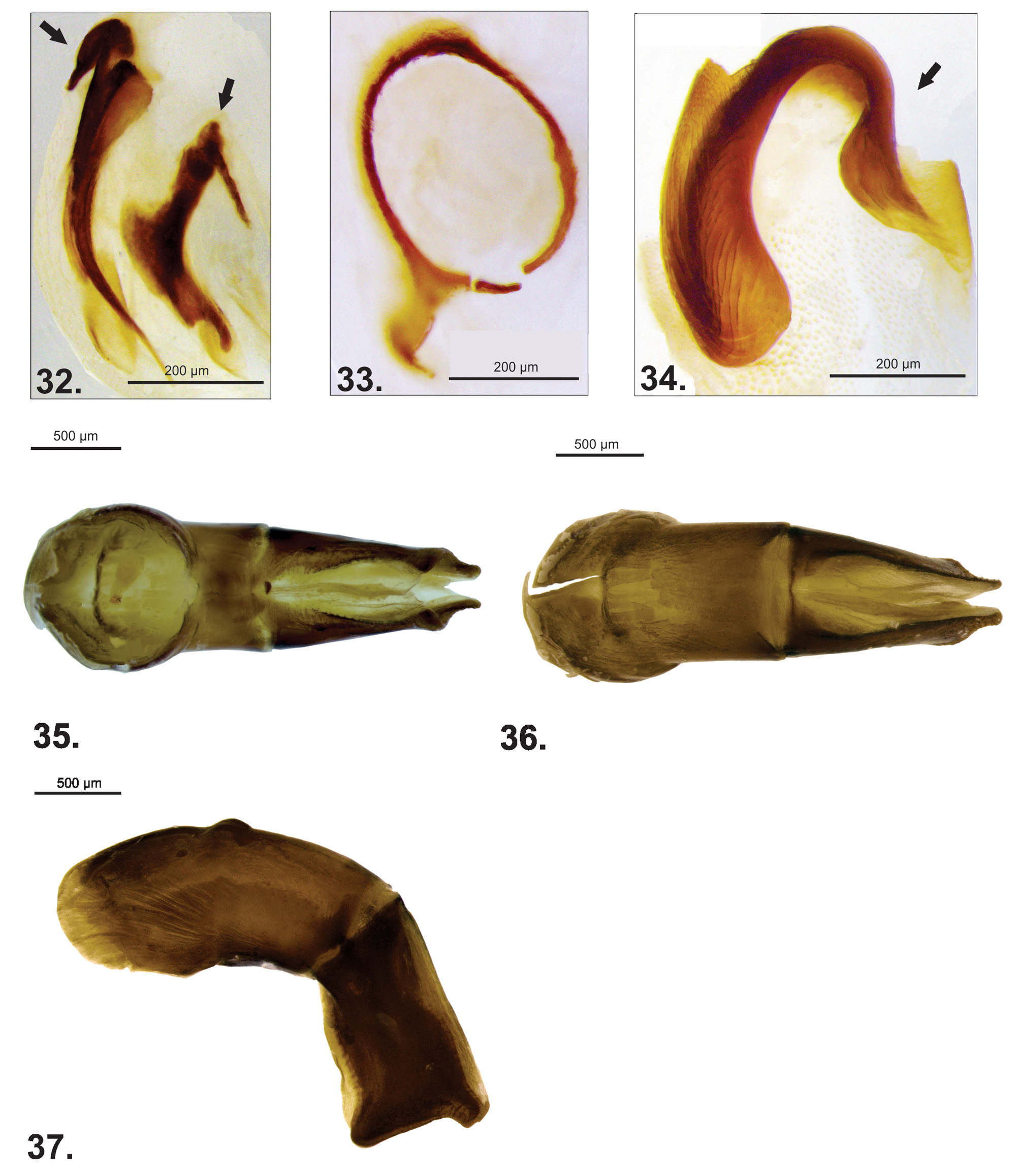
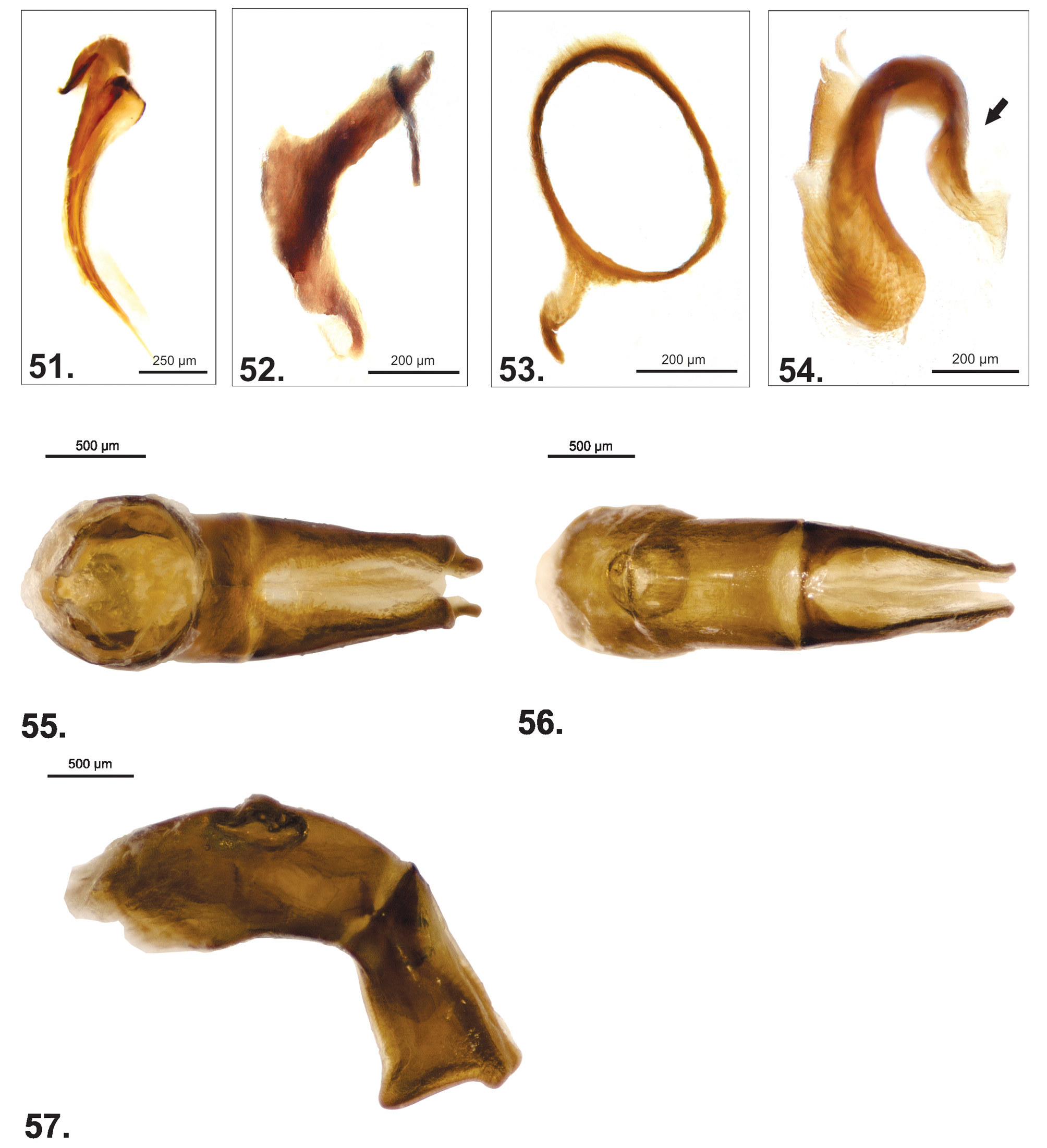
Description. Length. 7.2–10.4 mm. Head. Elyptical shape ( Fig. 1 View FIGURES 1 – 9 ); dark brown with greenish sheen ( Fig. 1 View FIGURES 1 – 9 ); anterior edge weakly margined and posterior with strong emargination; clypeofrontal and clypeogenal suture strongly indicated ( Fig. 1 View FIGURES 1 – 9 ). Clypeus with two distinct acute teeth, wider than longer, separated by shallow U-shaped emargination; each tooth with dorsal fovea and setae at its central portion. Adjacent edge of clypeal tooth straight. Anterior portion of gena, near the clypeogenal suture, with a small and almost inconspicuous tooth. Lateral edge of gena strongly convex. Dorsal surface of eyes narrow; dorsal interocular distance about twenty times width of eye; surface around eye with reddish or greenish luster. Pronotum. Disc dark brown, yellow, red or green; lateral portion dark brown ( Figs 16–25). Ventral surface brown or black; metasternal disc with strong greenish sheen. Pronotum moderately convex, twice wider than long, sides flattened and expanded. Anterior angles with margins rounded (> 90°). Lateral angles with acute tooth most visible ventrally ( Fig. 6 View FIGURES 1 – 9 ). Posterior angles with a small indentation at the adjacent edge ( Fig. 6 View FIGURES 1 – 9 ). Anterior edge of pronotum margined; posterior edge lacking conspicuous emargination. Margin between lateral and posterior angles approximately straight or slightly curved inwards. Lateral surface of pronotum with short setae and microtubercles distributed randomly. Pronotal disc glabrous and shiny, with median longitudinal depression on posterior portion; disc with different patterns of sleek bands surrounded by dense microtubercles ( Figs 16, 17, 18, 19, 22, 23, 24, 25 View FIGURES 18 – 25 ); otherwise disc completely covered by dense microtubercles ( Figs 20, 21 View FIGURES 18 – 25 ). Lateral pronotal fossae weakly impressed. Hypomera not excavated, with carina oblique, incomplete, not reaching lateral margin at anterior angle; carina extending at most two-thirds width of hypomera ( Fig. 2 View FIGURES 1 – 9 ). Mesosternum convex, about six times wider than long. Meso-metasternal suture nearly straight or slightly arcuate, never angulate medially. Metasternum convex, disc glabrous with reddish or greenish sheen. Elytra. Rounded; sides curved outward, lateral carina strong and complete. Elytra brown, opaque; lateral and base darker than surrounding surface, with greenish or reddish sheen. Nine elytral stria visible on disc, shining and thin; margins of striae close, often almost touching; foveiform punctures of elytral striae elongate. Elytral striae wider at base, with carinate margins clearly separated ( Fig. 3 View FIGURES 1 – 9 ). Seventh and eighth striae, at its anterior portion, narrowed than 1–6 striae. Elytral surface minutely granulate; short setae distributed randomly; surface with dense ocellate microtubercles, usually separated by distance equivalent to at least two times puncture diameter. Eighth interstria narrowed on anterior portion of elytra. Abdomen. Abdominal ventrites with short setae. Sixth ventrite longer than others. Pygidium vertical, longer than wide; apex rounded and margined; surface rugose, covered by dense microtubercles, distributed randomly. Pygidium and propygidium separated by carina. Legs. Profemur: anterior edge with emargination interrupted for row of long setae until almost two-thirds its length; ventral surface with short setae distributed randomly. Protibia: triangular, with three acute teeth on apical one-third and a row of small denticles at the outer margin ( Figs 4, 5 View FIGURES 1 – 9 ). Inner apical angle of protibia about 90°. Protarsus present. Meso and metafemur: middle and apex wider than at base; ventral surface with short setae; anterior margin with emargination interrupted for setae along its apical half. Meso and metatibia: cylindrical shape, weakly widened at apex, and slightly arched toward body. Secondary sexual characters. Male: protibial spur spatulate ( Fig. 4 View FIGURES 1 – 9 ). Female: protibial spur with bifurcate apex ( Fig. 5 View FIGURES 1 – 9 ). Females generally have the disc of pronotum completely glabrous and shiny, lacking ocellate punctures or microtubercles. Females are not readily diagnosed among the species of Hansreia . Therefore, it is difficult to tell them apart. However, to date, two species of Hansreia have not been found living at the same locality. Aedeagus. Parameres symmetric, simple, lacking ornamentation or setae; shorter than phallobase ( Figs 7 View FIGURES 1 – 9 , 29–31, 35–37, 42–44, 48–50, 55–57, 61–63). Endophallus: horseshoe-shaped sclerite arcuate ( Figs 9 View FIGURES 1 – 9 , 28 View FIGURES 26 – 28 , 34 View FIGURES 32 – 34 , 41 View FIGURES 38 – 41 , 47 View FIGURES 45 – 47 , 54 View FIGURES 51 – 54 , 60), Superior-right peripheral sclerite (SRP) circular, with a small lobe projected outward ( Figs 27 View FIGURES 26 – 28 , 33 View FIGURES 32 – 34 , 40 View FIGURES 38 – 41 , 46 View FIGURES 45 – 47 , 53 View FIGURES 51 – 54 , 59), Fronto-lateral peripheral sclerite (FLP) with irregular shape ( Figs 26 View FIGURES 26 – 28 , 32 View FIGURES 32 – 34 , 39 View FIGURES 38 – 41 , 45 View FIGURES 45 – 47 , 52 View FIGURES 51 – 54 , 58). Complex of axial and subaxial sclerites (A+SA) comma-shaped, with lateral lobe at anterior portion ( Figs 8 View FIGURES 1 – 9 , 26 View FIGURES 26 – 28 , 32 View FIGURES 32 – 34 , 38 View FIGURES 38 – 41 , 45 View FIGURES 45 – 47 , 51 View FIGURES 51 – 54 , 58).
Remarks. For an accurate identification of Hansreia species it is desirable to examine series of specimens from the same locality, since some diagnostic characters used here are not seen in all specimens analyzed. Despite this, so far, the valid species recognized here are geographically separate and their current distributions are congruent with some of the main endemism areas reported to dung beetles, terrestrial vertebrates and plants ( Haffer 1969; Cracraft 1985; Silva et al. 2002; Morrone 2014).
Hansreia species herein recognized are hypotheses tested based on both morphological and biogeographical evidence. Thus, this work is a starting point for further investigation. We state that a phylogeographical approach can be useful to test our species assumptions. Future studies can corroborate if the morphological variation mentioned here reflects the phylogeographic structure or resulted from ecological responses to different environmental conditions present in those Amazonian areas.
In order to facilitate specimen identification, we provide a set of diagnostic characters (external and male genitalia morphology) for each described species that should be used together for species determination. An identification key for the species was not included because it can lead to misidentification if all the diagnostic characters were not simultaneously analyzed during the identification process. For making direct identification easier, all diagnoses and geographical distributions are resumed in Table 1, that shall be used as an identification table to let to supposed species descriptions and figures.


No known copyright restrictions apply. See Agosti, D., Egloff, W., 2009. Taxonomic information exchange and copyright: the Plazi approach. BMC Research Notes 2009, 2:53 for further explanation.
|
Kingdom |
|
|
Phylum |
|
|
Class |
|
|
Order |
|
|
Family |
Hansreia Halffter & Martínez 1977
| Valois, Marcely, Vaz-De-Mello, Fernando Z. & Silva, Fernando A. B. 2015 |
Hansreia Halffter & Martínez 1977 : 64
| Medina 2001: 133 |
| Halffter 1977: 64 |