
Pleurodeles waltl, Michahelles, 1830
|
publication ID |
https://doi.org/ 10.1093/zoolinnean/zlab038 |
|
DOI |
https://doi.org/10.5281/zenodo.6535620 |
|
persistent identifier |
https://treatment.plazi.org/id/941D87F8-FF85-E258-FF48-FA8BD0327283 |
|
treatment provided by |
Plazi |
|
scientific name |
Pleurodeles waltl |
| status |
|
Pleurodeles waltl View in CoL ( Figs 3–5 View Figure 3 View Figure 4 View Figure 5 )
Smirnov et al. (2020) described cranial skeletogenesis of P. waltl and reported that ossification began soon after hatching. The results of Smirnov et al. (2020) and our results show a similar pattern of ossification. In the study by Smirnov et al. (2020), specimen body size was 5–7 mm at the hatching stage and 28–36 mm at the stage when metamorphosis occurs. However, in our study, these measurements were 8.5–9.2 mm and 48–54 mm, respectively. Given that the growth rate of salamanders is influenced strongly by external factors, such as temperature and food, this difference in the timing of ossification onset could be attributable to the different rearing environments ( Stewart, 1956).
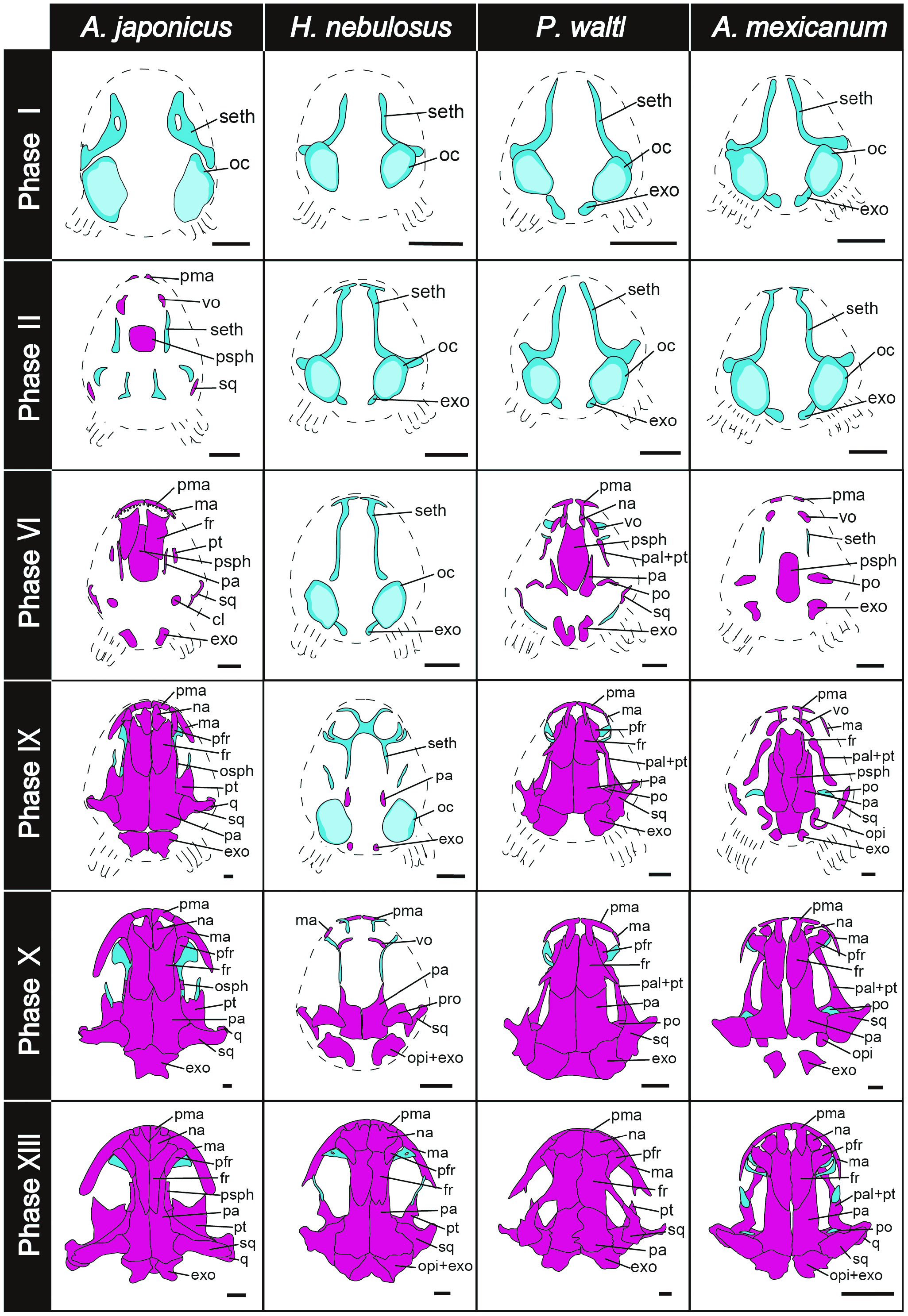
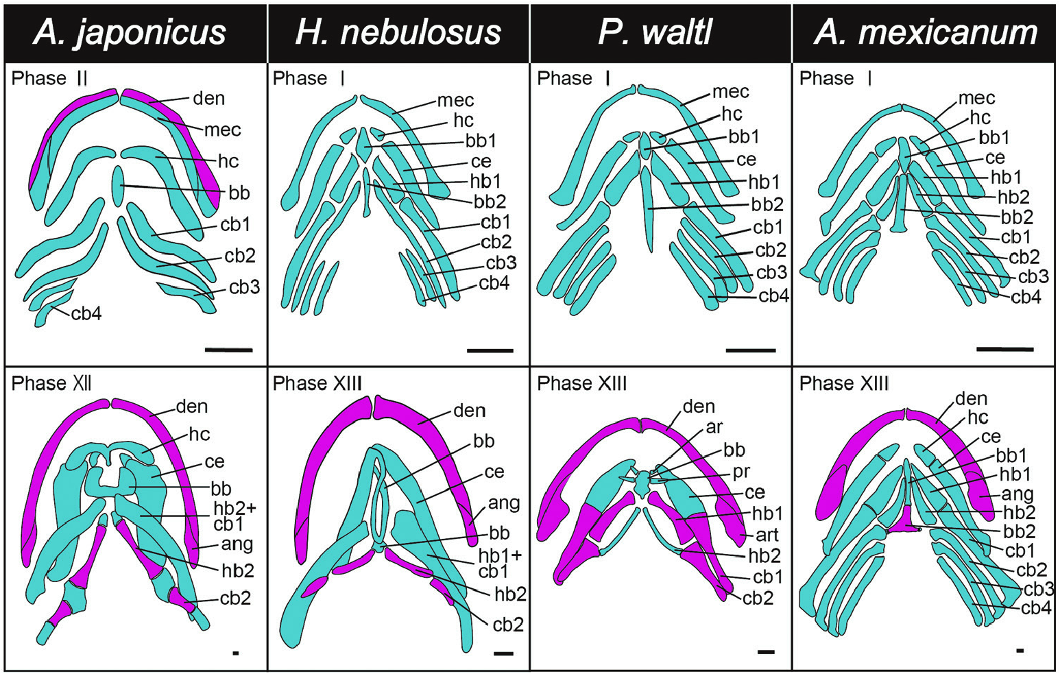
In phases I–V, ossification has not yet started. In the skull, the sphenethmoid, otic capsule and exoccipital are observed as cartilaginous elements. In the mandible and pharynx, the Meckel’s cartilage, the first and second basibranchials, ceratohyal, hypohyal, the first and second hypobranchials and the first to fourth ceratobranchials are detected as cartilaginous elements.
Ossification begins in phase VI. The premaxilla, vomer, prootic, squamosal, exoccipital, parasphenoid, dentary and angular begin to ossify. The palatine and pterygoid bones appear fused into a single bone.
In phase VII, ossification of the skull progresses rapidly, and all bones except the nasal and orbitosphenoid begin to ossify. In the mandible, the coronoid is ossified from the medial side.
In phase VIII, the nasal begins to ossify, meaning that all bones of the skull are ossified. In the hyobranchial skeleton, the second basibranchial starts to ossify.
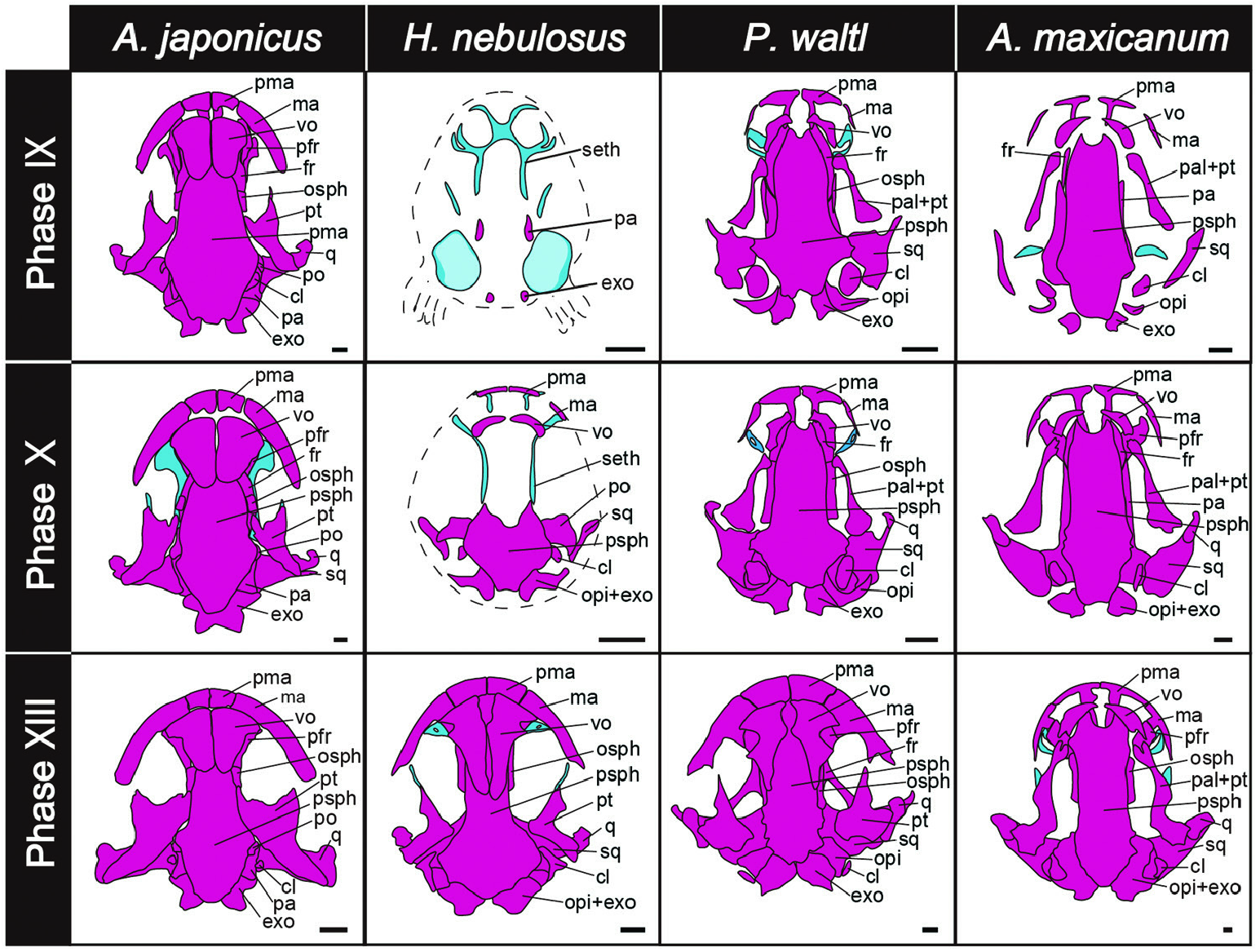
In phase IX, the posterolateral projection of the frontal and anteromedial projection of the squamosal are visible. The pterygoid is elongated in the anterior– posterior direction, forming a bony bar in the ventral side of the skull.
In phases X and XI, the third to fourth ceratobranchials degenerate.
In phases XII and XIII, the tips of the posterolateral projection of the frontal and the anteromedial projection of the squamosal fuse to form the frontosquamosal arch, a characteristic skull feature of the family Salamandridae . The maxilla is elongated posterolaterally and its posterior end is now bifurcated, possessing short lateral and medial projections. The bony bar formed by the formerly continuous anterior palatine and posterior pterygoid is now separated in the middle. In the mandible, the second basibranchial degenerates, and the previously cartilaginous ceratohyal, second ceratobranchial and second hypobranchial ossify.
No known copyright restrictions apply. See Agosti, D., Egloff, W., 2009. Taxonomic information exchange and copyright: the Plazi approach. BMC Research Notes 2009, 2:53 for further explanation.