
Ceriodaphnia nikolaii, Garibian & Andreeva & Kotov, 2023
|
publication ID |
https://doi.org/ 10.11646/zootaxa.5284.2.2 |
|
publication LSID |
lsid:zoobank.org:pub:834CD5F6-5FF2-46C3-880E-68D2CC7D9261 |
|
DOI |
https://doi.org/10.5281/zenodo.7930782 |
|
persistent identifier |
https://treatment.plazi.org/id/86578788-CB6C-FFFD-FF64-FF026035F794 |
|
treatment provided by |
Plazi |
|
scientific name |
Ceriodaphnia nikolaii |
| status |
sp. nov. |
Ceriodaphnia nikolaii sp. nov.
( Figs. 9–16 View FIGURE 9 View FIGURE 10 View FIGURE 11 View FIGURE 12 View FIGURE 13 View FIGURE 14 View FIGURE 15 View FIGURE 16 )
Etymology. The taxon is named after our teacher, Prof. Nikolai N. Smirnov, who initiated great progress in the studies of cladocerans in Eurasia and in the world and collected the type series.
Type locality. An un-named oxbow of the Lena River, City Yakutsk, Republic of Sakha (Yakutia), Russia (62.0˚N, 129.8˚E).
Type material. Holotype. A parthenogenetic female, MGU Ml 266 at the Collection of Zoological Museum of M. V. Lomonosov Moscow State University, Moscow, Russia.
Allotype. An adult male, MGU Ml 267.
Paratypes. Ten parthenogenetic females, six ephippial female and seven adult males, MGU Ml 268. Several parthenogenetic, ephippial females and males, AAK 1999-045.
Adult parthenogenetic female. General ( Figs. 9b,c,e,f View FIGURE 9 ). Body subovoid in lateral view, with maximum height in middle of valves. Dorsal margin interrupted by a depression in posterior head portion. Dorsal and ventral portion ovoid, poster-dorsal angle present, with small caudal spine. Postero-dorsal angle well-developed, sometimes represented by a short caudal spine, valve margin from the former to anterior margin uniformly rounded. In dorsal view, body subovoid and elongated.
Head ( Figs. 10a–g View FIGURE 10 ) small, ovoid, without rostrum, with a strong depression in posterior head half; small dorsal head pore in this depression. Dorsal head pore round; supraocular dome surround relatively big compound eye; ocellus minute. Labrum ( Fig. 10g View FIGURE 10 ) quadrangular, with a wide fleshy main body and large setulated labral plate. In dorsal view, fornices from rounded to slightly projected.
Valve large and ovoid ( Figs. 11a–f View FIGURE 11 ); ventral portion with long setae at inner margin ( Figs. 11a–b View FIGURE 11 ); postero-ventral portion with a row of short setae, short series of setules between them ( Fig. 11c View FIGURE 11 ); two relatively long setae in dorsal most portion ( Fig. 11d–f View FIGURE 11 ).
Abdomen ( Figs. 12b, h, i View FIGURE 12 ) short consisting of four segments, first segment having a long abdominal projection.
Postabdomen ( Figs. 12a–i View FIGURE 12 ) elongated, tapering distally. Ventral portion straight. Preanal margin from slightly to strongly projected and angled, postanal angle rounded. Postanal and anal margin bearing 8–9 teeths of different size. Preanal, anal and postanal portion covered by short series of minute setules. Postabdominal seta as long as postabdomen.
Postabdominal claw elongated, curved, with pointed tip ( Figs. 13a–h View FIGURE 13 ). Outer portion of dorsal margin with three successive pectens. First pecten with thin setules, second pecten consisting of twelve to twenty six stout teeth with variable size, and third pecten bearing numerous minute setules, not reaching the tip of claw.
Antenna I ( Figs. 10g,h View FIGURE 10 ) short and cylindrical. Antennular seta longer than antenna I body, mostly same in size with longest aesthetascs.
Antenna II ( Figs. 11g –i View FIGURE 11 ) with a narrow coxal part bearing two sensory setae. Basal segment of antenna II cylindrical elongated and covered by rows of small setules. Antennular branches long, approximately same in size, exopodite with four setulated segments, endopodite consist of three setulated segments. Antennal formula: 0-0-1- 3/1-1-3, second segment of exopodite bearing one short spine, distal part of basal segment with one short spine.
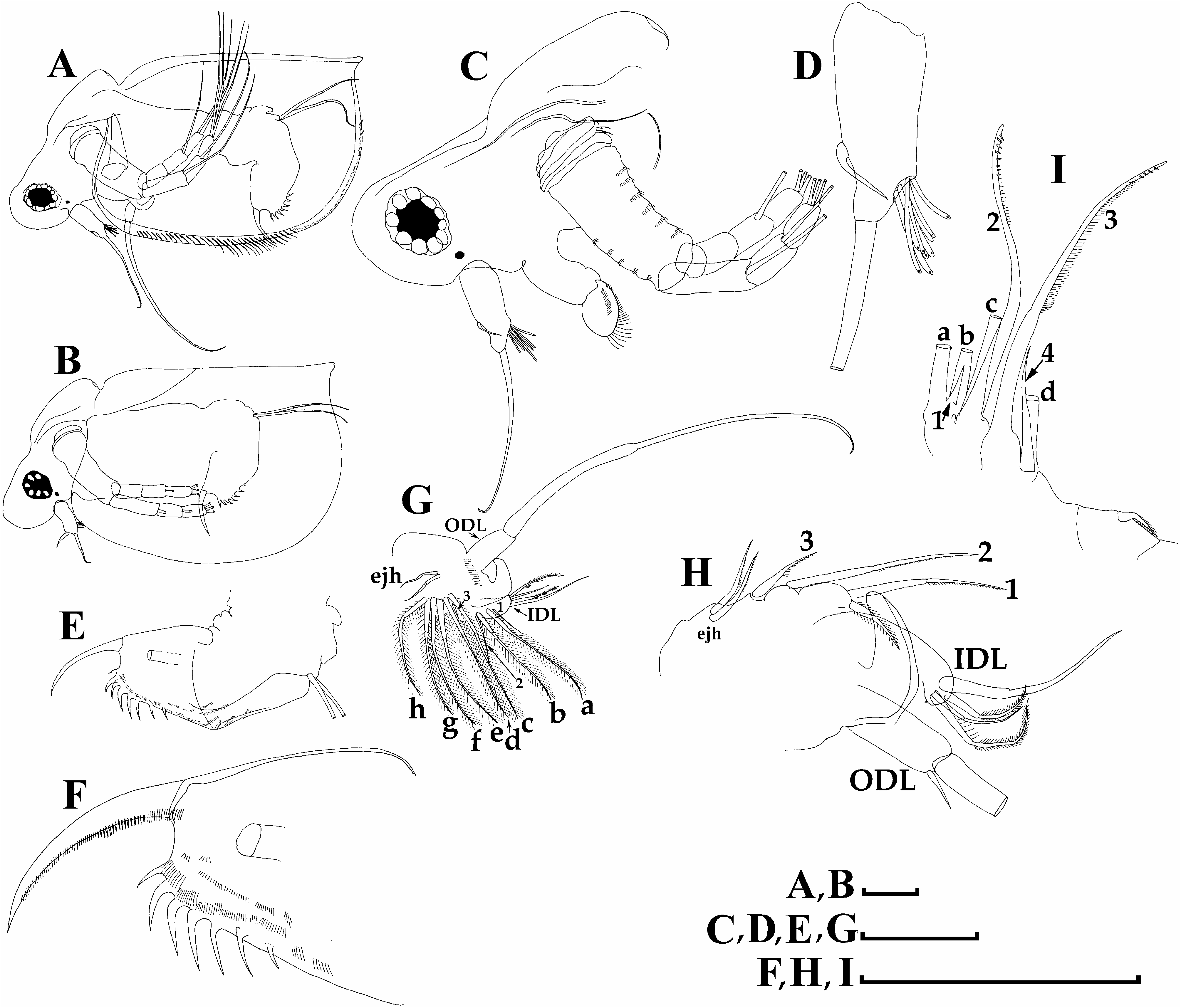
Limb I ( Figs. 14a View FIGURE 14 and 15a View FIGURE 15 ) with a bilaterally setulated accessory seta. Outer distal lobe ( ODL) bearing a very long, naked seta and a short seta setulated in distal part. Inner distal portion (endite 4) with a single anterior and two posterior setae of different size. Endite 3 with a single, long anterior seta (1) and two posterior setae (a–b); endite 2 with a single anterior seta (2) and two posterior setae (c–d); endite 1 with a single, short anterior seta (3) and four posterior setae (f–h). Two ejectors hooks subequal in size ( Figs. 14a View FIGURE 14 and 15a View FIGURE 15 ). Maxillar process absent.
Limb II ( Figs. 14b View FIGURE 14 and 15b–d View FIGURE 15 ) with an ovoid epipodite (epp) an elongated exopodite (ext) bearing two long bilaterally setulated setae. Endite 5 with a short anterior seta (1) and two long, bilaterally setulated posterior setae (a–b); endite 4 with a single long (longest one) stiff anterior setae (2) and a long posterior seta (c); endite 3 with a single short anterior seta (3) and no posterior setae; endite 2 with a short anterior seta modified into sensillum (4) and a single posterior seta (d). A seta on unclear homology near gnathobase. Endite 1(e1) = gnathobase with two rows of setae. Anterior row consists of four setae: setae 1 presented by a small sensillum; seta 2 very long and setulated; seta 3 shorter than seta 2; seta 4 short. Posterior row with eight setae of different morphology.
Limb III ( Figs. 14c View FIGURE 14 and 15e–h View FIGURE 15 ) with a subovoid and elongated prepipodite (pep) and ovoid epipodite (epp). Exopodite (ext) with two lateral (5–6) and four distal setae (1–4); distal segment of seta 1 and seta 2 thin, proximal part of seta 2 with a row of strong setules. Limb inner distal portion represented by five endites: endite 4 with a single anterior (2) and a single posterior (b) seta; endite 3 with a short anterior seta (3); endite 2 with a very short anterior seta (4); endite 1 (gnathobases) represented by a large lobe with a long distal anterior seta (1) and two small, stiff anterior setae (2–3), posterior portion represented by numerous long posterior setae.
Limb IV ( Figs. 14d View FIGURE 14 and 15i View FIGURE 15 ) with an elongated prepipodite (pep) and ovoid epipodite (epp). Exopodite (ext) bears two lateral setae (5–6) and four distal setae (1–4); distal segment of seta 1 thin. Inner limb portion with two setulated anterior setae (1–2); posterior portion bears numerous long posterior setae.
Limb V ( Figs. 14e View FIGURE 14 and 15j View FIGURE 15 ) with elongated epipodite (epp), exopodite (exp) triangular, with two distal setae of different size (1–2) and a large lateral seta (3). Inner portion of limb V with a setulated margin and a large distal seta.
Juvenile female ( Fig. 9d View FIGURE 9 ). Body subovoid and elongated; dorsal margin slightly convex; postero-dorsal and ventral portion round. Head round, with large compound eye and small ocellus. Dorsal portion with great depression between head and body; dorsal part of headshield with protruding dorsal head pore.
Ephippial female ( Figs. 9a View FIGURE 9 and 10a View FIGURE 10 ). Body subovoid, valves modified into ephippium with a single resting egg; in lateral view ephippium with expressed reticulation; ephippium without lateral projections.
Adult male ( Fig. 16a,c–i View FIGURE 16 ). Body subovoid and elongated, dorsal margin straight, postero-dorsal and ventral portion rounded. Postero-ventral angle rounded.
Head ( Fig. 16a,c View FIGURE 16 ) relatively small, without rostrum. Compound eye large and ocellus small. Dorsal portion of head shield in lateral view with high depression between head and body; dorsal head pore present. Labrum ( Figure 16c View FIGURE 16 ) as in parthenogenetic female.
Valve ( Fig. 16a View FIGURE 16 ) as in parthenogenetic female.
Abdomen ( Fig. 16e View FIGURE 16 ) with four setulated segments lacking any process, covered by minute setules.
Postabdomen ( Fig.16e,f View FIGURE 16 ) elongated, subrectangular; ventral margin convex.Dorsal margin convex, preanal angle projected. Postanal and anal portion with about seven strong teeth. Gonopore opens subdistally on postabdomen. Postabdominal claw as in female.
Antenna I ( Fig. 16c,d View FIGURE 16 ) cylindrical, long, with nine short aesthetascs. Sensory seta relatively short (shorter than longest aesthetascs) located subdistally on anttenna I body. Flagellum located distally, longer than antenna I body.
Antenna II ( Fig. 16c View FIGURE 16 ) same as in parthenogenetic female.
Limb I ( Fig. 16g,h View FIGURE 16 ) with a cylindrical outer distal lobe bearing a long seta and a short setulated seta. Inner distal lobe with a copulatory hook and four seta of different size. Morphology of endites mostly same as in female, but endite 5 with an additional seta.
Limb II ( Fig. 16i View FIGURE 16 ) endite 4 with long modified anterior seta (distal part bilaterally setulated); endite 3 and endite 2 bearing anterior seta longer than female anterior seta.
Juvenile male ( Fig. 16b View FIGURE 16 ). Body subovoid, dorsal margin relatively straight, postero-dorsal and ventral portion round. Head round, without rostrum; antenna I cylindrical with a long thick sensory seta on the basal part and a relatively short flagellum (same in size as antenna I body).
Size. Parthenogenetic female length 0.38–1.1 mm / height 0.23–0.8 mm; ephippial female length 0.88 mm / height 0.64 mm; male length 0.64–0.68 mm / height 0.35 mm.
Differential diagnosis. This taxon belongs to the C. dubia s.lat. species group and has four diagnostic characters mentioned in the Introduction. The main diagnostic character is the presence of larger teeth on the postabdominal claw, but their length is c.a. half the claw diameter in C. dubia group, while c.a. a claw diameter in C. reticulata group. Presence of these teeth as well as rudimentary stiff (anterior) setae on the inner-distal portion of limb II differentiate well C. nikolaii sp.nov. from C. laticaudata-rotunda group lacking such teeth and having relatively long setae on limb II.
In contrast to other taxa of the C. dubia group, parthenogenetic female of C. nikolaii sp.nov. has the preanal angle of postabdomen from slightly pronounced to greatly pronounced (similarly to that in Ceriodaphnia laticaudata ), while in other members of C. dubia s.lat. the preanal angle is smoothed or slightly projected. In addition, male postabdomen in C. nikolaii sp.nov. has an expressed preanal angle in contrast to. dubia s.l. with a completely smoothed preanal angle; the shortest seta of IDL has a size similar to that of two other setae, while in the latter the shortest seta is clearly shorter than other setae of IDL.
Distribution. C. nikolaii sp. nov. is known to date only from its type locality: an oxbow of the Lena River near city of Yakutsk. Perhaps it is an endemic species with a limited distribution (i.e. it is absent in any other numerous samples from Easter Siberia and Far East studied by the authors). Note that the majority of studied populations from Republic of Sakha (Yakutia) belonged to C. dubia s.l. instead of C. nikolaii sp. nov.


| V |
Royal British Columbia Museum - Herbarium |
No known copyright restrictions apply. See Agosti, D., Egloff, W., 2009. Taxonomic information exchange and copyright: the Plazi approach. BMC Research Notes 2009, 2:53 for further explanation.