
Ceriodaphnia dubia Richard, 1894
|
publication ID |
https://doi.org/ 10.11646/zootaxa.5284.2.2 |
|
publication LSID |
lsid:zoobank.org:pub:834CD5F6-5FF2-46C3-880E-68D2CC7D9261 |
|
DOI |
https://doi.org/10.5281/zenodo.7930776 |
|
persistent identifier |
https://treatment.plazi.org/id/86578788-CB62-FFE7-FF64-FF0266CEF737 |
|
treatment provided by |
Plazi |
|
scientific name |
Ceriodaphnia dubia Richard, 1894 |
| status |
s.l. |
Ceriodaphnia dubia Richard, 1894 View in CoL View at ENA s.l. from Northern Palaearctic
( Figs. 1–8 View FIGURE 1 View FIGURE 2 View FIGURE 3 View FIGURE 4 View FIGURE 5 View FIGURE 6 View FIGURE 7 View FIGURE 8 )
Richard 1894: 570–572, Fig. 6–8 View FIGURE 6 View FIGURE 7 View FIGURE 8 ; Lilljeborg 1901: 202–205, Pl. 28: Fig. 19–28 ( affinis ); Wagler 1937: 35 ( quadrangula var. affinis ); Behning 1941: 148–149, Fig. 57 ( affinis ); Šrámek-Hušek R. et al. 1962: 238–240, Fig. 85 ( affinis ); Manujlova 1964: 165–166, Fig. 64 ( affinis ); Scourfield & Harding 1966: 24, Fig. 56–57; Flössner 1972: 171–173, Fig. 80; Chiang & Du 1979: 139–140, Fig. 93; Ibrasheva & Smirnova 1983: 44–45 ( affinis ); Negrea 1983 a: 163–164, Fig. 67; Margaritora 1983: 42, Fig. 22B, B’, 23B, 24C; Margaritora 1985: 71–73, Fig. 30; Røen 1995: 139–141, Fig. 93; Alonso 1996: 186, 188, Fig. 82–I; Flössner 2000: 210–212, Pl. 79a–k; Hudec 2010: 134–136, Fig. 24 ( affinis ); Kotov et al. 2010: 187, Fig. 1 View FIGURE 1 ; Bledzki & Rybak 2016: 183 ( affinis ); Rogers et al. 2019: 669, Fig. 16.2.11 F View FIGURE 16 ; Korovchinsky et al. 2021: 69–71, Fig. 16 View FIGURE 16 : 1.
Material examined (all samples from Russia). Many parthenogenetic females, ephippial females and males from an oxbow of the Don River near village of Stogovskoy, Rostov Area, (49.73784˚N, 41.22782˚E), AAK M-4308; many pathenogenetic females, ephippial female and males from a puddle in Mongun-Taiyginskyi District, Republic of Tuva (50.092805˚N, 90.079438˚E), AAK 2022-097; few parthenogenetic females from a water in a metallic tank, city of Yakutsk, Republic of Sakha (Yakutia) (61.96021˚N, 129.6543˚E), AAK 2011-026; few parthenogenetic females from a nameless lake near the Federal Highway “Kolyma”, Republic of Sakha (Yakutia) (62.15591˚N, 130.9231˚E), AAK M-1949; few parthenogenetic female from a puddle near Chyoroktai Reservoir, Republic of Sakha (Yakutia) (62.05379˚N, 132.7742˚E), AAK M-1955; a single parthenogenetic female from lake Atyrdiakh , oxbow of the Handyga River , Republic of Sakha (Yakutia) (63.08686˚N, 134.0543˚E); few parthenogenetic females from a laboratory culture in the Institute of Inland Water Biology , Borok, Russia, AAK M-2883. All samples are kept at the working collection of the Laboratory of Aquatic Ecology and Invasions, A.N. Severtsov Institute of Ecology and Evolution, Moscow, Russia .
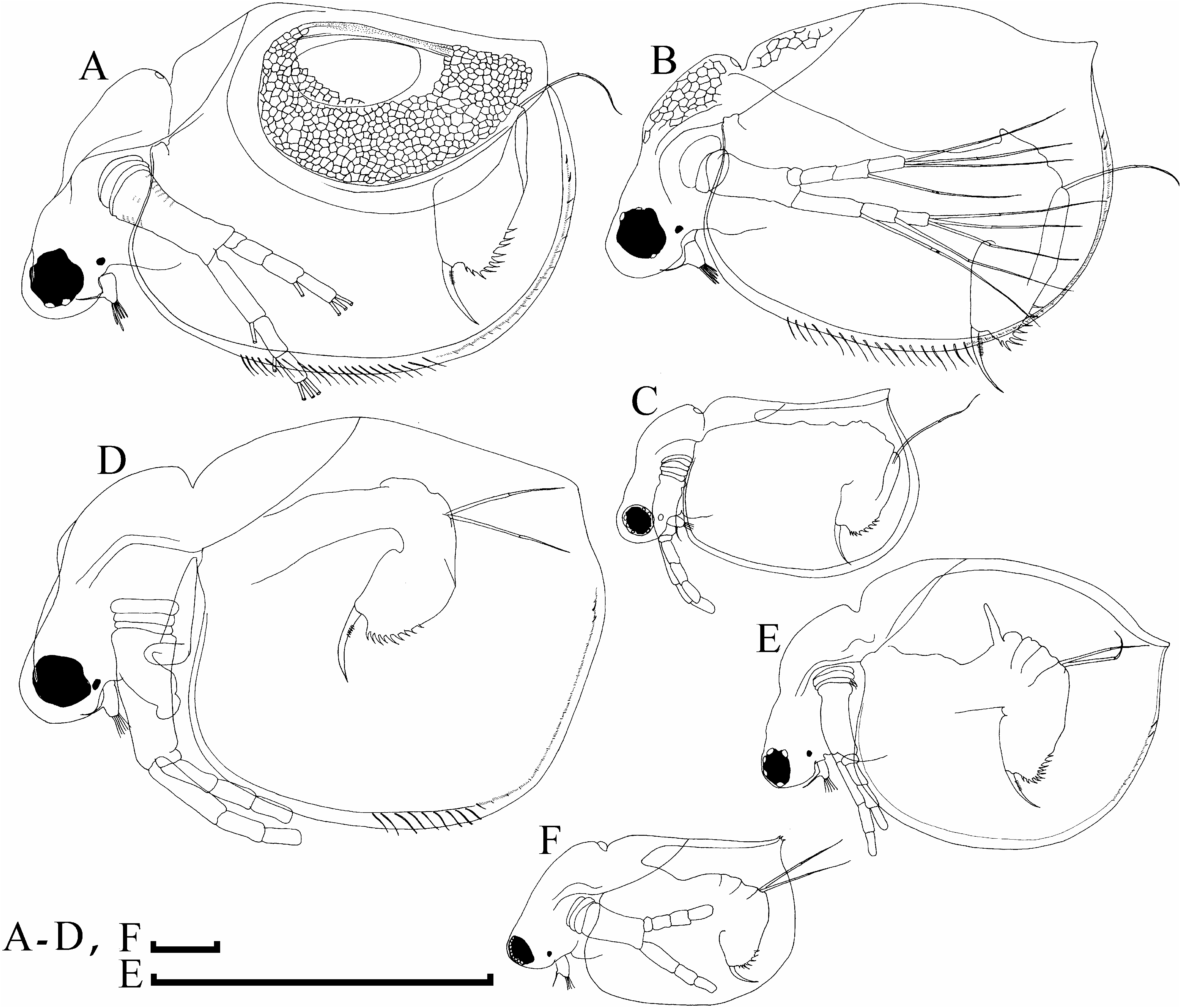
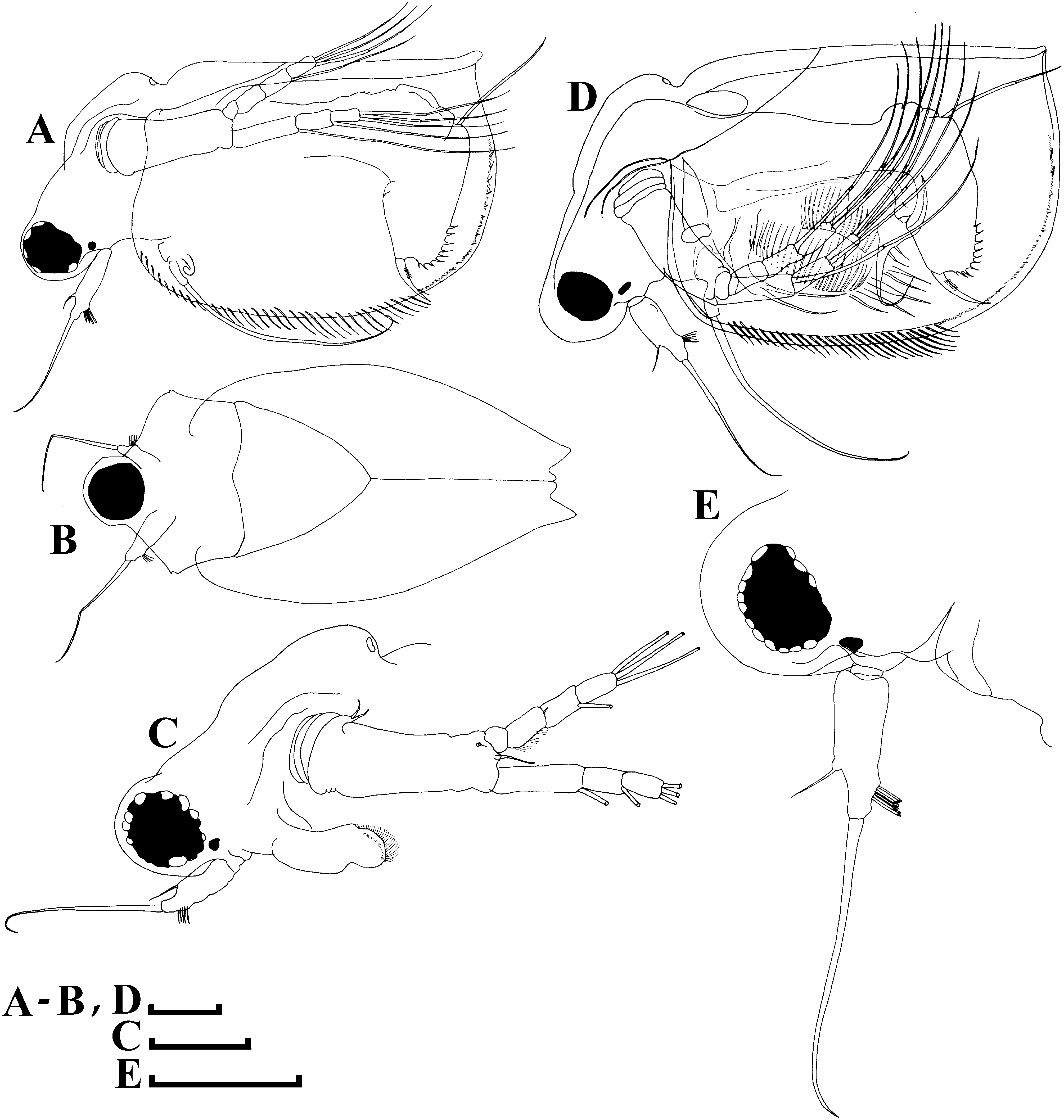
Redescription. Parthenogenetic female. General. In lateral view body ovoid, maximum height in middle of valves ( Figs. 1b, d, e View FIGURE 1 ). Dorsal margin interrupted by a depression in posterior head portion. Postero-dorsal angle well-developed, sometimes represented by a short caudal spine, valve margin from the anterior to posterior margin uniformly rounded. In dorsal view, body subovoid and elongated.
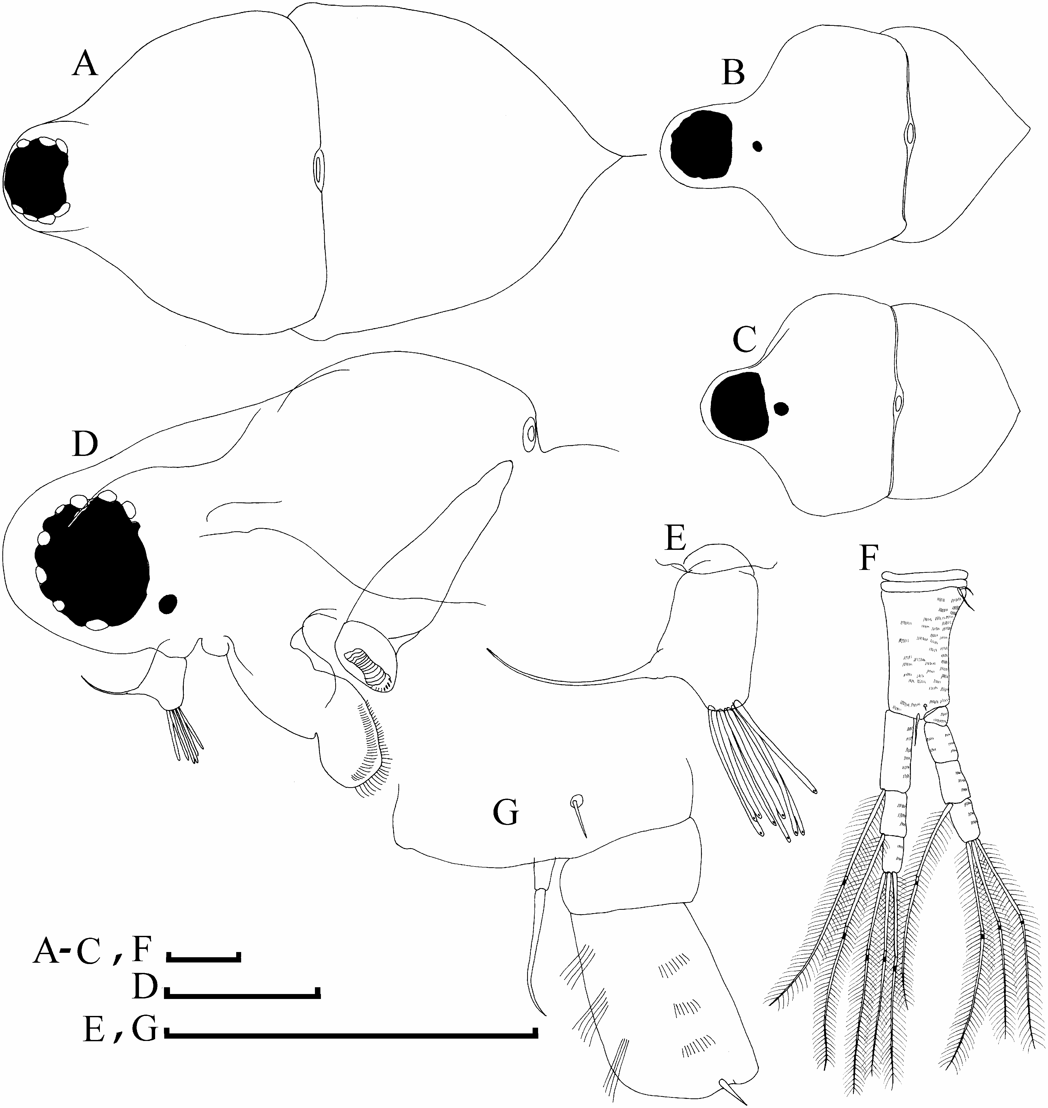
Head ( Figs. 2a–d View FIGURE 2 ) relatively small and round, rostrum very short. A strong depression in posterior head half, small dorsal head pore in this depression. Compound eye large, ocellus small, located near compound eye. Labrum ( Fig. 2d View FIGURE 2 ) with a main fleshy body and a large setulated distal lobe. In dorsal view, fornices from rounded to slightly projected (see also Alonso 1996: 187, Fig. 82; Hudec 2010: 135, Fig. 24 for illustrations of projected, sharp fornices).
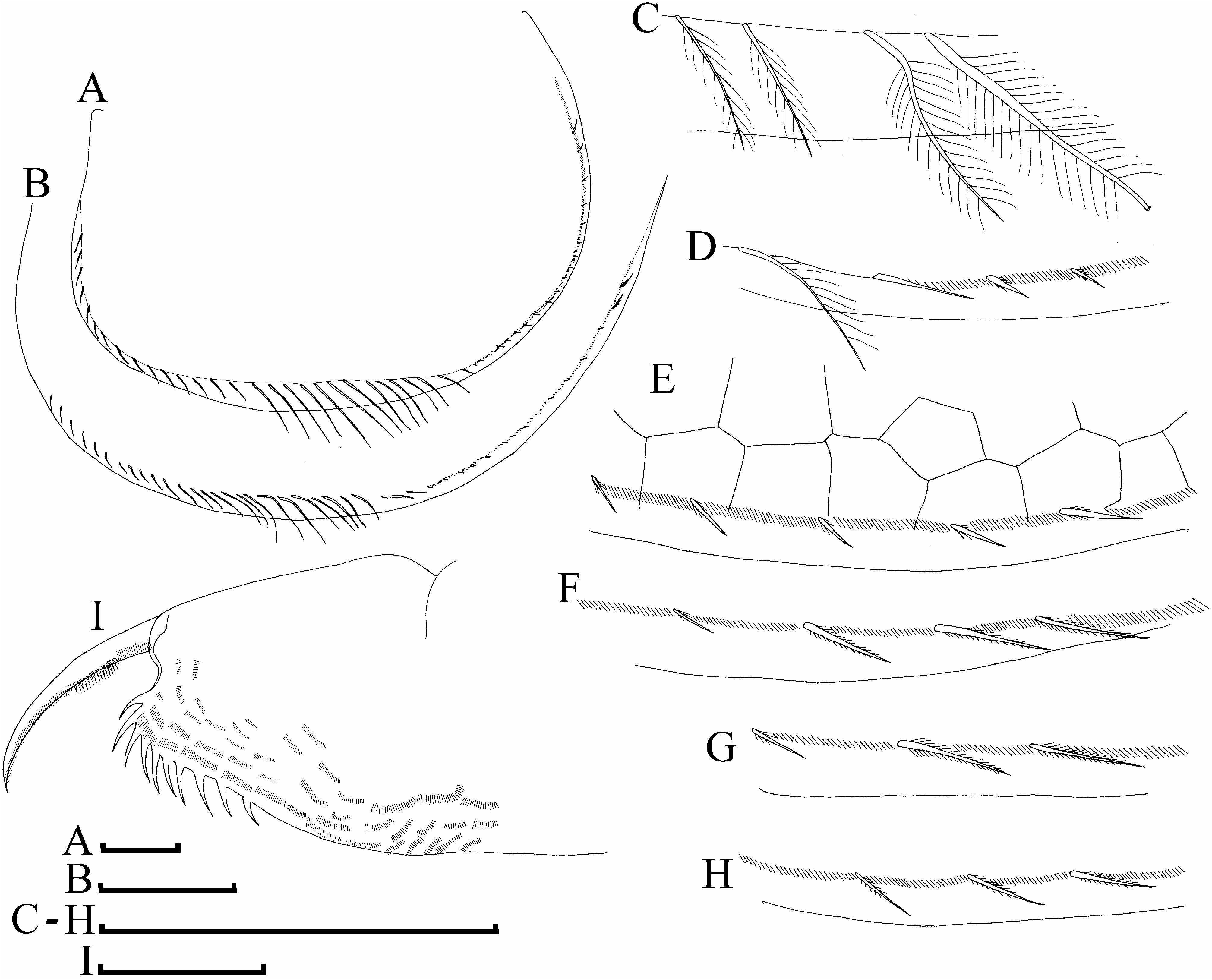
Valve large and ovoid ( Figs. 3a–h View FIGURE 3 ), its ventral portion with long setae at inner margin ( Figs. 3a–c View FIGURE 3 ), postero-ventral portion with a row of short setae, short series of setules between them ( Figs. 3d–h View FIGURE 3 ); two or three relatively long setae in dorsal most portion ( Figs. 3e–h View FIGURE 3 ).
Abdomen ( Figs. 4a–c, e View FIGURE 4 ) consists of four segments. First segment with a long process. Second, third and fourth segments setulated, lacking of processes.
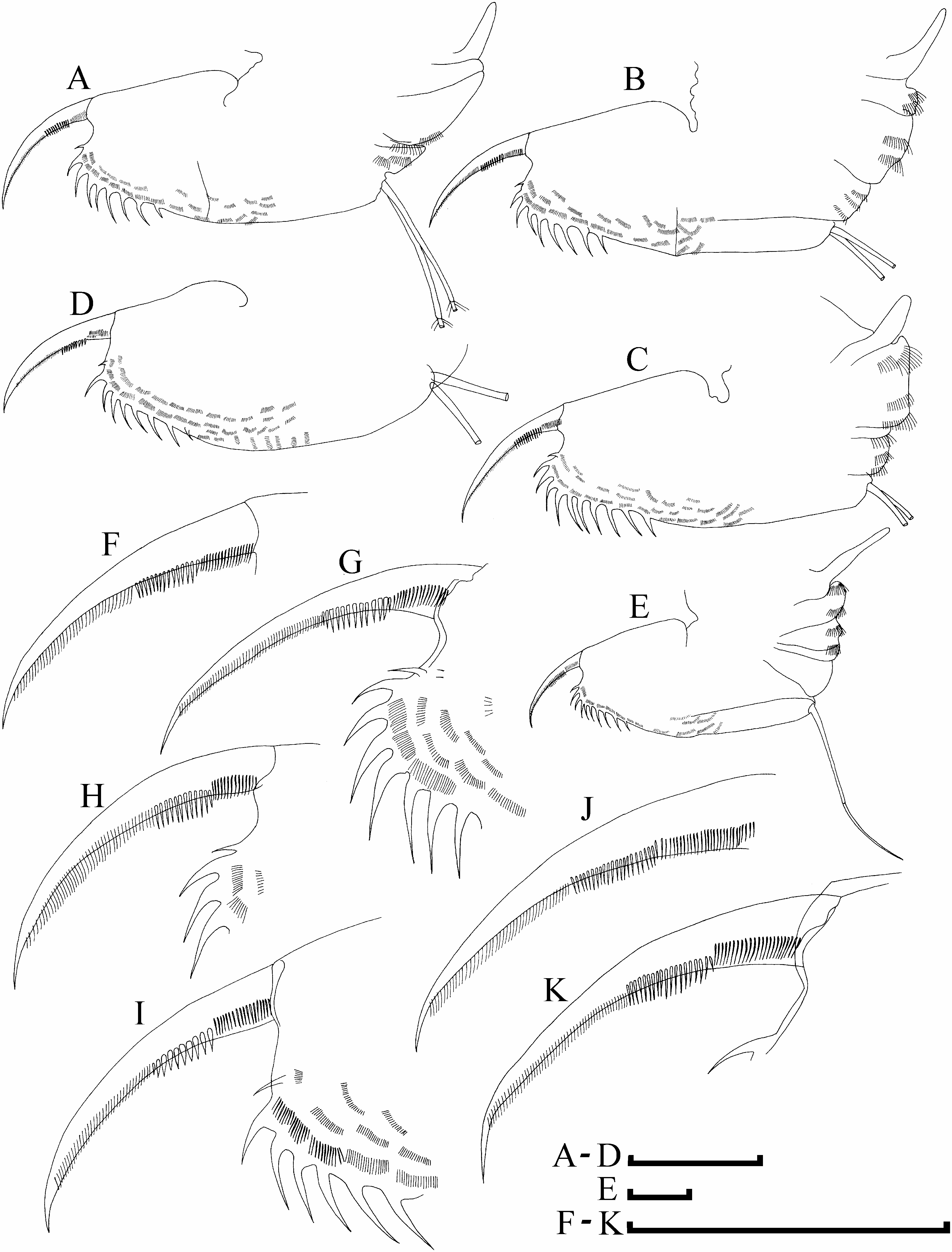
Postabdomen ( Figs. 3i View FIGURE 3 , 4a–e View FIGURE 4 ) elongated, with narrowing distal portion. Ventral margin straight. Preanal margin straight, preanal angle from fully smooth ( Fig. 4a View FIGURE 4 , see also Alonso, 1996: Fig. 82H) to little developed ( Fig. 4b View FIGURE 4 , see also Flössner, 1972: Fig. 80C) or even somewhat projected ( Fig. 4d View FIGURE 4 , See also Scourfield & Harding, 1966: Fig. 56), postanal angle rounded. Postanal margin straight, bearing nine to eleven pairs of teeths, their size continuously increasing distally, except for the most distal spine which is usually the shortest. Preanal, anal and postanal portions covered by short series of minute setules. Postabdominal setae long as postabdomen.
Postabdominal claw ( Figs. 4f–k View FIGURE 4 ) long, slightly curved, with pointed tip. On its outer side, three successive pectens along the dorsal margin. The first pecten consisting of thin setules; the second pecten composed of 10–22 stout teeth with different thickness among populations and even within a single population; the third pecten consists of minute fine setules, this row does not reach the tip of claw.
Antenna I ( Figs. 2d,e View FIGURE 2 ) cylindrical, relatively short (length c.a. 1.5 diameter); antennular seta slender, longer than antenna I body, arising distally. Nine aesthetascs of different size, the longest one longer than antenna I body.
Antenna II with a narrow coxopodite bearing two sensory setae ( Fig. 2f View FIGURE 2 ). Basal segment elongated, covered by rows of minute setules, its distal part having a posterior sensory seta and a short anterior spine ( Fig. 2g View FIGURE 2 ). Antennal branches elongated, subequal in size, exopodite with four segments, endopodite with three segments. Each branch segment covered by series of minute setules. Antennal setae formula: 0-0-1-3/1-1-3. The second segment of exopodite with a small, slender spine. Each swimming seta with proximal and distal segments bilaterally setulated and a chitinous insertion in distal segment near its connection with the proximal segment.
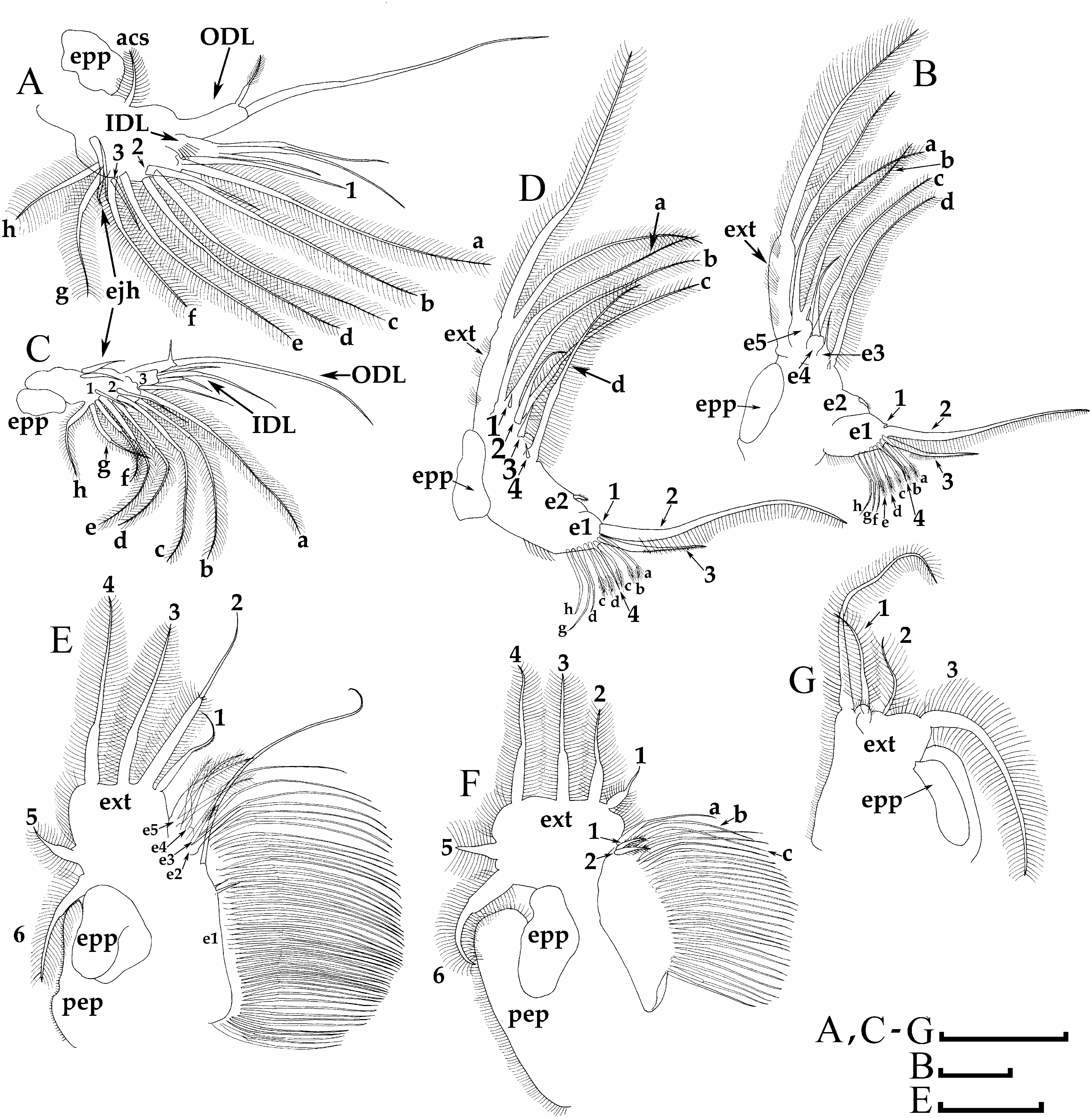
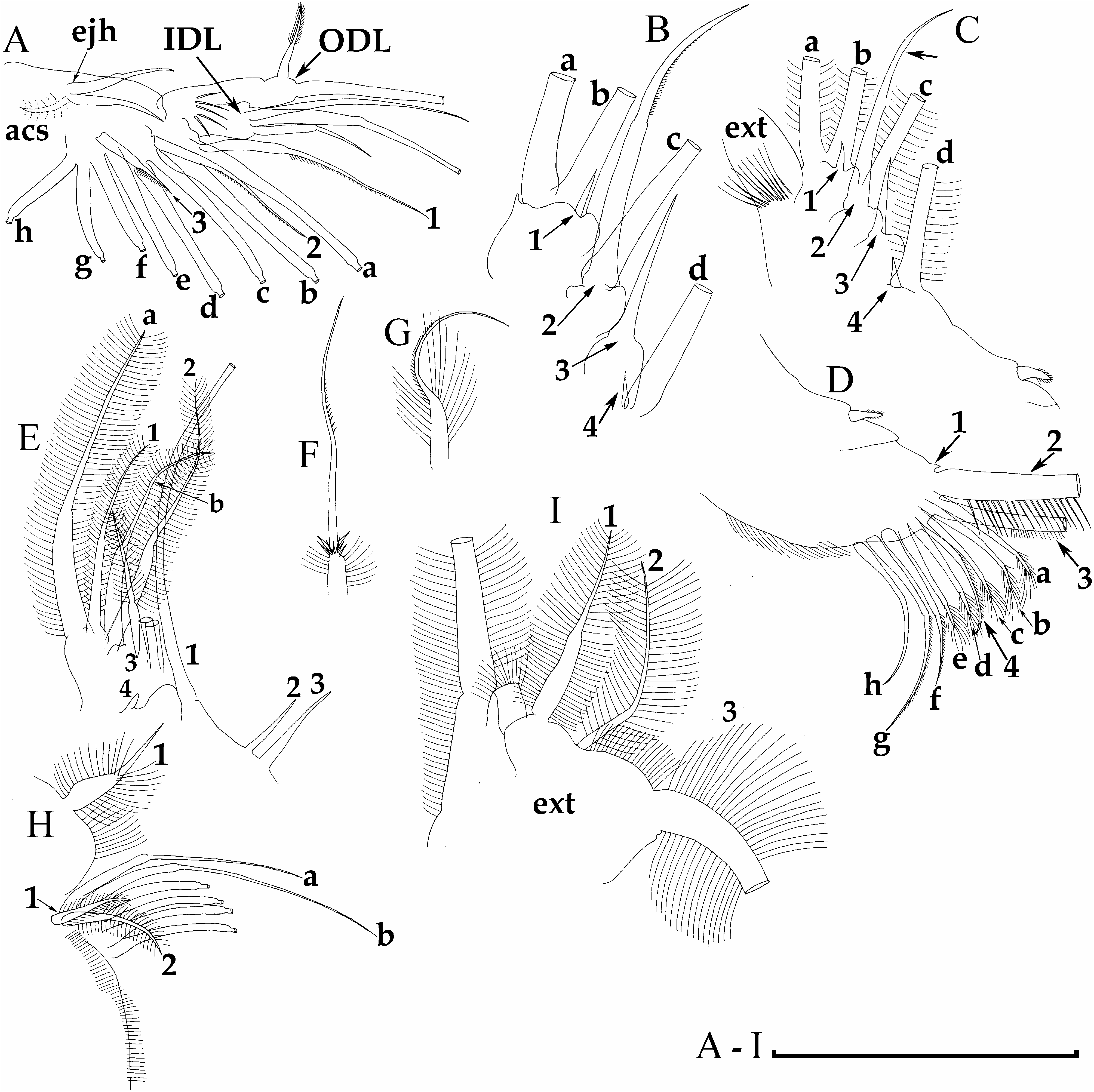
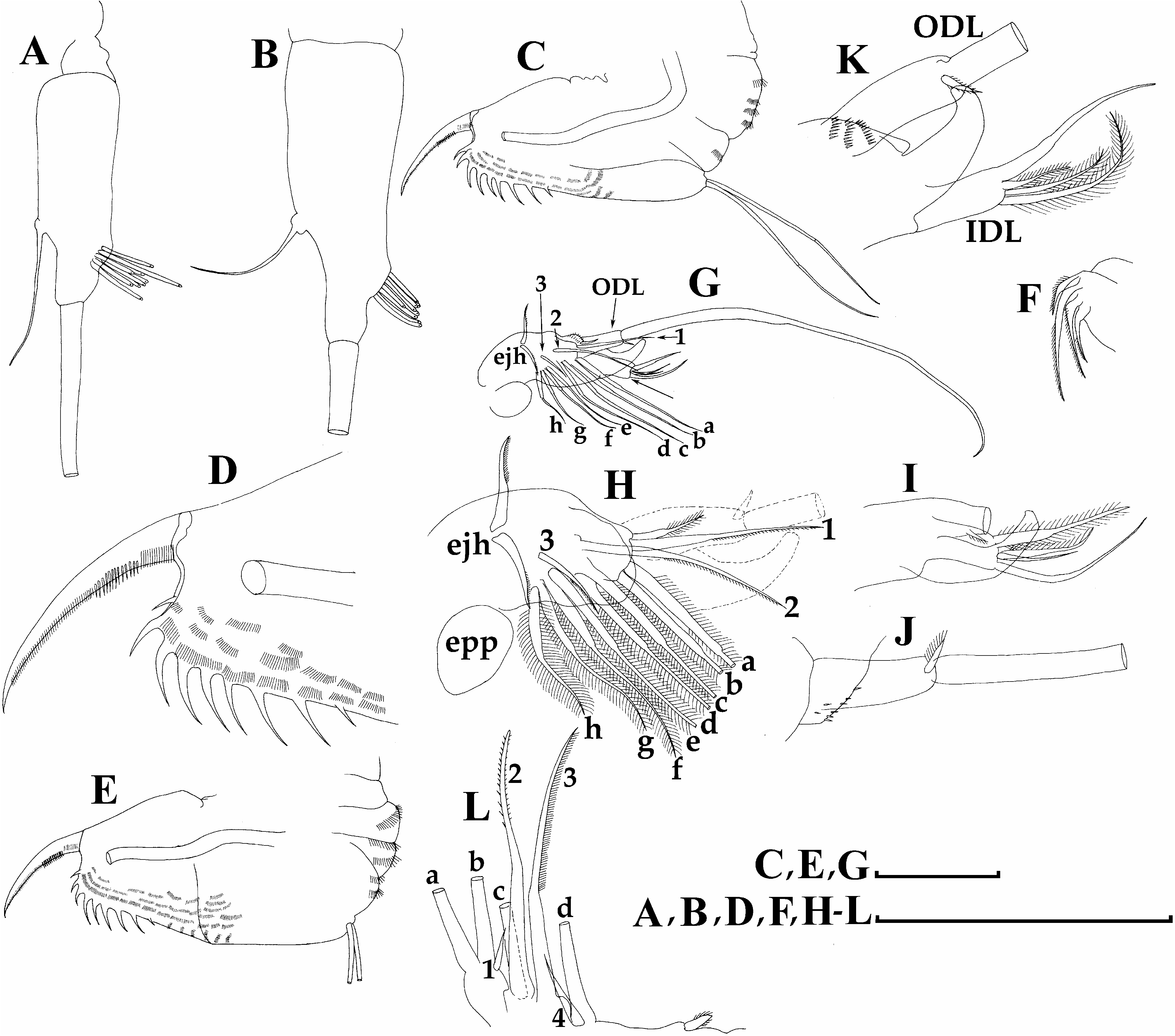
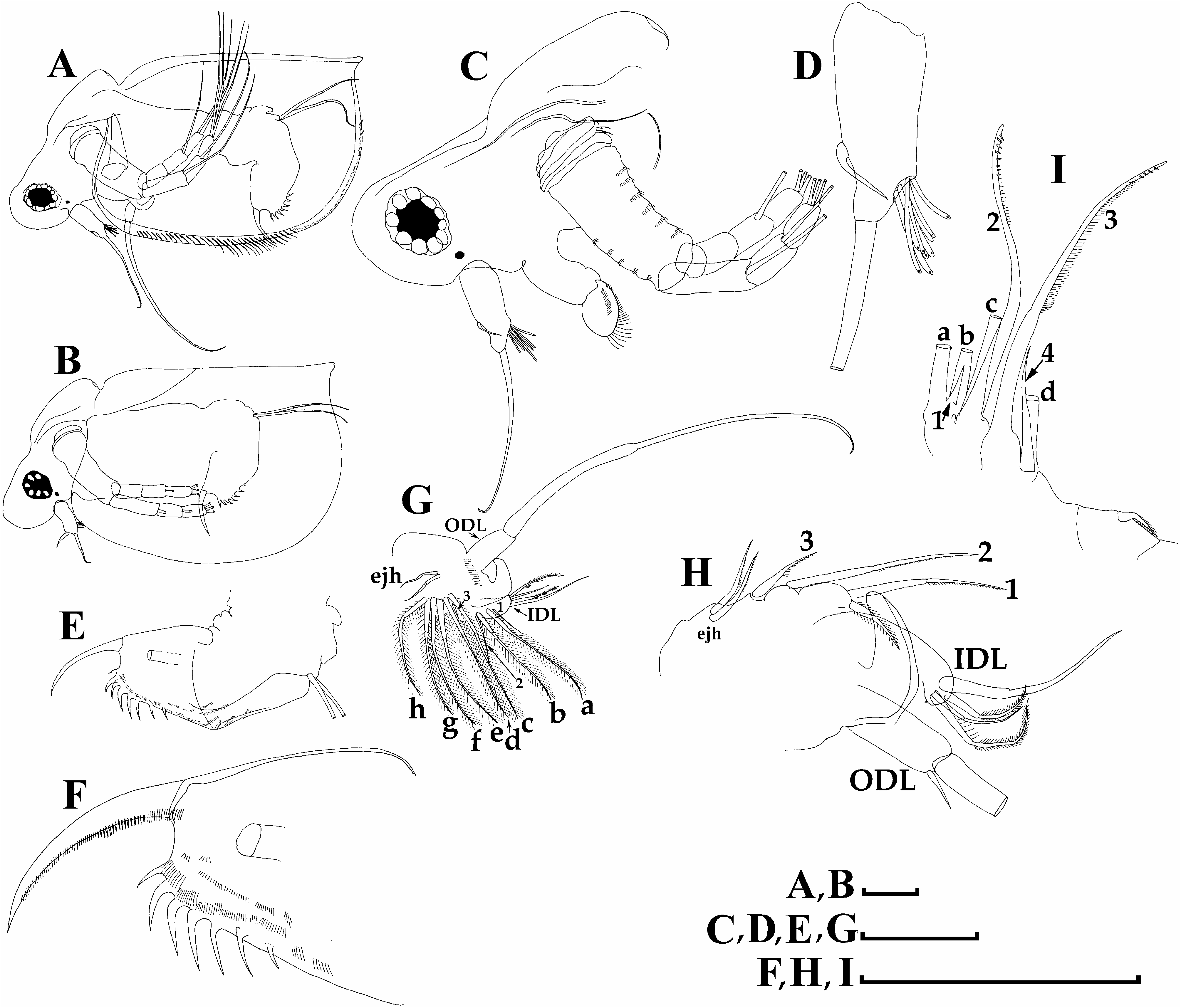
Limb I ( Figs. 5a, c View FIGURE 5 and 6a View FIGURE 6 ) with ovoid epipodite (epp) and a bilaterally setulated accessory seta (acs); outer distal lobe (ODL) bearing a long naked seta and a short seta setulated distally. Inner distal lobe (IDL) or endite 4 having three setae of different size: a single anterior and two posterior setae. Endite 3 having an anterior seta (1) and two posterior setae (a–b); endite 2 with a single anterior seta (2) and two posterior setae (c–d); endite 1 with a single anterior seta (3) and four posterior setae (e–h). Two ejectors hooks relatively small, subequal in size. Maxillar process absent.
Limb II ( Figs. 5b, d View FIGURE 5 and 6b–d View FIGURE 6 ) with ovoid epipodite (epp), exopodite (ext) elongated, with two long setae of different size. Endite 5 (e5) with a short stiff anterior seta (1) and two soft, long posterior setae (a–b); endite 4 (e4) with one relatively long, stiff anterior seta (2) and a long posterior seta; endite 3 (e3) with one relatively short, stiff anterior seta (3) shorter than anterior seta of endite 3, but longer than anterior seta of endite 5, and a long posterior seta (c); endite 2 (e2) with a short anterior seta (3) and no posterior setae. A seta on unclear homology near gnathobase. Endite 1(e1) = gnathobase with two rows of setae. Anterior row with four setae; seta 1 represented by a small sensillum; seta 2 very long, with long setules; seta 3 long but shorter than seta 2; seta 4 short, naked. Posterior row of gnathobase with eight setae of different morphology (a–h).
Limb III ( Figs. 5e View FIGURE 5 and 6e–g View FIGURE 6 ) with an elongated preepipodite (pep) and subovoid epipodite (epp). Exopodite (ext) subovoid and flat with two lateral setae (5–6) and four distal setae (1–4). Distal segments of seta 1 and seta 2 slender, seta 2 with a row of strong setules on proximal part of distal segment. Limb inner distal portion consist of five endites: endite 5 ( Fig. 5e View FIGURE 5 : e5) with a single anterior ( Fig. 6e View FIGURE 6 : 1) and a single posterior (a) seta; endite 4 with a single anterior (2) and a single posterior (b) seta; endite 3 with a short anterior seta (3); endite 2 with a very short anterior seta (4); endite 1 (gnathobases) represented by a large lobe with a long distal anterior seta (1) and two small, stiff anterior setae (2–3), posterior portion represented by numerous long posterior setae.
Limb IV ( Figs. 5f View FIGURE 5 and 6h View FIGURE 6 ) with an elongated prepipodite (pep) and a subovoid epipodite (epp). Exopodite (ext) subovoid, flat, bearing two lateral setae (5–6) and four distal setae (1–4). Distal segment of seta 1 very thin, naked. Limb inner distal portion with two soft anterior setae (1–2); inner margin of a gnathobase represented by numerous posterior setae.
Limb V ( Figs. 5g View FIGURE 5 and 6i View FIGURE 6 ) with elongated epipodite (epp), exopodite (exp) triangular, with two distal setae of different size (1–2) and a large lateral seta (3). Inner portion of limb V with a setulated margin and a large distal seta.
Juvenile female ( Figs. 1c,f View FIGURE 1 ). Body subovoid, relatively elongated. Dorsal margin almost straight, postero-dorsal margin with a small caudal spine, sometimes with few minute terminal denticles; ventral margin ovoid, postero-ventral angle smooth. Head rounded, compound eye relatively large, completely occupies distalmost body portion, ocellus minute. Head in lateral view with a strong depression, dorsal head pore protruded.
Ephippial female ( Fig. 1a View FIGURE 1 ). Body mostly same as in parthenogenetic female, dorsal portion of valves modified into ephippium with a single resting egg. Ephippium subovoid in lateral view, with a clear reticulation, without any lateral projections.
Adult male( Figs. 7a,d View FIGURE 7 ). Body subovoid, dorsal margin straight; postero-dorsal angle well-developed, sometimes represented by a short caudal spine. Ventral margin ovoid, postero-dorsal angle rounded.
Head ( Figs. 7a–e View FIGURE 7 ) rounded large, rostrum absent; compound eye relatively large, occupies most part of the distal head extremity; ocellus present. In lateral view headshield having a strong depression with a minute dorsal head pore. In dorsal view, headshield with mostly triangular fornices ( Fig. 7b View FIGURE 7 ). Labrum ( Fig. 7c View FIGURE 7 ) same as in parthenogenetic female.
Valve ( Figs. 7a,d View FIGURE 7 ) similar to that in parthenogenetic female.
Abdomen ( Figs. 8c,e View FIGURE 8 ) with four setulated segments lacking any processes, covered by minute setules.
Postabdomen ( Figs. 8c,e View FIGURE 8 ) elongated, subrectangular; ventral margin convex. Dorsal margin from straight to slightly convex. Preanal angle not expressed or slightly projected; postanal portion with nine strong spines of different size. Gonopore opens subdistally on postabdomen. Postabdominal claw ( Fig. 8d View FIGURE 8 ) long with three pectens, tip pointed; pectens as in parthenogenetic female.
Antenna I ( Figs. 7c,e View FIGURE 7 and 8a,b View FIGURE 8 ) long, cylindrical, with nine short aesthetascs of different size. Sensory seta long (longer than the longest aesthetascs), located subdistally of antenna I body. Flagellum located distally, very long (longer than antenna I body).
Antenna II ( Fig. 7c View FIGURE 7 ) as in parthenogenetic female.
Maxilla ( Fig. 8f View FIGURE 8 ) bearing four stiff setulated setae of different size.
Limb I ( Figs. 8g –k View FIGURE 8 ) with outer distal lobe bearing a single, very long seta and a short setulated seta. Inner distal lobe (IDL) modified, bearing a copulatory hook and four seta of different size. Among them, two anterior seta: one seta as in female, and the second an additional seta. The rest of limb I as in female.
Limb II ( Fig. 8i View FIGURE 8 ) in general as in female, with few modifications: endite 4 with a long anterior seta 2 bilaterally armed by short setules; endite 3 with anterior seta 3 longer than in female; endite 2 with anterior seta 4 longer than in female.
Size. Parthenogenetic female length 0.44–1.25 mm, height 0.24–0.84 mm; ephippial female length 0, 82 mm, height 0.55 mm; male length 0.69–0.81 mm, height 0.35–0.42 mm.
Taxonomic comments. C. dubia s.str. was described from Lake Toba, Sumatra, Indonesia ( Richard 1894). Unfortunately, its type material is lost ( Kotov & Ferrari 2010). In this article we cannot relate the morphological identity of C. dubia s.l. from Northern Eurasia with that from Indonesian populations, which it is a task of further studies. But we did not find any morphological evidence for the existence of more than one species in northern Eurasia (except for a single Yakutian population, see below). Note that Ceriodaphnia affinis Lilljeborg, 1901 was described from Europe, but its validity could be checked only by a comparison with Indonesian populations. To resolve the issue of the exact species status, a redescription of the population from Lake Toba and a full-scale revision of all populations belonging to the C. dubia s.l. species complex should be carried out in the future.
Distribution and ecology. To date, C. dubia s.l. needs to be regarded as a widely distributed (almost cosmopolitan) taxon ( Korovchinsky et al. 2021).
No known copyright restrictions apply. See Agosti, D., Egloff, W., 2009. Taxonomic information exchange and copyright: the Plazi approach. BMC Research Notes 2009, 2:53 for further explanation.