
Diadema africanum, Rodríguez, Adriana, Hernández, José Carlos & Clemente, Sabrina, 2013
|
publication ID |
https://doi.org/10.11646/zootaxa.3636.1.6 |
|
publication LSID |
lsid:zoobank.org:pub:FF3A24CC-6545-4B77-83C5-2503143E7F16 |
|
DOI |
https://doi.org/10.5281/zenodo.6145098 |
|
persistent identifier |
https://treatment.plazi.org/id/84171C00-D224-F62E-FC94-7BDA102AF91E |
|
treatment provided by |
Plazi |
|
scientific name |
Diadema africanum |
| status |
sp. nov. |
Diadema africanum View in CoL sp. nov.
Figs 5 View FIGURE 5 –10, tables 1–3.
Diagnosis. Test and spines typically black with a red tinge and a turquoise sheen when viewed in direct sunlight. The iridophore pattern occurs as bold blue lines down either side of the naked interambulacral areas, as a pentamerous ring around the apical disc and as lines along some plate boundaries. Apical disc is hemicyclic with arch-shaped depressions on the denuded genital plates in both adults and juveniles. Gonopores measure 33% of the length of the genital plates. The periproctal cone is black with no markings. The Peristome is proportionally large measuring 40–60% of the test’s horizontal diameter. Spines are verticillate and hollow with distal barbs. Verticilations are formed of urn-shaped solid wedges that are visible when the spines are viewed in transverse section. These wedges typically number twenty in ambulacral spines and twenty-four in interambulacral spines. Only tridentate and triphyllous pedicellariae are present. The tridentate pedicellariae occur as a single form with reasonably broad, curved valves, with almost straight edges, that can either be smooth or serrated, with an expanded distal gripping region. The blades of the valves meet only along the upper fifth of their length. The head of each pedicellaria is supported by a long muscular neck, attached to a mid-length stalk.
Holotype: TFMCBMEQ/00232 in the ‘Museo de Ciencias Naturales de Tenerife’ (TFMC), Santa Cruz de Tenerife, Canary Islands, Spain.
Paratypes: TFMCBMEQ /00233, TFMCBMEQ /00234, TFMCBMEQ /00235 in the ‘Museo de Ciencias Naturales de Tenerife’ (TFMC), Santa Cruz de Tenerife, Canary Islands, Spain.
Other material. Twenty-one specimens in the Zoological Collection in the ‘Departamento de Biología Animal (Ciencias Marinas)’, Universidad de La Laguna, Tenerife, Canary Islands.
Etymology. Species name refers to the geographical distribution of the species on the western coasts of Africa. It is distributed on islands and continental coasts along the African continental shelf.
Ecology. Diadema africanum is an important macro-herbivore on subtropical and tropical rocky reefs off the West African coasts. In the eastern Atlantic islands, and particularly in Madeira and the Canary Islands, the species is distributed throughout the islands at densities that can reach more than 12 individuals / m2 (Brito et al. 1984; Alves et al. 2001; Hernández 2006; Clemente 2007; Hernández et al. 2008). The species can therefore dramatically reduce the abundance of non-crustose macroalgae resulting in the formation of sea urchin-dominated barren grounds (Hernández et al. 2008). This phenomenon is especially relevant in Madeira, Salvage and the Canary Islands, where macroalgal beds represent the main ecosystem. Diadema africanum also occurs on coral dominated reefs in Cape Verde and Sâo Tome, where sea urchin control of macroalgae via grazing is beneficial for coral settlement and growth. Only a few studies have looked at the abundance and ecological role of D. africanum along the tropical western African coasts (e.g. John et al. 1977; 1992), with further investigations needed.
Distribution. Diadema africanum occurs in the Eastern Atlantic islands, from Madeira Islands to the Guinean Gulf including Salvage Islands, Canary Islands, Cape Verde Islands (Hernández et al. 2008), and Sâo Tome Island (Lessios et al. 2001). It has also been also recorded in continental areas of Ghana (John et al. 1977, 1992) and in Ngor Island, Senegal (P. Wirtz, pers. com).
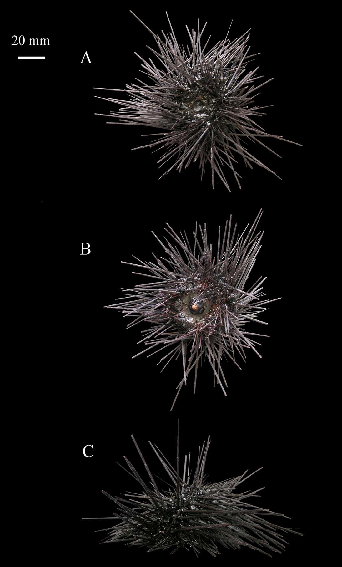
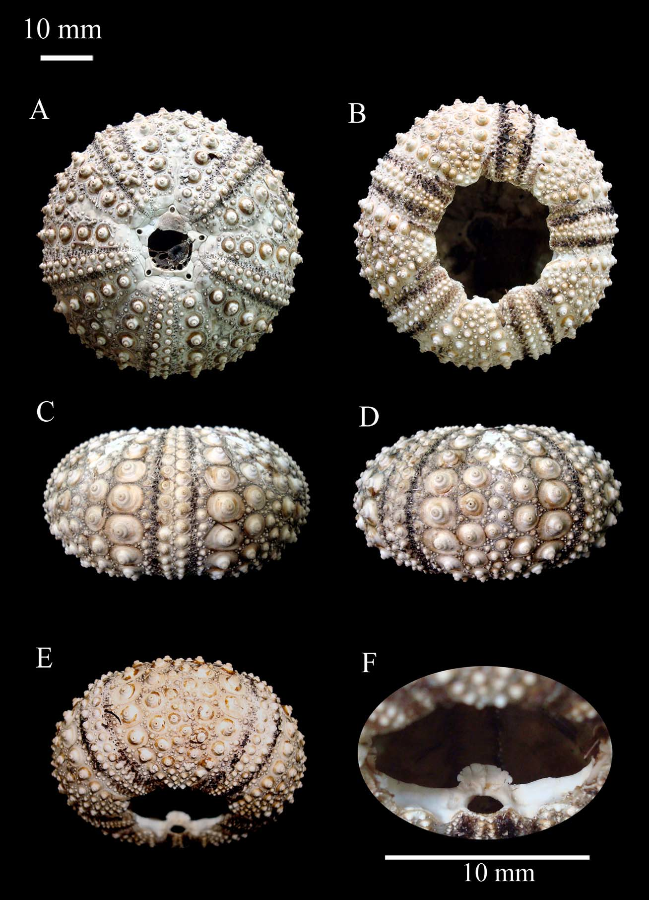
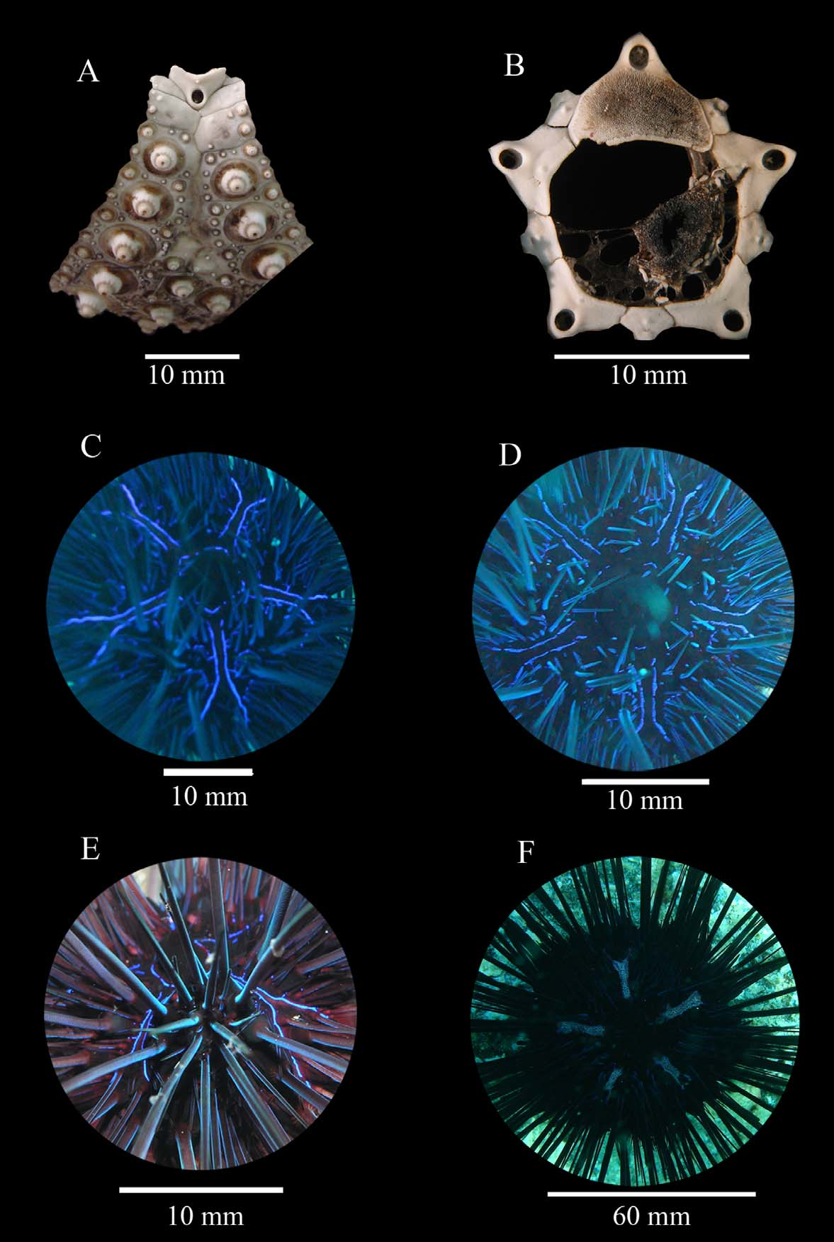
Description. The test is hemispherical, with a horizontal diameter of 66.73 mm and a vertical diameter of 32.78 mm in the paratype and 61.23 mm h.d. and 31.36 mm v.d. in the holotype. The base colour of the test epithelium is black with a red tinge ( Fig. 5 View FIGURE 5 A–C), while the denuded test is white ( Fig. 6 View FIGURE 6 A–E). The apical system measures 18.14 mm in the paratype and is hemicyclic with ocular plates II and III exsert ( Fig. 6 View FIGURE 6 A). The genital plates are wider than long, and have up to four small tubercles along their inner-edge (Table 1& Fig. 7 View FIGURE 7 A–B) with large genital pores that measure 33% of the genital plate length ( Fig. 7 View FIGURE 7 A). The ocular plates are pentagonal with a small pore located at the top of the plate and have from one to three small tubercles along the centre of the plate ( Fig. 7 View FIGURE 7 B).
On the naked test, distinct arch-shaped depressions are present on the genital plates of both adults and juveniles (Table 5 & Fig. 7 View FIGURE 7 B). These depressions correspond to the corners of the pentamerous apical ring of iridophores seen in living sea urchins ( Fig. 7 View FIGURE 7 C–E). The iridophore pattern is bold and bright on the apical system when viewed in sunlight. The periproct is 11.26 mm wide in the paratype (approximately 16% of the test’s horizontal diameter) and has a small black periproctal cone that has no platelets or markings on the skin ( Fig. 5 View FIGURE 5 A).
The ambulacra are slightly raised aborally ( Fig. 6 View FIGURE 6 C) and measure 30% of the width of the interambulacra ( 8.65 mm) at the ambitus in the paratype. They have two rows of large primary tubercles and an offset inner series of small tubercles that are perforated and crenulated, with non-conjugate pore-pairs and phyllodes developed adorally.
The interambulacra are broader than the ambulacra at the ambitus ( Fig. 6 View FIGURE 6 C–D), with 5–6 series (the inner median series is more offset in some specimens giving the impression of two series) of 14 primary tubercles. Tubercles are perforate and crenulate and have areoles of moderate size ( Fig. 7 View FIGURE 7 A). During the day blue lines of iridophores can be seen down either side of the naked median regions.
The peristome is subcircular ( Fig. 6 View FIGURE 6 B) and in the paratype it measures 26.9 mm in diameter, 40% of the test’s horizontal diameter. The peristomal membrane is black with a red tinge and has five pairs of buccal tube feet ( Fig. 5 View FIGURE 5 B), with abundant triphyllous pedicellariae. The auricles are robust and have high processes ( Fig. 6 View FIGURE 6 F).
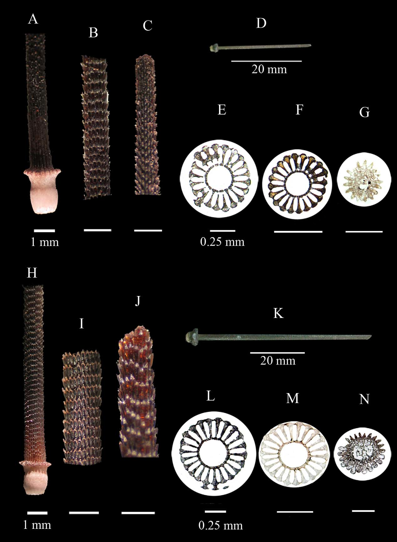
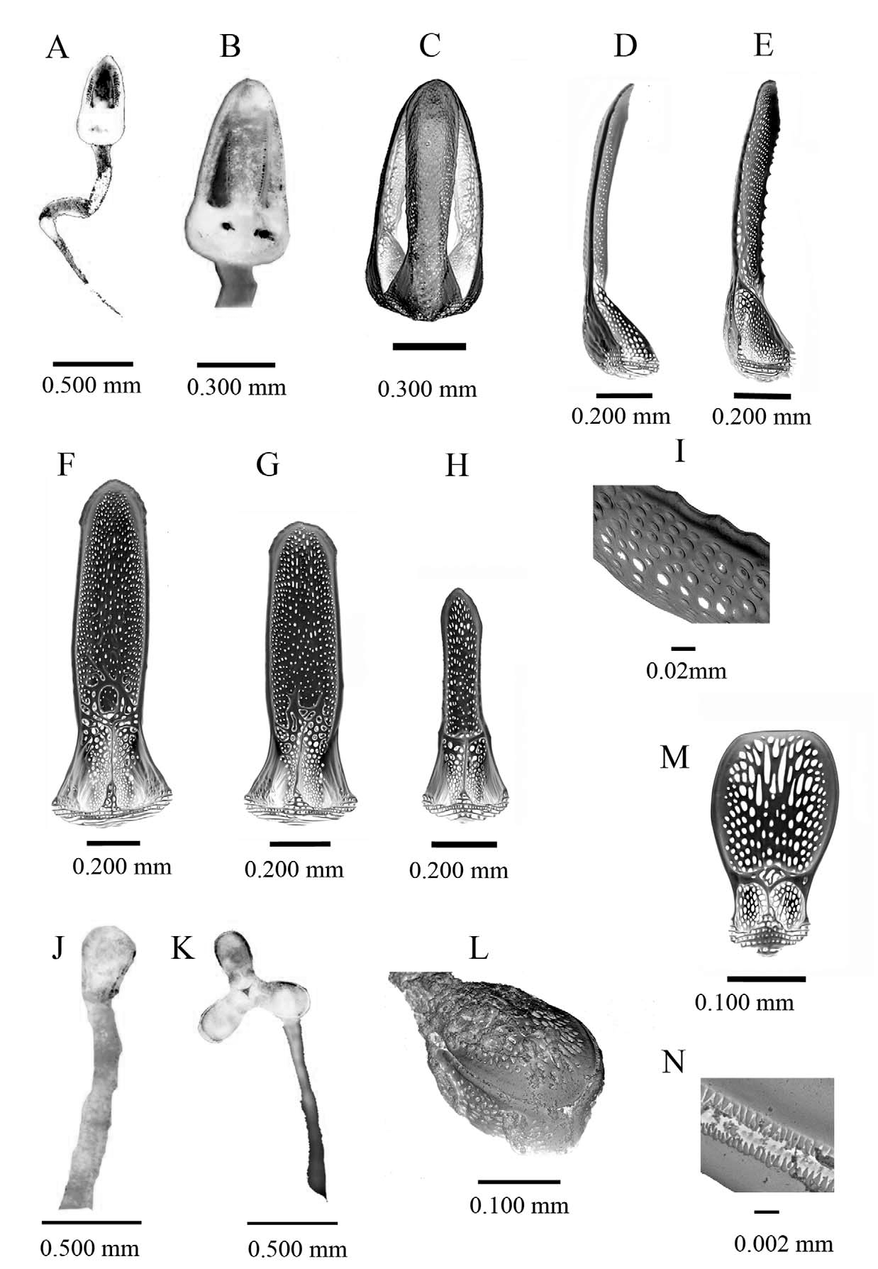
In adults the spine epithelium is black with a red tinge and a turquoise sheen when viewed in direct sunlight. In juveniles, spines are banded red and white, which is typical of the genus. Ambulacral spines measure 43.21 mm in length in the paratype (28.83% of the test horizontal diameter). These spines are strongly verticillate with barbs pointing distally ( Fig. 8 View FIGURE 8 A–D). Proximally these spines have a mean diameter of 0.89 mm that decreases distally ( Fig. 8 View FIGURE 8 E–G). The axial cavity comprises 28.8% of the horizontal diameter, which increases to 35.22% in the distal region ( Fig. 8 View FIGURE 8 E–G). The solid wedges are urn-shaped, and typically number twenty comprising 63.42% of the spine’s horizontal diameter in the proximal section.
Interambulacral spines ( Fig. 8 View FIGURE 8 H–N), are long and slender, with a maximum diameter of 2 mm proximally, and taper distally ( Fig. 8 View FIGURE 8 K–N). These spines measure 58.62 mm in the paratype. In transverse section the axial cavity comprises 29.92% of the horizontal diameter proximally, but increases to 39.17% in the distal region (Table 2; Fig. 8 View FIGURE 8 L–N). The solid wedges are urn-shaped and typically number twenty-four, 62.57% of the of the spine’s horizontal diameter in the proximal section.
Both tridentate and triphyllous pedicellariae are present in D. africanum ( Fig. 9 View FIGURE 9 ). No ophicephalous pedicellariae were found, either true ophicephalous or of the claviform type, which have been reported in other species of Diadema (Mortensen, 1940) . Only a single form of tridentate pedicellaria is found in this species ( Fig. 9 View FIGURE 9 A–I), which are abundant orally and aborally, but particularly around the periproct and around the peristome. This form of tidentate pedicellaria has a long, muscular neck on a long stalk, which allows a high degree of movement ( Fig. 9 View FIGURE 9 A). The valves of the pedicellariae are moderately curved and only meet along the upper fifth of their length. The blade of the valves has almost straight edges that are either smooth or serrated with an expanded distal gripping region ( Fig. 9 View FIGURE 9 D–I). Triphyllous pedicellariae are more abundant than the tridentate form and are distributed all over the sea urchin test, but occur in particularly large numbers around the peristome. Their valves are small and broad ( Fig. 9 View FIGURE 9 J–N), with numerous small peripheral teeth that form two rows along the edges of the valves, which interlock when the valves are closed ( Fig. 9 View FIGURE 9 L–N).
Comparisons between D. antillarum and D. africanum using a PCA ordination based on 29 morphological characters of the test, spines and pedicellariae are illustrated in Fig. 10. Overall differences observed in the ordination are supported by the R statistic associated with the ANOSIM test (Global R-statistic=1, p<0.5), which shows that D. africanum is morphologically distinct from D. antillarum . The PCA shows differences between groups of individuals. Mean diameter of axial cavity (% spine diameter) (3) and the percentage of the spine’s diameter comprised of solid wedges (4) are variables that are highly correlated with axis 1, which means that the two species highly differ on this variables. Diadema africanum specimens have wider diameters of spines and a larger portion of the spine comprised of solid wedges. The peripheral gripping area as % of distal peripheral area (mm2) (18) and the diameter of gonopores (% of genital plate height) (25) are negatively correlated with axis 1, meaning that D. africanum shows reduced peripheral gripping area and narrower gonopores than D. antillarum . There are also intraspecific variations on horizontal test diameter (mm) (21) and vertical test diameter (mm) (22) for both species, which can be seen in the ordination plot as vertical spread of the points following axis 2.
FIGURE 10. Principal Components Analyses (PCA) showing the first 2 axes that explain the 76.1 of variability (54.9 first axis, 21.3 second axis and 7.3 third axis) based on 29 quantitative variables including morphological characters of the test, spines and pedicellariae in D. antillarum (two specimens: neotype plus another individual) and D. africanum ( 17 specimens). The numbers correspond to: 1–5 spine characters; 6–20 pedicellarial characters and 21–29 test characters: 1. Spine diameter; 2. Number of solid wedges; 3. Mean diameter of axial cavity (% spine diameter); 4. Percentage of the spine’s diameter comprised of solid wedges; 5. Percentage of the spine’s diameter comprised by the foraminated ring; 6. Number of types of pedicellariae; 7. Length of distal region (mm); 8. Width of distal region (mm); 9. Length of proximal region (mm); 10. Width of proximal region (mm); 11. Total area of distal region (mm2); 12. Internal area of distal region (mm2)¸13. Peripheral area of distal region (that does not grip; mm2); 14. Peripheral gripping area (mm2); 15. Total area of proximal region (mm2);16. Adductor muscle insertion area (mm2); 17. Keel and peripheral area of proximal region (mm2); 18. Peripheral gripping area as % of distal peripheral area (mm2), 19. Height of teeth (mm); 20. Width of teeth (mm); 21. Horizontal test diameter (mm); 22. Vertical test diameter (mm); 23. Number of tubercles on genital plate; 24. Height to width (at widest point) ratio of genital plate; 25. Diameter of gonopores (% of genital plate height); 26. Apical system (% of horizontal test diameter); 27. Periproct (% of horizontal test diameter); 28. Peristome (% of horizontal test diameter).; 29. Ambulacra % of interambulacra (at ambitus).
No known copyright restrictions apply. See Agosti, D., Egloff, W., 2009. Taxonomic information exchange and copyright: the Plazi approach. BMC Research Notes 2009, 2:53 for further explanation.
|
Kingdom |
|
|
Phylum |
|
|
Class |
|
|
Order |
|
|
Family |
|
|
Genus |