
Micromalthus debilis, LECONTE, 1878
|
publication ID |
https://doi.org/10.1111/j.1096-3642.2009.00549.x |
|
DOI |
https://doi.org/10.5281/zenodo.10545467 |
|
persistent identifier |
https://treatment.plazi.org/id/7F51878C-7E7F-FFBB-48C6-FBF9D1B61AAE |
|
treatment provided by |
Valdenar |
|
scientific name |
Micromalthus debilis |
| status |
|
MICROMALTHUS DEBILIS LECONTE, 1878 View in CoL
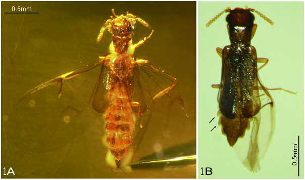
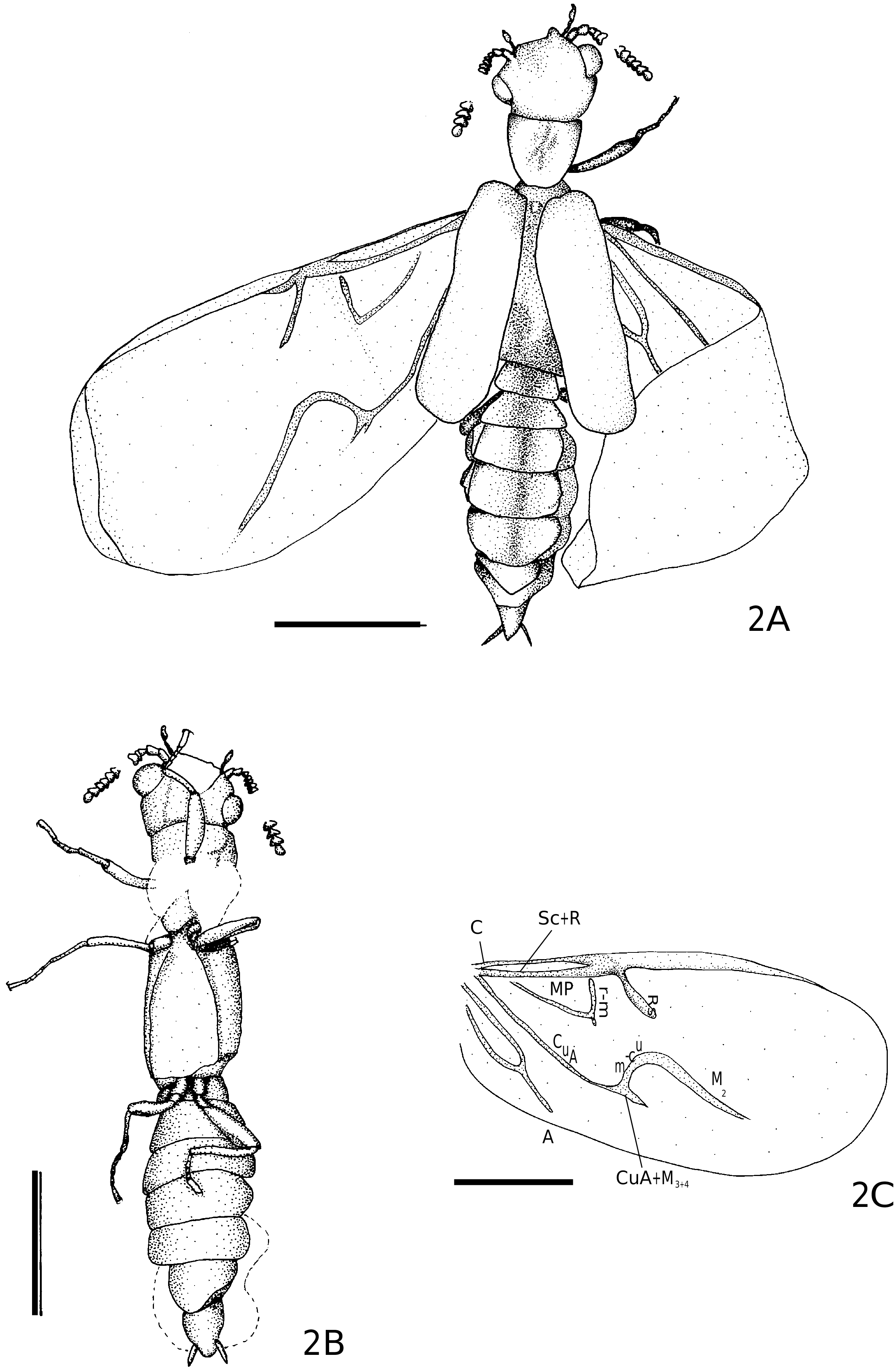
Figures 1A View Figure 1 , 2A–C View Figure 2
DESCRIPTION OF DOMINICAN AMBER SPECIMENS
A detailed description of specimen MTEC226 ( Figs 1A View Figure 1 , 2A–C View Figure 2 ) is given as representative of the Dominican amber specimens.
The fossil is embedded in a very clear piece of Dominican amber of light amber colour. There is a fissure running from one corner of the amber piece to the tip of the abdomen.
With a few restrictions, the beetle is completely visible from both dorsal and ventral surfaces. It is intact with the exception of both antennae, which are broken after the fifth (left) and fourth (right) antennomere. Both hind wings are outstretched. The left one lies flat and shows the complete venation, whereas the right one is unnaturally folded at the radial hinge. Both elytra are partially closed and translucent ( Fig. 1A View Figure 1 ). Overall, the animal is 2.18 mm long. In comparison with the figured extant specimen ( Fig. 1B View Figure 1 ), the amber inclusions seem to have a very long abdomen. The length of the abdomen is very variable both in extant and fossil specimens. The abdominal segments are often more contracted in males than in females. For example, this may be because of physiological conditions, e.g. the developmental stage of eggs in the reproductive system of the female. Therefore, the overall length, especially of females, can be very variable. Further measurements are given in Table 2.
Head
The head is turned to the left and directed slightly upward. Of the mouthparts, only the protruding maxillary palps are visible. Each terminal palpomere is enlarged and bears a large sensory area with long rod-shaped sensilla. A white foggy substance covers the remaining mouthparts. Both antennae are complete but broken, the left antenna behind the fifth antennomere and the right antenna behind the fourth antennomere. The distal parts of the antennae are separated by the lengths of approximately three (right) to five (left) antennomeres from the more proximal segments. It is possible that the distal parts of the antennae stuck to the resin and were separated when the beetle attempted to free itself. The antennae are 11-segmented. The two basal antennomeres are distinctly larger than the third. Antennomeres 3 to 11 increase in size. The terminal antennomere is similar in size to the pedicel. The head is approximately 0.26 mm long and 0.38 mm wide. Each antenna is about 0.29 mm long without the gap.
Thorax
The pronotum is 0.27 mm wide at the anterior margin and 0.18 mm wide at the posterior margin. The ventral sides of the prothorax and mesothorax are obscured by a white foggy substance so that neither the prosternal area nor the procoxae or trochanters are visible ( Fig. 2B View Figure 2 ). The tibiae are about two-thirds as long and about half as wide as the femora. Only in the hind legs are the tibiae nearly as long as the femora. All tarsi are five-segmented. The terminal tarsomere bears two claws and is nearly as long as the remaining tarsomeres combined. The metathorax is slightly longer than the prothorax and mesothorax combined. The metacoxae are inserted at the posterior margin of the metathorax. They stand close together and are somewhat cylindrical in shape. The elytra are very translucent and partially opened. Their surface is smooth and without recognizable pubescence. It is not possible to distinguish any details on the dorsal sides of the mesothorax and metathorax. The alae are nearly completely extended. The distal half of the right wing is turned upward and inward so that its tip is directed toward the abdomen. The left wing is nearly fully outstretched. Only the apex is slightly crumpled. Combining features from both hind wings, it is possible to reconstruct the complete venation ( Fig. 2C View Figure 2 ).
Abdomen
The abdomen is completely collapsed with distinctly concave tergites, which are obliquely pressed against the sternites, so that the pleural membranes on the right side of the body can be seen in dorsal view. The abdomen has seven visible segments. None of the abdominal tergites have pubescent fovae, which, in combination with the long styli visible at the tip of the abdomen, indicates that the specimen is a female.
No known copyright restrictions apply. See Agosti, D., Egloff, W., 2009. Taxonomic information exchange and copyright: the Plazi approach. BMC Research Notes 2009, 2:53 for further explanation.