
Pseudodira Gordon, 1975
|
publication ID |
https://doi.org/ 10.5281/zenodo.5372914 |
|
publication LSID |
lsid:zoobank.org:pub:1AFFE36D-921D-49D3-B765-0DD398FCF5D2R |
|
persistent identifier |
https://treatment.plazi.org/id/6E5D87FA-FFCB-FF90-FE12-445DFD35FE89 |
|
treatment provided by |
Marcus |
|
scientific name |
Pseudodira Gordon, 1975 |
| status |
|
Pseudodira Gordon, 1975: 207 . Type species: Pseudodira clypealis Gordon, 1975 , by monotypy.
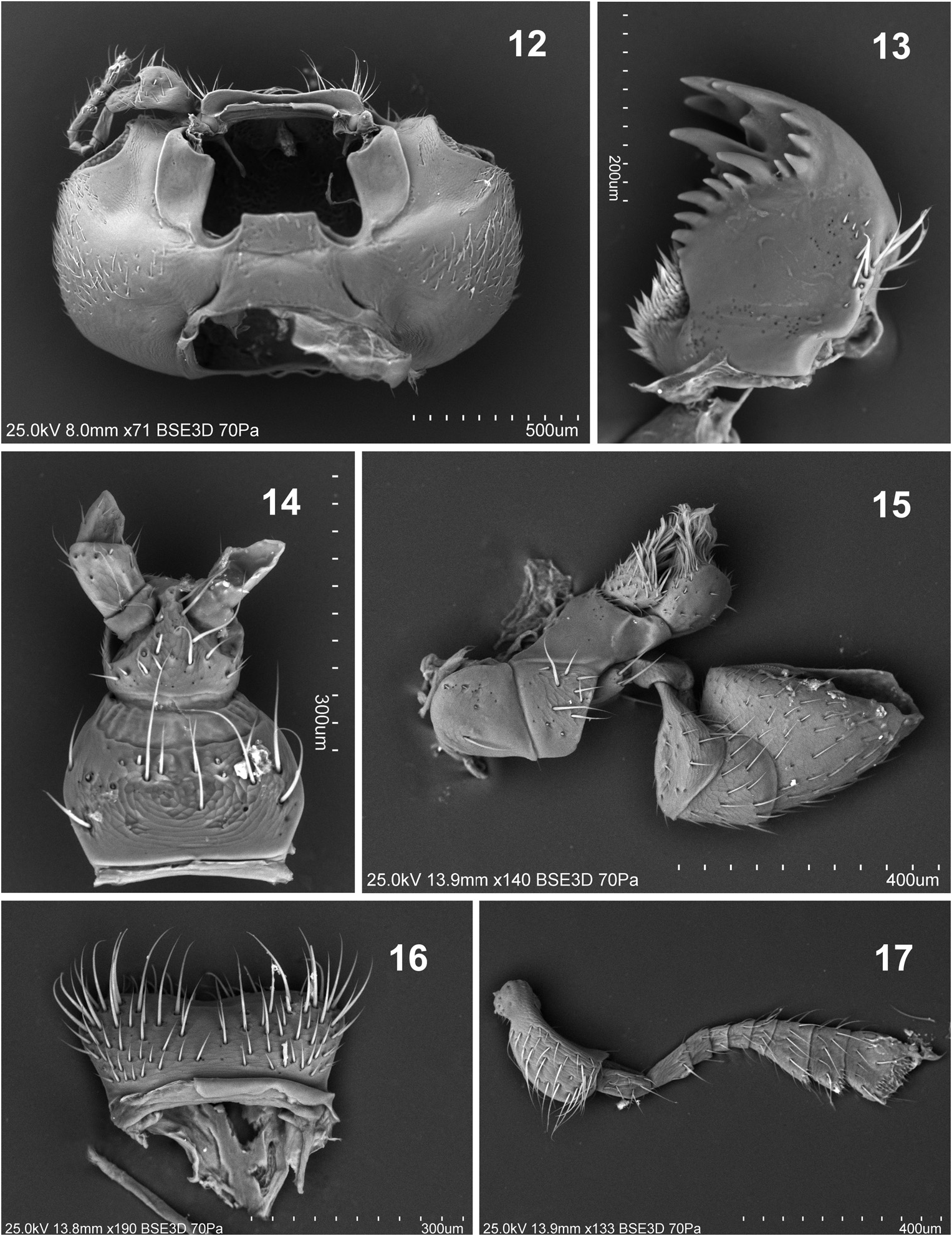
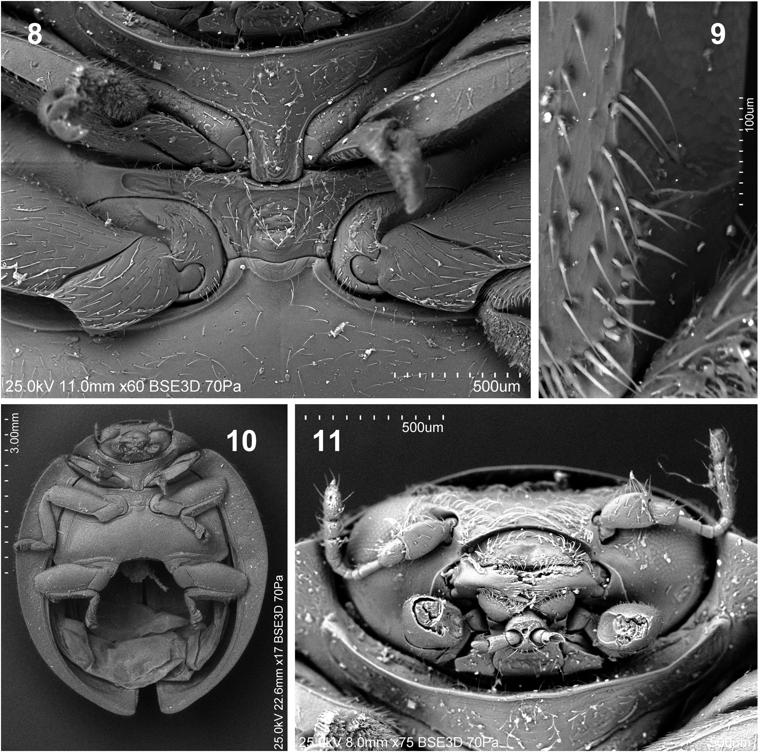
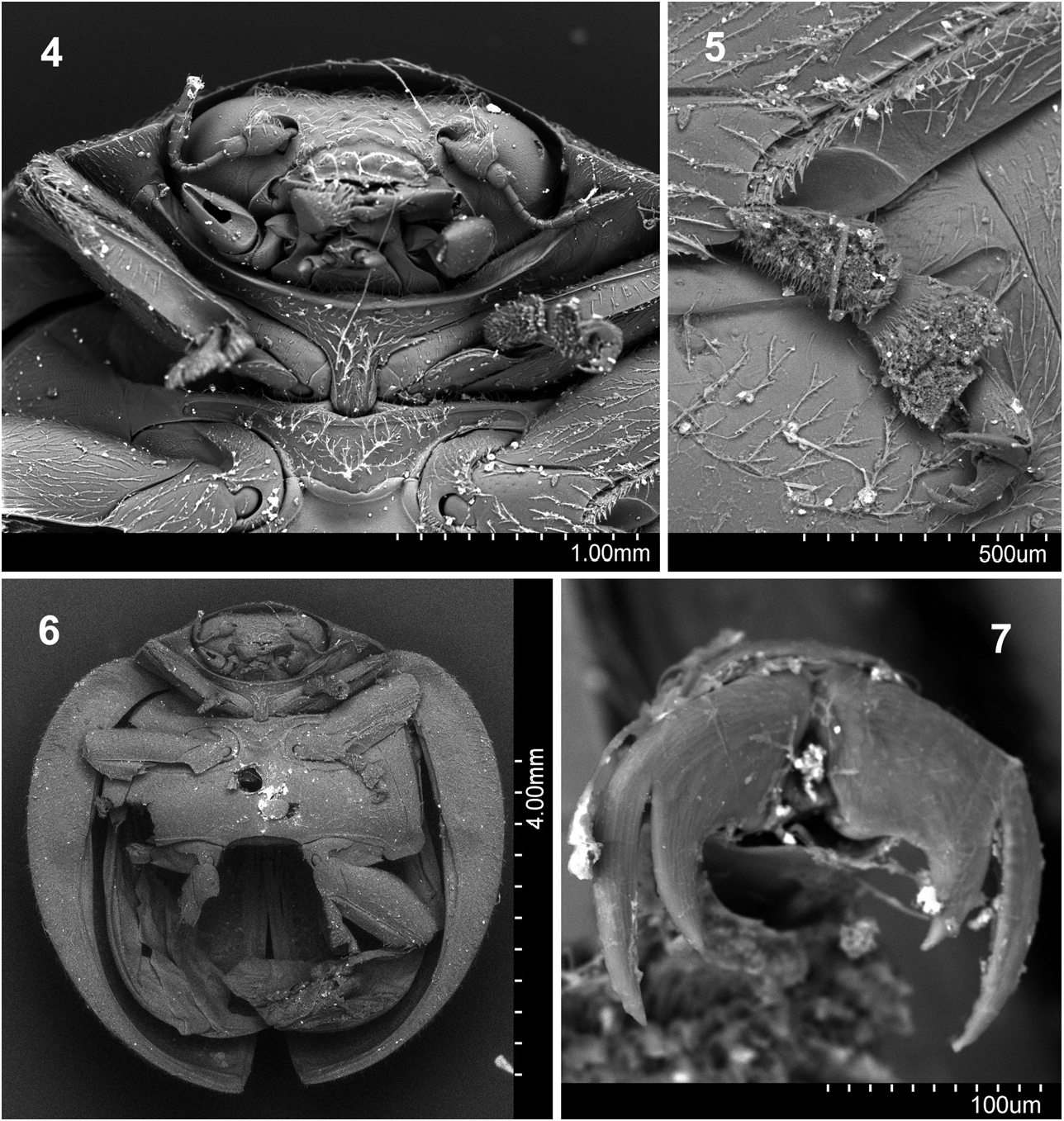
Diagnosis. Pseudodira can be characterized by: i) the incisor edge of the mandibles with large denticles continuing as a sinuate line to the apex of the mandible on the dorsal surface ( Fig. 13 View Figs 12–17 ); ii) the anterior margin of the metaventrite emarginated and distinctly bordered, the border with distinct incisions in the anterior angles between the midcoxae ( Fig. 8 View Figs 8–11 ); iii) the presence of a carina on the apices of the mid and hind tibiae ( Fig. 5 View Figs 4–7 ). The last character is shared with the South American genera Damatula Gordon, 1975 and Lorma Gordon, 1975 .
Redescription. Size: length 5.8–7.5 mm, width 5.0– 6.4 mm. Body oval, strongly convex. Dorsum covered with dense pubescence.
Head retracted in prothorax ( Fig. 11 View Figs 8–11 ). Eyes finely faceted; interocular distance 0.63–0.70 times as wide as head across eyes. Gular sutures deep, separated, reaching half length of gula ( Fig. 12 View Figs 12–17 ). Antenna short, less than width of the head; 10- ( P. clypealis ) or 11-segmented (other two species); scape large, swollen, along external surface regularly convex; pedicel distinctly narrower than scape; antennal club 3-segmented, asymmetrical; penultimate antennomere distinctly shorter than terminal one; terminal segment subquadrate ( Fig. 17 View Figs 12–17 ). Antennal grooves absent. Clypeus with anterior margin emarginate. Labrum slightly emarginate apically ( Fig. 16 View Figs 12–17 ). Mandible mesal and apical surfaces strongly serrated, apical third with strong prominent teeth; molar part absent; prostheca well developed, setose ( Fig. 13 View Figs 12–17 ). Maxilla with cardo subquadrate; mediastipes and basistipes distinctly separated ( Fig. 15 View Figs 12–17 ), lacinia simple, broad, and short with its apical surface covered with long setae and its mesal surface covered with irregularly arranged short pubescence. Galea round-oval; apex densely pubescent. Terminal maxillary palpomere large and securiform. Mentum ( Fig. 14 View Figs 12–17 ) transverse; sides rounded; anterior margin weakly arcuate posteriorly. Prementum oval, ligula reduced. Labial palps narrowly separated, originated subapically on prementum; apical palpomere shorter and narrower than penultimate one; basal palpomere transverse, about two times shorter than penultimate.
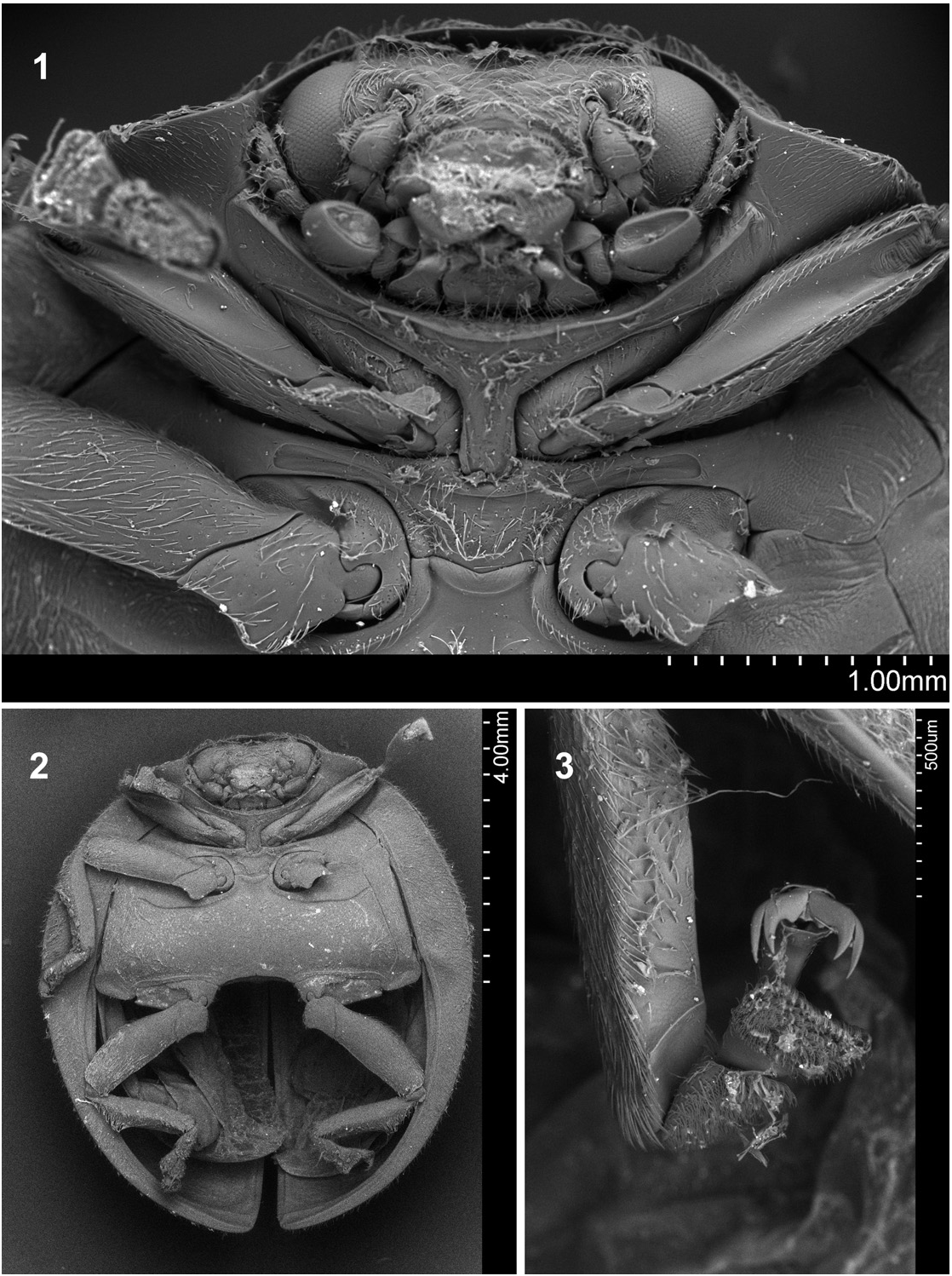
Prothorax. Anterior angles weakly produced anteriorly. Lateral margin not upturned. Prothoracic hypomeron smooth, setose. Prosternal process complete and less than 0.5 width of mesoventral process; surface smooth ( Figs 1 View Figs 1–3 , 4 View Figs 4–7 ) or with small tubercle ( Fig. 8 View Figs 8–11 ), sparsely setose, margined laterally. Prosternum in front of coxa shorter than coxal longitudinal diameter at the same position, with anterior margin slightly produced. Procoxal cavity with anterior border marginated.
Meso- and metathorax. Mesoventrite with raised anterior border. Mesoventral process flat ( Figs 1 View Figs 1–3 , 4 View Figs 4–7 ) or with small tubercle ( Figs 8 View Figs 8–11 ). Meso-metaventral junction with suture visible, angulate posteriorly. Scutellum triangular. Lateral margins of elytra not or slightly explanate; often visible from above; surface with double size punctation. Elytral epipleuron almost complete, reduced only at apex; with distinct foveae to receive tips of mid and hind femora; inner margin with border area throughout narrow and border line fading before base of elytron. Metaventral postcoxal lines contiguous, antero-lateral angles (between midcoxae) deeply incised ( Figs 1 View Figs 1–3 , 4 View Figs 4–7 , 8 View Figs 8–11 ); laterally complete, slightly arcuate.
Legs. Trochanters angulately produced posteriorly. Femora with distinct, complete grooves to receive tibiae. Fore tibiae with complete groove on outer edge. Mid and hind tibiae with oblique carina on outer edge near apex ( Figs 3 View Figs 1–3 , 5 View Figs 4–7 , 9 View Figs 8–11 ). Protibia with single apical spur; mid and hind tibia with two spurs. Tarsus 4-segmented, pseudotrimerous; claws double, base weakly swollen ( Fig. 7 View Figs 4–7 ).
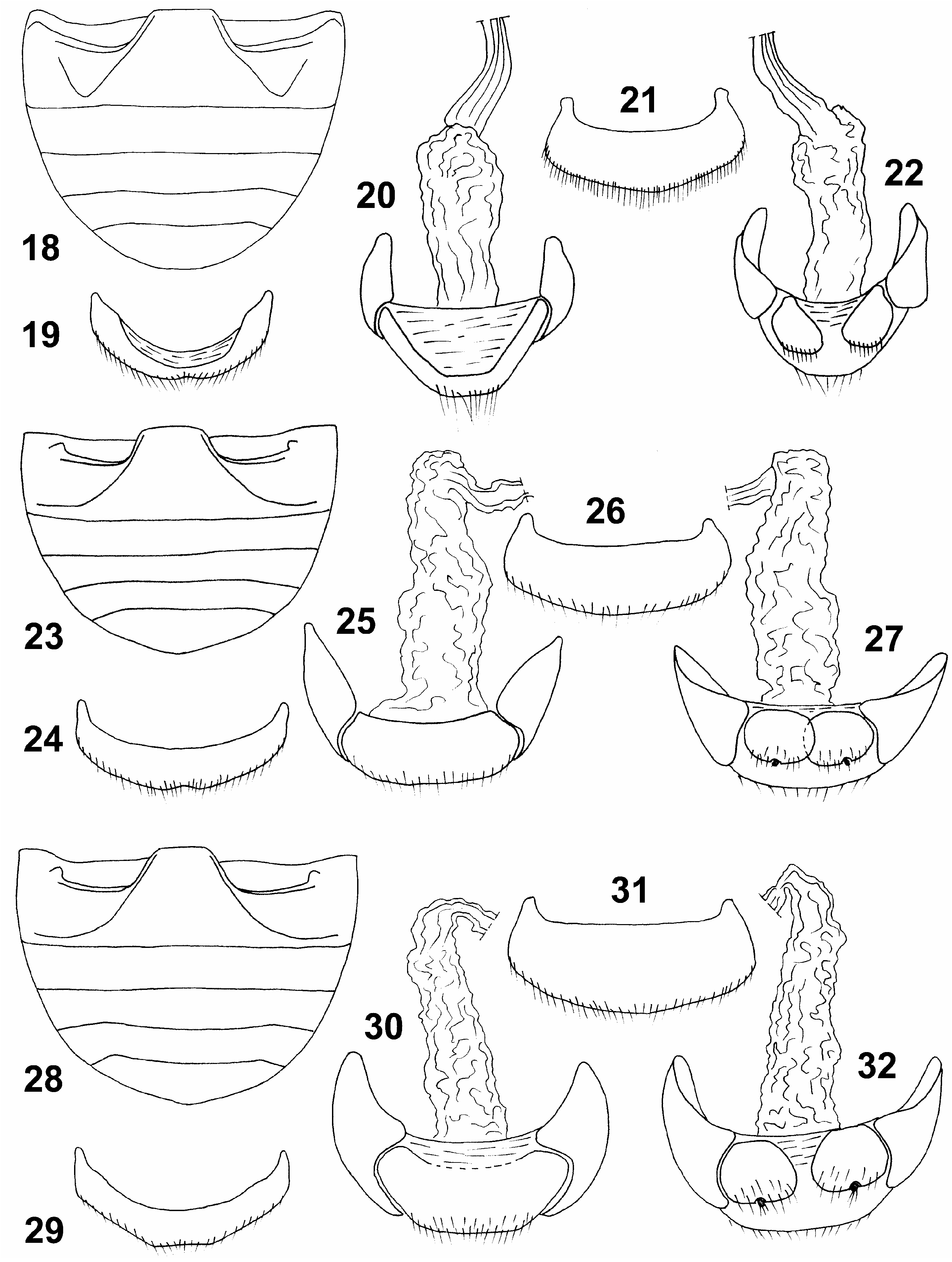
Abdomen. Abdominal postcoxal lines separated medially, V-shaped ( Fig. 18 View Figs 18–32. 18–22 ) or parallel to posterior margin of ventrite 1 ( Figs 23, 28 View Figs 18–32. 18–22 ), well developed but incomplete; without additional oblique line. Females with 5 ventrites, ventrite 5 broadly rounded; abdominal sternite VIII not divided longitudinally in middle. Female tergite 8 rounded ( Figs 21, 26, 31 View Figs 18–32. 18–22 ).
Male genitalia. Known only for P. clypealis but not examined in the present study; see figures in GORDON & ALMEIDA (1986).
Female genitalia. Coxites more or less oval in outline; ventral surface of coxite smooth. Bursa copulatrix simple, ending with common oviduct; infundibulum, sperm duct and spermatheca absent.
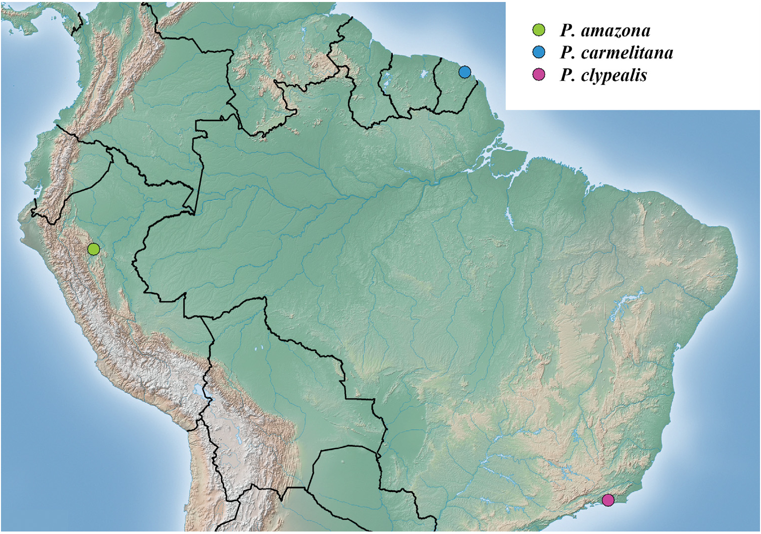
Distribution. Brazil, French Guiana, and Peru ( Fig. 36 View Fig ).
No known copyright restrictions apply. See Agosti, D., Egloff, W., 2009. Taxonomic information exchange and copyright: the Plazi approach. BMC Research Notes 2009, 2:53 for further explanation.
|
Kingdom |
|
|
Phylum |
|
|
Class |
|
|
Order |
|
|
Family |
Pseudodira Gordon, 1975
| Szawaryn, Karol 2015 |
Pseudodira
| GORDON R. D. 1975: 207 |