
Tartessus iridescens, Gnezdilov, 2020
|
publication ID |
https://doi.org/ 10.11646/zootaxa.4852.3.6 |
|
publication LSID |
lsid:zoobank.org:pub:63EF0EAF-35CE-407C-9718-FCF6ABEC067E |
|
DOI |
https://doi.org/10.5281/zenodo.4501536 |
|
persistent identifier |
https://treatment.plazi.org/id/601087DD-9E29-FFB0-FF25-FC5CF2E5FE7B |
|
treatment provided by |
Plazi |
|
scientific name |
Tartessus iridescens |
| status |
sp. nov. |
Tartessus iridescens View in CoL sp. n.
( Figs 1–18 View FIGURES 1–6 View FIGURES 7–9 View FIGURES 10–18 )
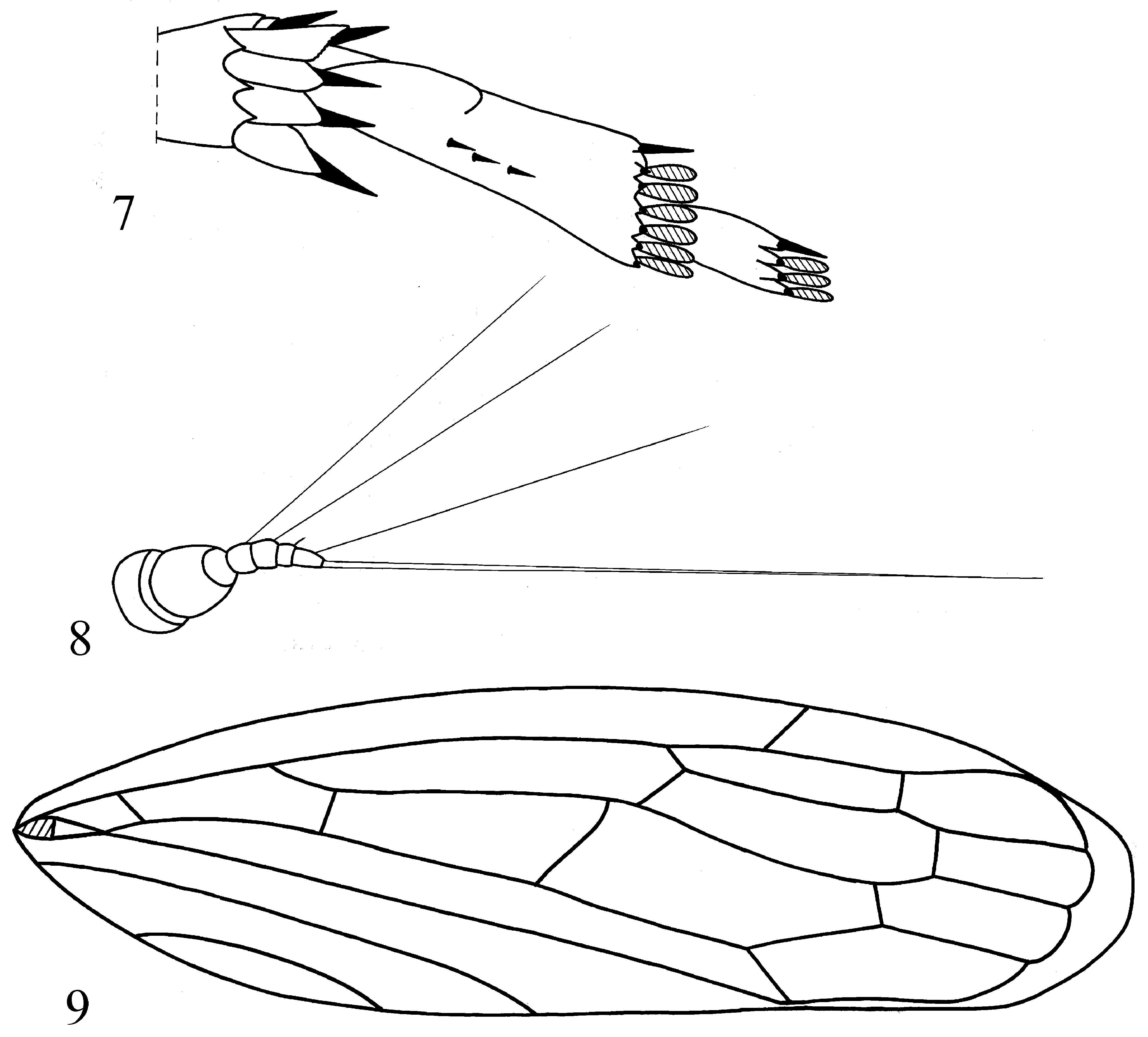
Description ( Figs 1–9 View FIGURES 1–6 View FIGURES 7–9 ). Vertex wide and very short at midline, with transverse carina on its anterior margin between ocelli ( Fig. 2 View FIGURES 1–6 ). Frontoclypeus large, cross striated in its upper part above the pedicels ( Fig. 3 View FIGURES 1–6 ). Scape short, cylindrical. Pedicel almost twice longer, barrel-shaped. First, second, and fourth segments of antennal flagellum each with long seta (more than half as long as whole flagellum); third segment with short seta ( Fig. 8 View FIGURES 7–9 ). Rostrum short, almost reaching fore coxae, only apical part of second segment and whole third segment visible. Pronotum large, with anterior margin strongly convex between eyes; cross striated medially and posteriorly. Forewings with 3 anteapical and 4 apical cells, wide appendix, and long clavus—2/3 of wing length ( Fig. 9 View FIGURES 7–9 ). Basal cell of fore wing in shape of isosceles triangle. Hind femur with 2 + 2 macrosetae apically. Hind tibia with four longitudinal rows of setae and 7 apical spines ( Figs 4 View FIGURES 1–6 , 7 View FIGURES 7–9 ). First tarsomeres of fore and middle legs with sparse macrosetae ventrally. First metatarsomere 1.5–2 times as long as second one, with keel-shaped ventral projection proximally and 2–3 short macrosetae in middle row ventrally. First metatarsomere with 7 apical spines bearing 6 platellae. Second metatarsomere with 4 apical spines bearing 3 platellae ( Fig. 7 View FIGURES 7–9 ). Arolium of pretarsus sharply and deeply notched medially (in dorsal view). Each claw with 3 long setae.
Coloration ( Figs 1–5 View FIGURES 1–6 ). Vertex light yellow greenish ( Fig. 2 View FIGURES 1–6 ). Genae black under eyes. Frontoclypeus orange yellowish, with upper part light green yellowish, with wide transverse black stripe running below ocelli ( Figs 1, 3 View FIGURES 1–6 ). Lower part of face and rostrum light yellow. Pronotum orange yellow anteriorly, with its lower angles light green. In some specimens head and pronotum generally ochre yellow, with transverse black stripe under ocelli and black genae under the eyes. Mesonotum light brown yellowish. Thoracic episterna and epimera with large black areas. Forewings light brown, with green tint. Median plate black behind the basal cell. Forewing veins dark brown to black; costal margin with black stripe in proximal part of the wing; costal cell dark in proximal half ( Fig. 1 View FIGURES 1–6 ). Hind wings greyish. Fore and middle femora light yellow. Hind femora yellow brownish. Fore and middle tibiae light yellow, with greenish distal parts. Hind tibiae light yellow greenish, with dark brown to black stripe on inner margin in its proximal half; setal basements black. Tarsomeres light brown yellowish; metatarsomeres with dark brown basal parts and spines; platellae brown. Claws dark brown to black dorsally. Abdominal sternites from light brown yellowish or light yellow (in some females) to dark brown (sternites IV–VII) in some males. Male abdominal sternite VIII sometimes with black area below hind margin. Abdominal tergites dark brown to black, with light brown yellowish hind margins. Male subgenital plates dark brown except yellowish basal parts. Male pygofer dark brown. Female pygofer yellowish, with large black area on each side; valvulae blackish, except yellowish basally ( Fig. 5 View FIGURES 1–6 ).
Male genitalia ( Figs 6 View FIGURES 1–6 , 10–18 View FIGURES 10–18 ). Anal tube wide and long, narrowing apically (in dorsal view) ( Fig. 11 View FIGURES 10–18 ), with two long, tapered processes arising basad and extended distad on each side (in lateral view) ( Fig. 10 View FIGURES 10–18 ). Pygofer lobes triangular (in lateral view), with a large horn-shaped process turned upwards at apex ( Fig. 12 View FIGURES 10–18 ). Genital valve with hind margin deeply notched medially ( Fig. 13 View FIGURES 10–18 ). Subgenital plates wide and long, gradually narrowing apically (in ventral view) ( Fig. 15 View FIGURES 10–18 ), each with short row of nearly ten thick setae apically and with hair-shaped sparse microsetae on most of ventral surface and with apical area of hair-shaped dense microsetae dorso-laterally ( Fig. 14 View FIGURES 10–18 ). Style with apical part narrowing to acute apex and with two subapical setae ( Fig. 16 View FIGURES 10–18 ). Connective short and wide. Aedeagal base strongly convex (in lateral view) ( Fig. 18 View FIGURES 10–18 ). Aedeagal shaft straight, narrow, narrowing apically, with pair of short curved apical processes extended anterolaterad; gonopore near shaft middle ( Fig. 17 View FIGURES 10–18 ).
Female genitalia ( Fig. 5 View FIGURES 1–6 ). Sternite VII hind margin sharply concave medially. Ovipositor valvulae slightly surpassing pygofer lobes (in lateral view).
Total length. Males—8.0– 9.4 mm. Females—10.0–11.0 mm.
Etymology. The species name is derived from the Latin “ iridéscens ” means iridescent and refers to the multicoloured, head, pro- and mesonotum.
Type material. Holotype: male, Vietnam, Ðồng Nai Province , Cat Tien National Park, 11˚25΄ N 107˚25΄ E, 8.VI.2012, A.F. Emeljanov leg. Paratypes: Vietnam: Ðồng Nai Province : Cat Tien National Park : 1 male, 1 female, 4. V.2012, forest, A.F. Emeljanov leg.; 1 female, 11˚25΄ N 107˚25΄ E, 8. VI.2012, A.F. Emeljanov leg.; 3 males, 9. V.2012, forest, A.F. Emeljanov leg.; 1 female, 11˚27΄ N 107˚24΄ E, 26.XI.2012, V. M. Gnezdilov leg. ; 1 male, 1 female, 13. V.2012, on light, D.E. Shcherbakov leg.; 2 females, Bau Sau Lake , 13. V.2012, D.E. Shcherbakov leg. ; Ðồng Nai Province : Nature Reserve Ma Da : 1 female, 28. V.2012, A.F. Emeljanov leg.; 2 males, Strakhov path, 20. V.2012, A.F. Emeljanov leg. ; 1 female, Strakhov path, 20. V.2012, D.E. Shcherbakov leg. ; 1 female, 22. V.2012, on light, D.E. Shcherbakov leg.; 2 males, 6 females, Strakhov path, 29. V.2012, A.F. Emeljanov leg. ; Bà R ịa-ũng Tàu Province, Binh Chau-Phuoc Buu National Park, 5 km E Bung Rieng village : 1 male, 10˚31.156΄ N 107˚28.478΄ E, 5. VI.2014; 1 male, 10˚30.314΄ N 107˚29.351΄ E, 8. VI.2014; 1 male, 10˚30.770΄ N 107˚28.860΄ E, 9. VI.2014; 1 male, 1 female, 10˚30.927΄ N 107˚28.326΄ E, 10. VI.2014, all leg. V.M. Gnezdilov .
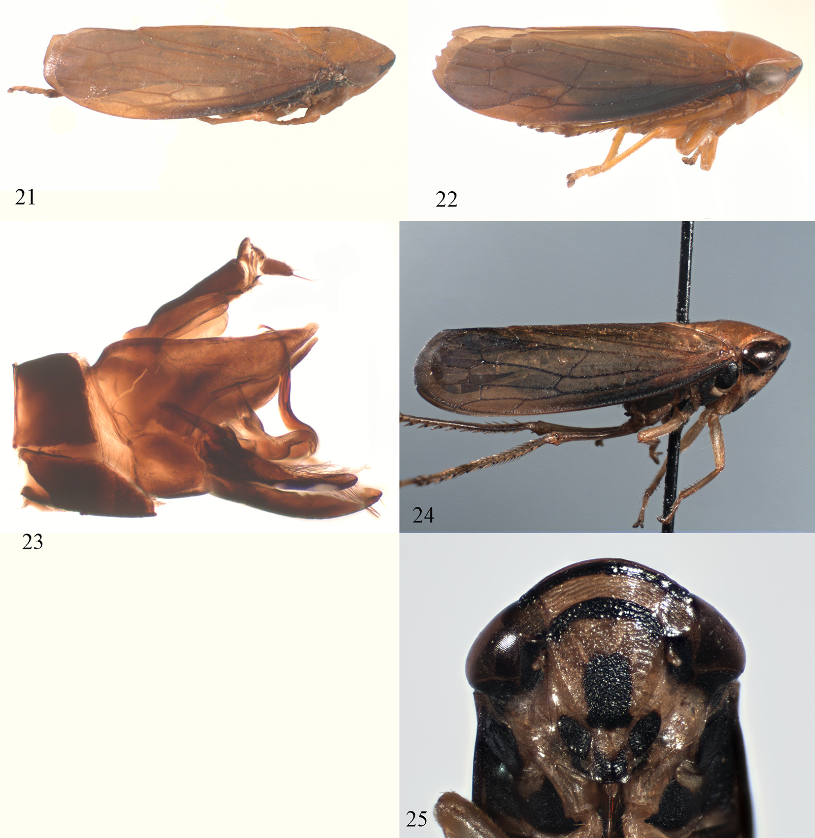
Biology. In Binh Chau-Phuoc Buu National Park the species was collected by sweeping in dry Dipterocarpus forest, often from low trees, e.g. Ficus sp., at forest edges ( Figs 19, 20 View FIGURES 19–20 ). It was collected by light trapping in Cat Tien National Park.
Distribution. Southeastern Vietnam: Ðồng Nai and Bà Rịa-ũng Tàu Provinces.
Differential diagnosis. The new species is closely related to T. malayus ( Stål, 1859) sensu Evans (1981) based on the male pygofer lobes with horn-shaped processes on the corners, the aedeagal base convex, and the aedeagal shaft with pair of short apical processes ( Evans 1981, figs 33B, 33C), but distinctly differs by the different shape of the lateral processes of the male anal tube, slender style apex, gonopore near the middle of the aedeagal shaft, and face with only one black stripe ( Fig. 3 View FIGURES 1–6 ), while T. malayus sensu Evans (1981 , fig. 33A) has several black markings on the face ( Fig. 25 View FIGURES 21–25 ).


| V |
Royal British Columbia Museum - Herbarium |
| VI |
Mykotektet, National Veterinary Institute |
| R |
Departamento de Geologia, Universidad de Chile |
No known copyright restrictions apply. See Agosti, D., Egloff, W., 2009. Taxonomic information exchange and copyright: the Plazi approach. BMC Research Notes 2009, 2:53 for further explanation.