
Selenops muehlmannorum, Jäger, Peter & Praxaysombath, Bounthob, 2011
|
publication ID |
https://doi.org/ 10.5281/zenodo.203045 |
|
DOI |
https://doi.org/10.5281/zenodo.6194484 |
|
persistent identifier |
https://treatment.plazi.org/id/5C75E043-FFBB-FFCD-FF23-1D51FD092E59 |
|
treatment provided by |
Plazi |
|
scientific name |
Selenops muehlmannorum |
| status |
sp. nov. |
Selenops muehlmannorum View in CoL spec. nov.
Figs 1–14 View FIGURES 1 – 7 View FIGURES 8 – 14
Type material. Holotype: Male, Laos, Champasak Province, Muang Pathoumphone, 2.5 km S of Pakse, Vat Phou Salao, N 15°05´38.8´´, E 105°48´34.6´´, 149 m altitude, secondary forest, dry bed of stream, rocks, by hand, at night, 23 November 2009, P. Jäger & S. Bayer leg. ( SMF).
Paratypes: 1 male, 1 female ( SMF), same data as for holotype (tissue sample for molecular analysis available—SD 696: male, two legs; SD 680: female, 1 leg).
Further material examined: 1 juvenile ( SMF), same data as for holotype.
Etymology. The species is named in honour of family Mühlmann from Germany for supporting the systematic research, description of biodiversity and nature conservation in Laos; noun in genitive case plural.
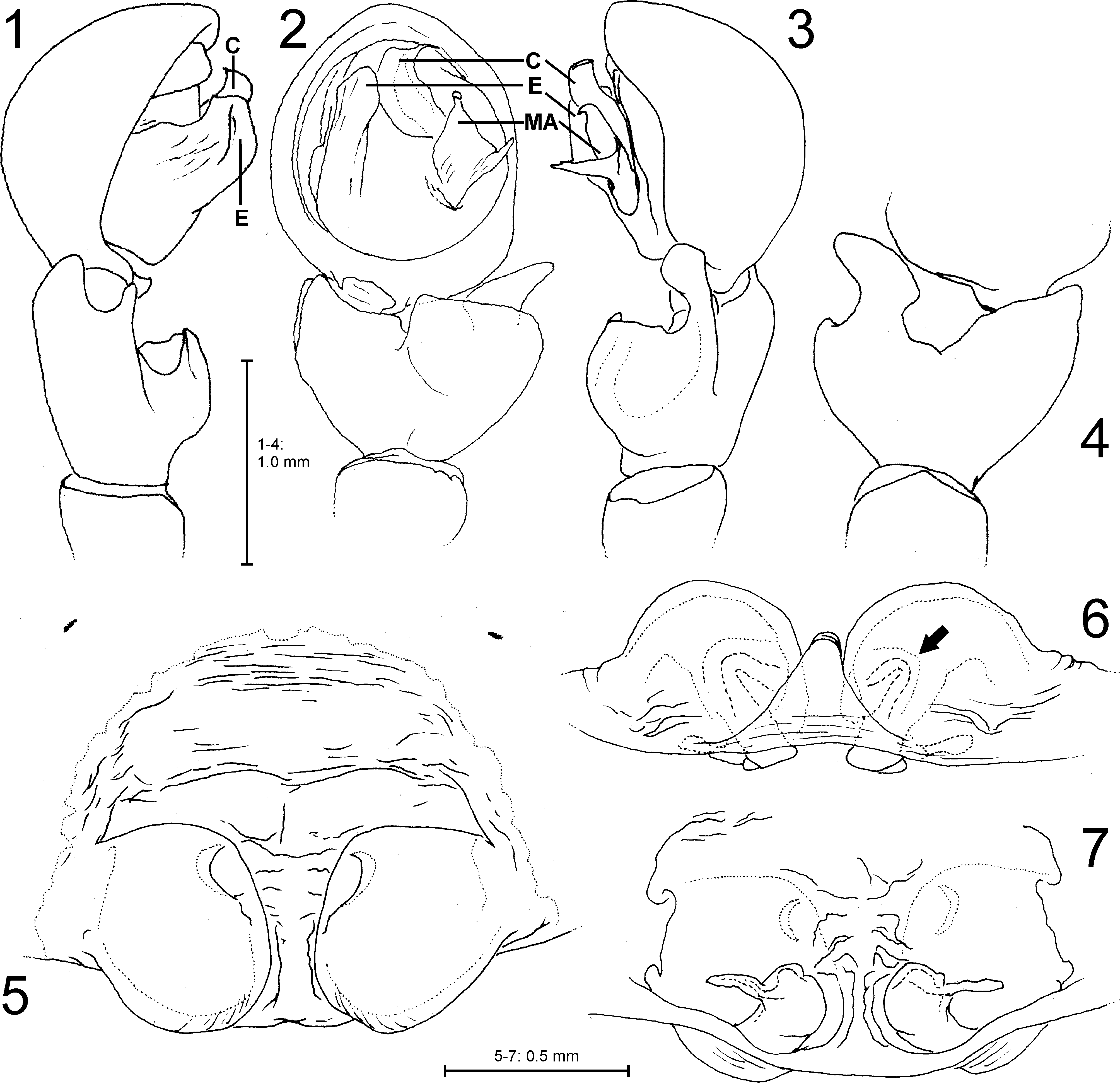
Diagnosis. Males can be recognised by having 1. a massive RTA making palpal tibia distinctly wider than long, 2. median apophysis with two apices, the distal one hook-shaped, the retrolateral one tapering, and 3. embolus broad and blunt ( Figs 1–4 View FIGURES 1 – 7 ). Female epigyne similar to that of S. ollarius Zhu, Sha and Chen, 1990 (see Zhu et al. 1990), but 1.
lateral lobes closer together and wider, 2. epigynal field wider than long and without undulated epigynal furrows ( Figs 5– 7 View FIGURES 1 – 7 , 13 View FIGURES 8 – 14 ).
Description. Male (holotype, with data of paratype in parentheses): PL 4.7 (3.7), PW 5.2 (4.3), AW 2.5 (1.9), OL 5.4 (5.0), OW 3.9 (3.3). Eye diametres: AME 0.31, ALE 0.21, PME 0.30, PLE 0.46. Eye interdistances: AME–AME 0.19, AME–PME 0.10, ALE–PLE 0.21. Leg and pedipalpus measurements: pedipalpus 5.1 (1.4, 0.8, 1.3, -, 1.6); leg I 19.9 (5.5, 2.5, 5.6, 4.4, 1.9); leg II 22.5 (6.6, 2.5, 6.6, 5.0, 1.8); leg III 22.1 (7.2, 2.3, 6.2, 4.6, 1.8); leg IV 20.0 (6.8, 2.1, 5.2, 4.2, 1.7); leg formula 2341. Spination: Femur I p110, d111, r001, II–III d111; Tibia I r110, v2221, II r100, v2220, III v1200, IV 1100; Metatarsus I v120, II v220, III v200. Chelicerae with 3 anterior and 2 posterior teeth.
Palp as in diagnosis. Tibia short, RTA with short and broad ventral part and slender dorsal branch. Cymbium subcircular. Embolus arising in 8-o’clock-position, median apophysis in 4-o’clock-position, and conductor 2-o’clockposition; the latter with concave retrolateral margin ( Figs 1–4 View FIGURES 1 – 7 ).
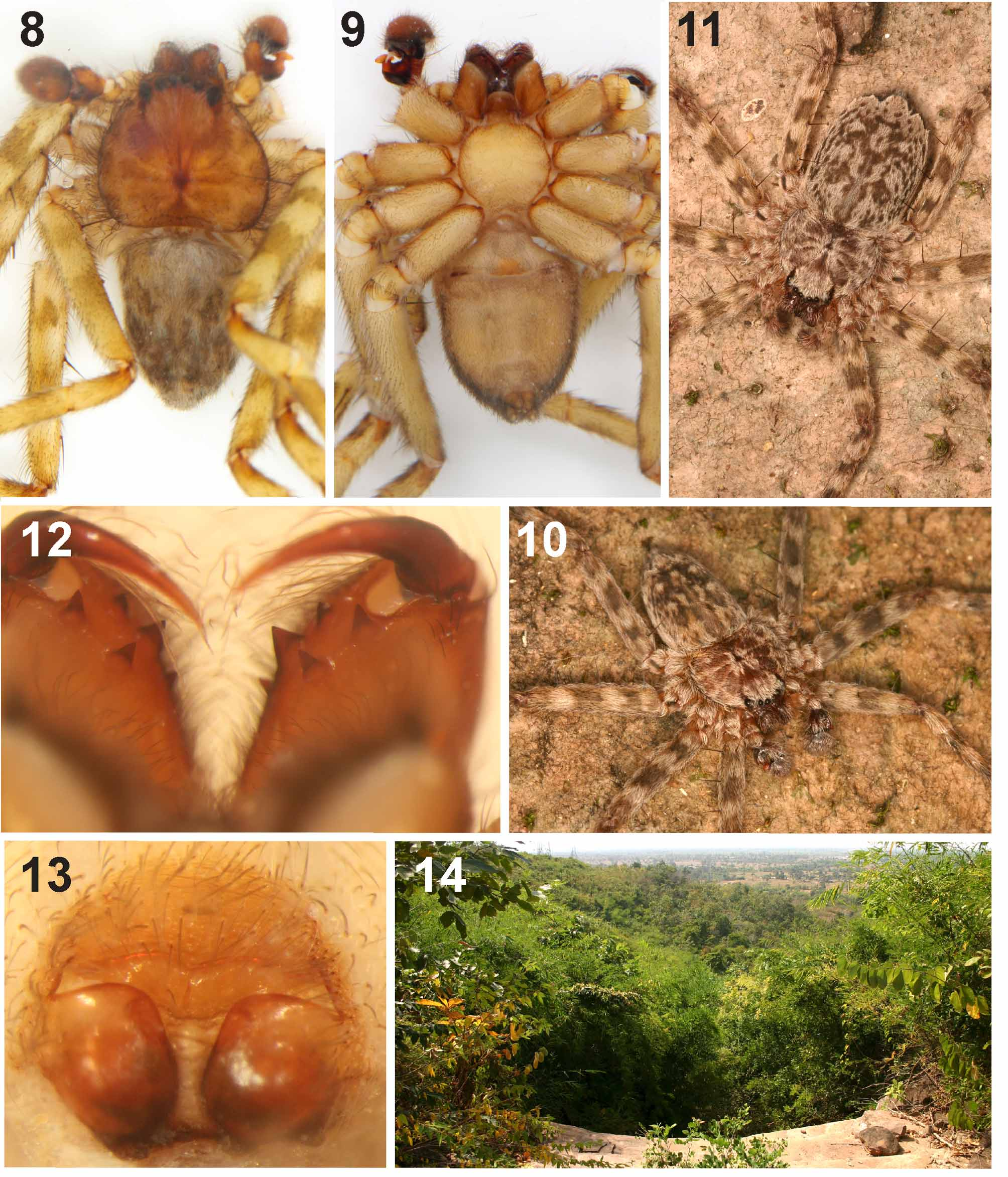
Colouration in ethanol ( Figs 8–9 View FIGURES 8 – 14 ). Dorsal prosoma light reddish brown, indistinct pattern of marginal and central patches. Ventral prosoma and opisthosoma yellowish brown without pattern. Legs yellowish brown; femora and tibiae with each two dark annulate patches, the latter not continued ventrally; distal femora with small dark patch. Dorsal opisthosoma mottled with dark and bright irregular patches. For colour pattern of living specimens see Fig. 10 View FIGURES 8 – 14 .
Female (paratype): PL 4.0, PW 4.4, AW 2.1, OL 6.3, OW 4.1. Eye diametres: AME 0.28, ALE 0.18, PME 0.29, PLE 0.40. Eye interdistances: AME–AME 0.20, AME–PME 0.06, ALE–PLE 0.24. Leg and pedipalpus measurements: pedipalpus 3.9 (1.1, 0.7, 1.0, -, 1.1); leg I 13.7 (4.1, 1.7, 4.0, 2.7, 1.2); leg II 16.3 (5.2, 2.0, 4.6, 3.1, 1.4); leg III 16.2 (5.6, 1.7, 4.4, 3.2, 1.3); leg IV 14.3 (5.1, 1.5, 3.8, 2.6, 1.3); leg formula 2341. Spination (as in male, only exceptions given): Femur I p1100, d111; Tibia I V2220, II v2220, III v1100, Metatarsus I v220, III v100. Chelicerae with 3 anterior and 2 posterior teeth ( Fig. 12 View FIGURES 8 – 14 ). Palpal claw with 11 teeth. Copulatory organ as in diagnosis. Lateral lobes each with one median pocket, and strongly arched in posterior view. Internal duct system short and simple, in posterior view with a distinct Vshaped part (arrow) ( Figs 5–7 View FIGURES 1 – 7 , 13 View FIGURES 8 – 14 ).
Colouration as in male, but pattern of dorsal prosoma more distinct and ventral opisthosoma grey. For colour pattern of living specimens see Fig. 11 View FIGURES 8 – 14 .
Natural history. The holotype of Selenops muehlmannorum spec. nov. has been collected on a wall of a staircase of a temple (Vat Phou Salao), both paratypes from rocks in a dry stream bed in a forest ( Fig. 14 View FIGURES 8 – 14 ).
Distribution. Known only from the type locality.
| SMF |
Forschungsinstitut und Natur-Museum Senckenberg |
No known copyright restrictions apply. See Agosti, D., Egloff, W., 2009. Taxonomic information exchange and copyright: the Plazi approach. BMC Research Notes 2009, 2:53 for further explanation.