
Dendromonocotyle colorni Chisholm, Whittington & Kearn, 2001
|
publication ID |
https://doi.org/10.5281/zenodo.183100 |
|
DOI |
https://doi.org/10.5281/zenodo.5670112 |
|
persistent identifier |
https://treatment.plazi.org/id/5C258787-FF85-1A64-FF49-FCE2FC63FA10 |
|
treatment provided by |
Plazi |
|
scientific name |
Dendromonocotyle colorni Chisholm, Whittington & Kearn, 2001 |
| status |
|
Dendromonocotyle colorni Chisholm, Whittington & Kearn, 2001 View in CoL ( Figs. 2 View FIGURE 2 B, 4F, 8–10)
Type-host: Himantura uarnak (Forsskål) .
Additional host: H. gerrardi (Gray) .
Type locality: Eilat Underwater Observatory, Israel.
Additional locality: uShaka Sea World, Durban, South Africa.
Site on host: Dorsal skin surface.
Materials examined: SAMCTA 29450 (7 vouchers), AHC 29171-29177 (6 vouchers), BMNH 2007.2.21.5-10 (6 vouchers).
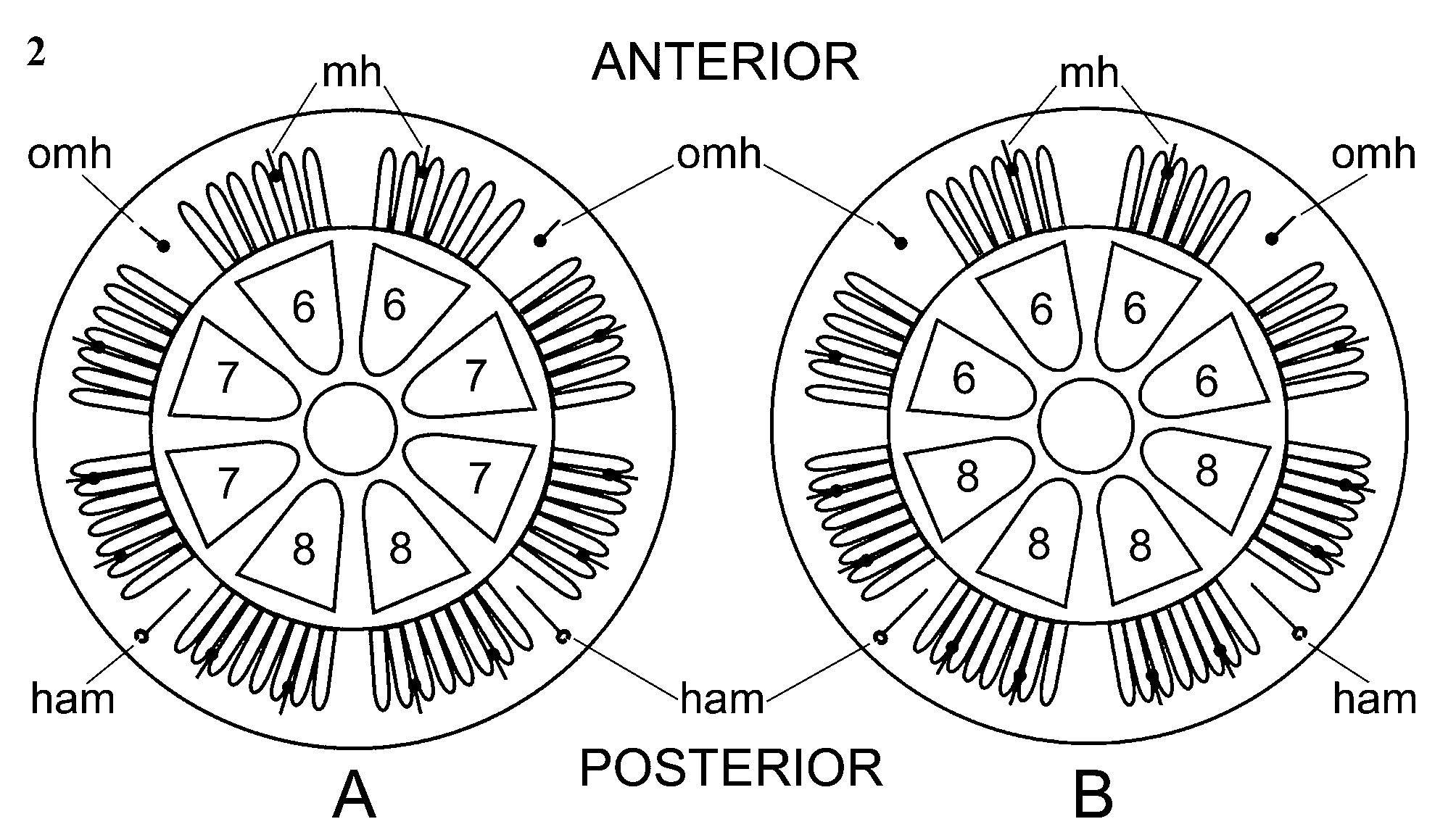
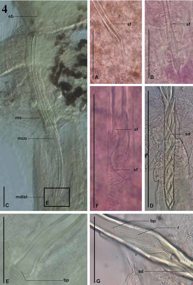
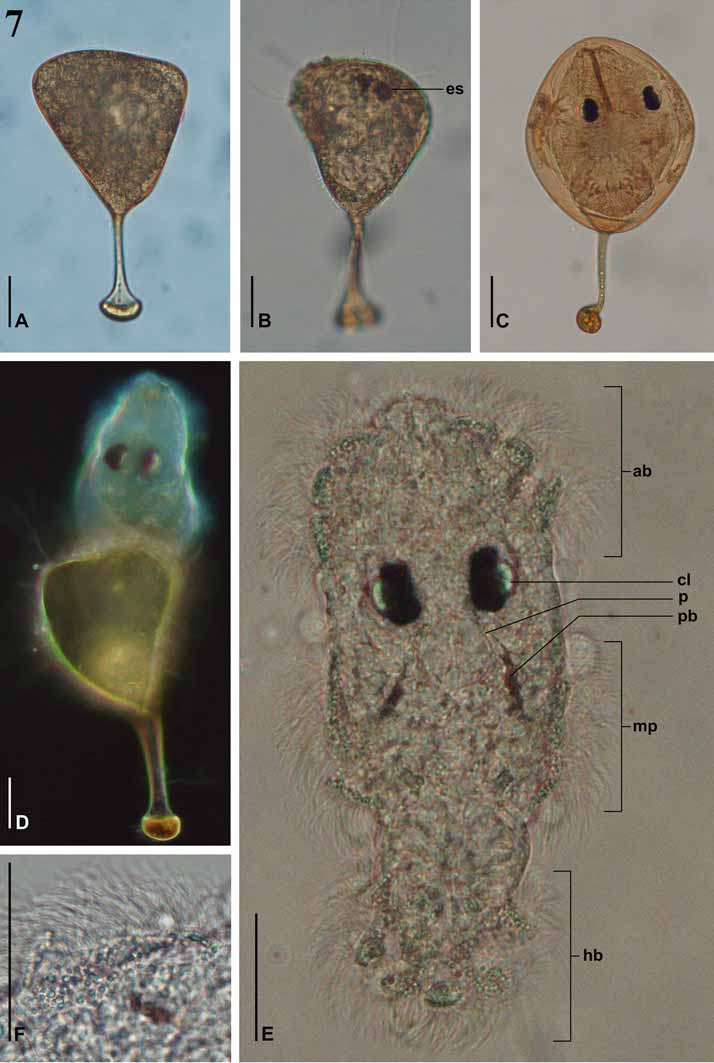
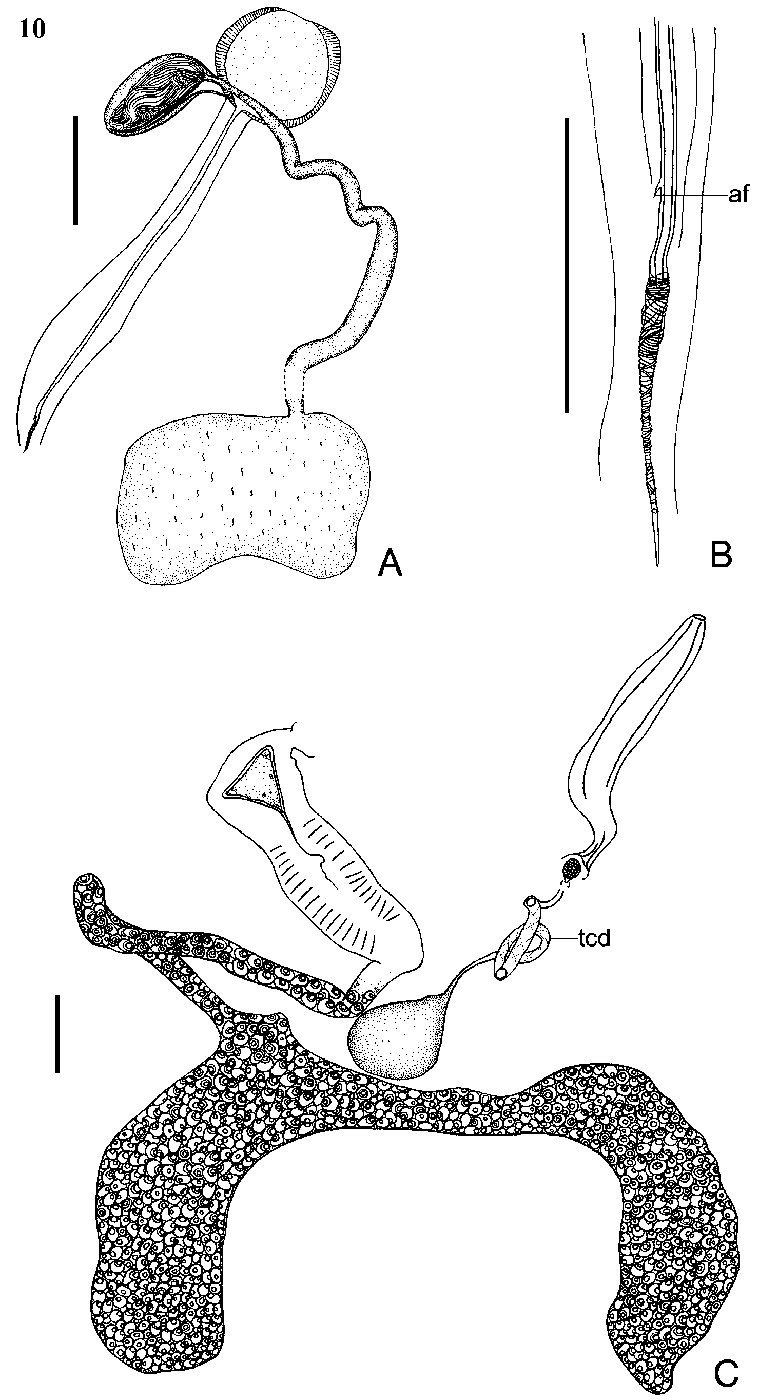
Redescription. Based on 20 flattened adult specimens. Total body length 3255 ± 532.8 (2080–4300, n = 20). Body-proper 2510 ± 485.1 (1500–3520, n = 20) long, and 2136 ± 486.3 (1150–3200, n = 20) maximum width ( Fig. 8 View FIGURE 8 A). Haptor 2195 ± 376.8 (1330–2800, n = 20) in diameter ( Fig. 7 View FIGURE 7 B), with 8 peripheral, and 1 central loculus. Hamuli present ( Figs 8 View FIGURE 8 B, 8I, 9A), 512 ± 4.6 (46–59, n = 13) long. Fourteen marginal hooklets ( Fig. 8 View FIGURE 8 J) 11 ± 1.4 (10–15, n = 13) long, distributed in marginal valve between every 4 papillae. Haptoral rim with 56 marginal haptoral papillae each armed with 3 or 4 sclerites ( Figs 8 View FIGURE 8 G, 8H, 9B, 9C). Terminal papillary sclerite as illustrated ( Figs 8 View FIGURE 8 H, 9B, 9D). Outer-ring septal sclerites distinct ( Fig. 8 View FIGURE 8 C) and radial septal sclerites ( Fig. 8 View FIGURE 8 D) larger than papillary sclerites ( Fig. 8 View FIGURE 8 G), but smaller than terminal papillary sclerite ( Fig. 8 View FIGURE 8 H). Tripartite sclerites observed at junction of radial septa and inner-ring septum and at junction of radial septa and outer ring septum ( Figs 8 View FIGURE 8 E, 8F, 9C). Anterior and anterolateral loculus pairs each with 6 associated marginal haptoral papillae. Posterolateral and posterior loculus pairs each with 8 associated marginal haptoral papillae ( Figs 2 View FIGURE 2 B, 8B). Mouth ventral, subterminal, subterminal groove anterior to mouth ( Fig. 8 View FIGURE 8 A). Six distinct anterior gland duct openings open laterally on anterior end of worm as illustrated ( Figs 8 View FIGURE 8 A, 9E). One pair of eyespots, situated dorsally to pharynx. Pharynx 286 ± 45.2 (189–368, n = 20) long, and 179 ± 26.7 (129–229, n = 20) wide. Dendritic intestinal caecum anterordorsal to pharynx, extending to both posterior marginal lobes of body proper ( Fig. 8 View FIGURE 8 A). Intestinal caecum highly pigmented. Testis singular with vas deferens originating from the left side of testis, dorsal to the ovary. Testis 231 ± 36 (170–280, n = 8) long, and 454 ± 71.6 (287–620, n = 20) wide. Vas deferens extends anterodorsally to vagina, with 2 small loops, 1 on each side of vagina. Vas deferens crosses ventrally over posterior portion of ejaculatory bulb and swells to form seminal vesicle, which narrows and enters ejaculatory bulb distally. Ejaculatory bulb 179 ± 30 (136–231, n = 20) long, and 194 ± 30.6 (143–235, n = 20) wide. Male copulatory organ sclerotised ( Fig. 10 View FIGURE 10 A), 804 ± 31.9 (751–852, n = 18) long, 9 ± 0.5 (9–10, n = 20) wide. Accessory flange present at distal end of male copulatory organ ( Figs 4 View FIGURE 4 F, 10B). Muscular sheath 57 ± 8.8 (50–75, n = 18) wide, covers entire length of male copulatory organ ( Fig. 10 View FIGURE 10 A). Ovary bi-lobed 360 ± 53.4 (240–430, n = 14) long, and 715 ± 139.6 (420–980, n = 16) wide, immediately anterior to testis with lobes extending to either side of testis ( Figs 8 View FIGURE 8 A, 10C). Single ovarian branch loops right intestinal caecum dorsoventrally. Ootype 314 ± 54.4 (212–387, n = 20) long, ventral to male copulatory organ. Tetrahedral egg ( Fig. 10 View FIGURE 10 C) side length 74 ± 10.3 (55–90, n = 7); filament 158 ± 32.3 (110–210, n = 9) long extending from 1 pole of egg. Mehlis glands not observed. Vaginal pore on left side of body at level of anterior portion of ejaculatory bulb ( Fig. 8 View FIGURE 8 A). Vagina narrows to form distinctly coiled translucent duct, which narrows to enter ovoid seminal receptacle anterior to ovary ( Fig. 10 View FIGURE 10 C). Seminal receptacle 141 ± 24.4 (105– 175, n = 13) long, and 106 ± 15.2 (87–140, n = 13) wide. No spermatophores observed. Vitellaria obscured by pigmented intestinal caecum.
Remarks. All stingrays in the exhibit were collected from immediate local wild population in Durban Bay in 2003. Dendromonocotyle colorni was only observed on the hosts when an outbreak occurred months later in the aquarium exhibit. No local wild populations of H. uarnak or H. gerrardi have been examined for D. colorni . Furthermore, we could not determine whether the infection of D. colorni on H. uarnak and H. gerrardi represents a natural association between closely related hosts, or an artificially created host-parasite interaction between similar host species under lower-than-normal spatial arrangements and increased stress levels in captivity.
The D. colorni collected in South Africa differ slightly from those collected in Israel but we do not deem these minor differences to be enough to warrant erection of a separate species. The marginal haptoral papillae of the South African material are sometimes armed with 4 sclerites (including the terminal papillary sclerite) whereas the specimens from Israel only have 3. Tripartite sclerites were found at the junction of the radial septum and the outer-ring septum as well as inner-ring septum ( Figs 8 View FIGURE 8 E, 8F, 9C) in the South African specimens whereas tripartite sclerites were only seen at the junction of the radial septa and inner ring septum from those specimens from Israel ( Chisholm et al. 2001a).
We only found 1 pair of eyespots the South African specimens, not 2, as indicated by Chisholm et al. (2001a), however, it is likely that the second pair was obscured by the dark pigment in the intestinal caeca. The vas deferens in the South African specimens crosses the posterior end the ejaculatory bulb not the anterior end as illustrated by Chisholm et al. (2001a) for specimens from Israel.

No known copyright restrictions apply. See Agosti, D., Egloff, W., 2009. Taxonomic information exchange and copyright: the Plazi approach. BMC Research Notes 2009, 2:53 for further explanation.
|
Kingdom |
|
|
Phylum |
|
|
Class |
|
|
Order |
|
|
Family |
|
|
Genus |