
Spegazzinia camelliae N. Suwannarach, J. Kumla & S. Lumyong, 2021
|
publication ID |
https://doi.org/ 10.11646/phytotaxa.483.2.4 |
|
persistent identifier |
https://treatment.plazi.org/id/5C1087BB-FFEA-1308-23AF-545BFAFEFE34 |
|
treatment provided by |
Marcus |
|
scientific name |
Spegazzinia camelliae N. Suwannarach, J. Kumla & S. Lumyong |
| status |
sp. nov. |
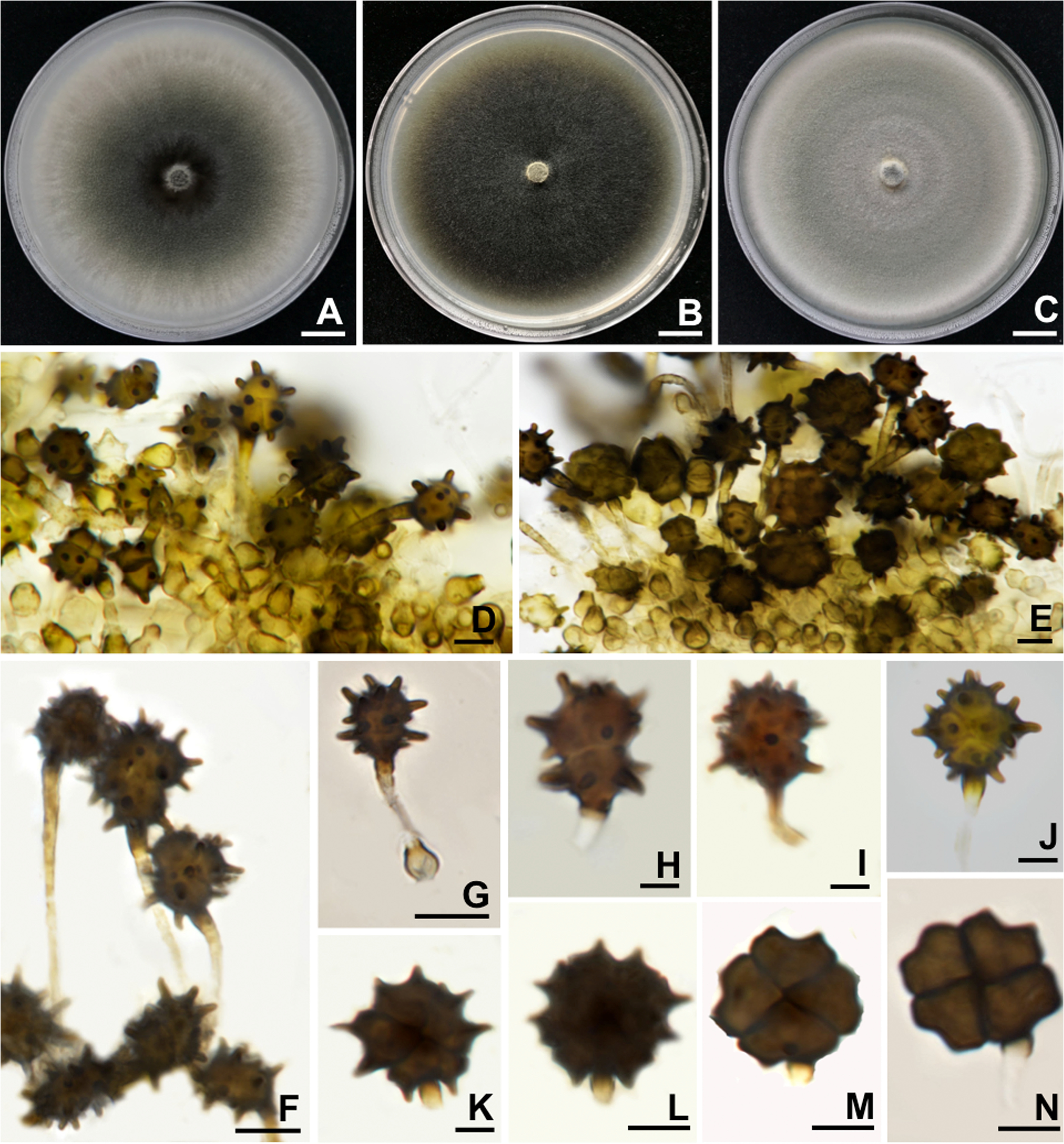
Spegazzinia camelliae N. Suwannarach, J. Kumla & S. Lumyong View in CoL , sp. nov. Figure 2 View FIGURE 2 .
MycoBank number: 837969
Facesoffungi number: FoF 09467
Etymology: The specific epithet “ camelliae ” refers to generic name of the host plant, Camellia .
Holotype: TBRC13888 View Materials
Endophytic associated with leaves of Camellia sinensis var. assamica . Sexual morph: Undetermined. Asexual morph: Hypomycetous. Conidia forming were observed on PDA, MEA and OA within two weeks. Mother cells of conidiophores doliiform or subspherical, brown and 5–7 × 3–4.5 μm ( Fig. 2 View FIGURE 2 , D, E). Conidiophores secession basauxic, usually short to long bearing two types of conidia referred to here as α and β types. Conidiophores of α conidia up to 15–50 × 2–2.5 μm, pale brown or dark golden brown, rough-walled, hyaline at bottom near the conidiophore mother cell, pale brown at middle, dark golden brown at top near conidial cells, erect or flexuous, narrow and long, generally unbranched but rarely branched. Conidiophores of β conidia 5–7.5 × 2–2.5 μm short, erect, unbranched, hyaline. Conidiogenous cells holoblastic at the apex of conidiophore. Conidia of two kinds: α conidia 4-celled, subglobose, brown to dark brown, 9–15 × 8–12 μm, with conspicuous spines 2–3.5 μm long, scattered ( Fig. 2 View FIGURE 2 , F – J); β conida 9–15 × 6–14 μm, 4-celled, pale brown to dark brown, subglobose, flattened in one plane, cuciately septate, smooth ( Fig. 2 View FIGURE 2 , K – N) .
Culture characteristics:— Colonies growing on PDA, MEA and OA reached a diameter of 45−50, 50−55 and 45−55 mm after one week at 25°C ( Fig. 2 View FIGURE 2 , A−C). Colonies on PDA and MEA appear to be raised, moderately dense with undulated margins, middle grey, periphery brownish grey and olive green at the immature stage; and reverse white to greyish white. Colonies on OA were greyish white, unevenly raised with a rough surface, moderately dense, radially striated at the center with crenulated margins and reverse white to greyish white. Hyphae hyaline when young and becoming brown to dark brown with age, smooth to slightly verruculose, 2–3 μm wide.
Material examined:— THAILAND, Skad, Pua District , Nan Province, 19°15’42”N 101°0’16”E, elevation 994 m), isolated as an endophyte from leaves of Camellia sinensis var. assamica , September 2017, N GoogleMaps . Suwannarach, dried culture: SDBR-CMU328 .
Note:— Spegazzinia camelliae is distinguished from S. intermedia , S. parkeri Sivasith. (1974: 427) , S. subbramanianii ( Bhat 1994: 131) and S. xanthorrhoeae Subram (1988: 67) which only produce only β conidia ( Table 2) ( Ellis 1971; Sivasithamparam 1974; Bhat 1994; Whitton et al. 2012). The production of the 8-celled α conidia in S. cruciata Whitton, K.D. Hyde & McKenzie (2012: 290) separates it from S. camelliae ( Whitton et al. 2012) . The α conidia in S. camelliae (9–15 × 8–12 µm) are smaller than in S. affinis J. Mena & T. Cantillo (2017: 295) (16–22 × 12–24 µm), S. deightonii (18–28 × 17–29 µm), S. musae Samarakoon, Phookamsak, Wanas., Chomnunti & K.D. Hyde (2020: 27) (15–22.7 × 14.5–20.5 µm) and S. radermacherae (18–22 × 17.5–20 µm) ( Ellis 1971; Hughes 1953b; Mena-Portales et al. 2017; Jayasiri et al. 2019; Samarakoon et al. 2020). The larger size of the β conidia in S. affinis , S. bromeliacearum , S. flabellata S.M. Leão & Gusmão (2010: 414) , S. lobulata Thrower (1954: 362) , S. radermacherae and S. sundara Subram. (1956: 78) can be used to distinguish them from our new species ( McLennan et al. 1954; Subramanian 1956; Leão-Ferreira & Gusmão 2010; Mena-Portales et al. 2017; Crous et al. 2019; Jayasiri et al. 2019) ( Table 2). The α and β conidia in S. camelliae are similar to S. neosundara Thambug. & K.D. Hyde (2017: 724) and S. tessarthra . However, the spines on the surface of the S. camelliae (2–3.5 µm long) are shorter than S. neosundara (2–8 µm long) and S. tessarthra (up to 10 µm long) ( Saccardo 1886; Whitton et al. 2012; Thambugala et al. 2017).
| E |
Royal Botanic Garden Edinburgh |
| F |
Field Museum of Natural History, Botany Department |
| J |
University of the Witwatersrand |
| K |
Royal Botanic Gardens |
| N |
Nanjing University |
No known copyright restrictions apply. See Agosti, D., Egloff, W., 2009. Taxonomic information exchange and copyright: the Plazi approach. BMC Research Notes 2009, 2:53 for further explanation.