
Pseudolyciella pallidiventris (Fallén 1820), Fallen, 1820
|
publication ID |
https://doi.org/10.11646/zootaxa.3780.3.1 |
|
publication LSID |
lsid:zoobank.org:pub:170F4E3F-847E-48F7-AF68-AA0E4BC7936A |
|
DOI |
https://doi.org/10.5281/zenodo.6131195 |
|
persistent identifier |
https://treatment.plazi.org/id/501F0803-ED12-2E24-FF53-FB61FAB2239A |
|
treatment provided by |
Plazi |
|
scientific name |
Pseudolyciella pallidiventris (Fallén 1820) |
| status |
|
Pseudolyciella pallidiventris (Fallén 1820)
The living larvae soft and translucent, with marked welts and crevices. The third instar larvae large and dorsoventrally flattened; massive lobes arises from second and third thoracic and abdominal segments. Distal parts of Malpighian tubules thin, filled with white matter.
Egg ( Figs 18–20 View FIGURES 15 – 23 ). Length 0.78–0.81 mm. The shape of egg asymmetrical from lateral view, with the micropylar pole up-turned. The posterior tubercle small, with few openings. The chorion with numerous ridges; towards the egg's poles the ridges become exaggerated with zig-zag margin; the transversal ribs indicated; small pits between the ribs and ridges sometimes indicated.
First instar ( Figs 105–108 View FIGURES 105 – 110 ). Length 1.26–2.37 mm. The peristomal cirri very slender and long. The ventral organ with two digitiform processes, the lobe laterally to the processes reduced. The integument of body segments covered by fine wrinkles, ventral side smooth. The thoracic segments without spines. The abdominal segments with spinous creeping welts, but the spines on dorsal creeping welts flat and triangular, resembling folds of the skin rather than spines. Only one (lateral) pair of processes developed on the anal division. Sprouts of the posterior spiracles short, compact; the peristigmatic tufts elongated.
Cephaloskeleton ( Fig. 171 View FIGURES 168 – 173 ). Length 0.31–0.33 mm. The distal part of mouth hooks with 5–7 teeth; stalk parts hyaline, slender and closely adherent one to another. The epistomal sclerite elongated, two times as long as broad. The ventral cornua sclerotised along dorsal margin. Other features as in previous species.
Second instar ( Figs 109, 110 View FIGURES 105 – 110 ). Length 2.19–3.58 mm.
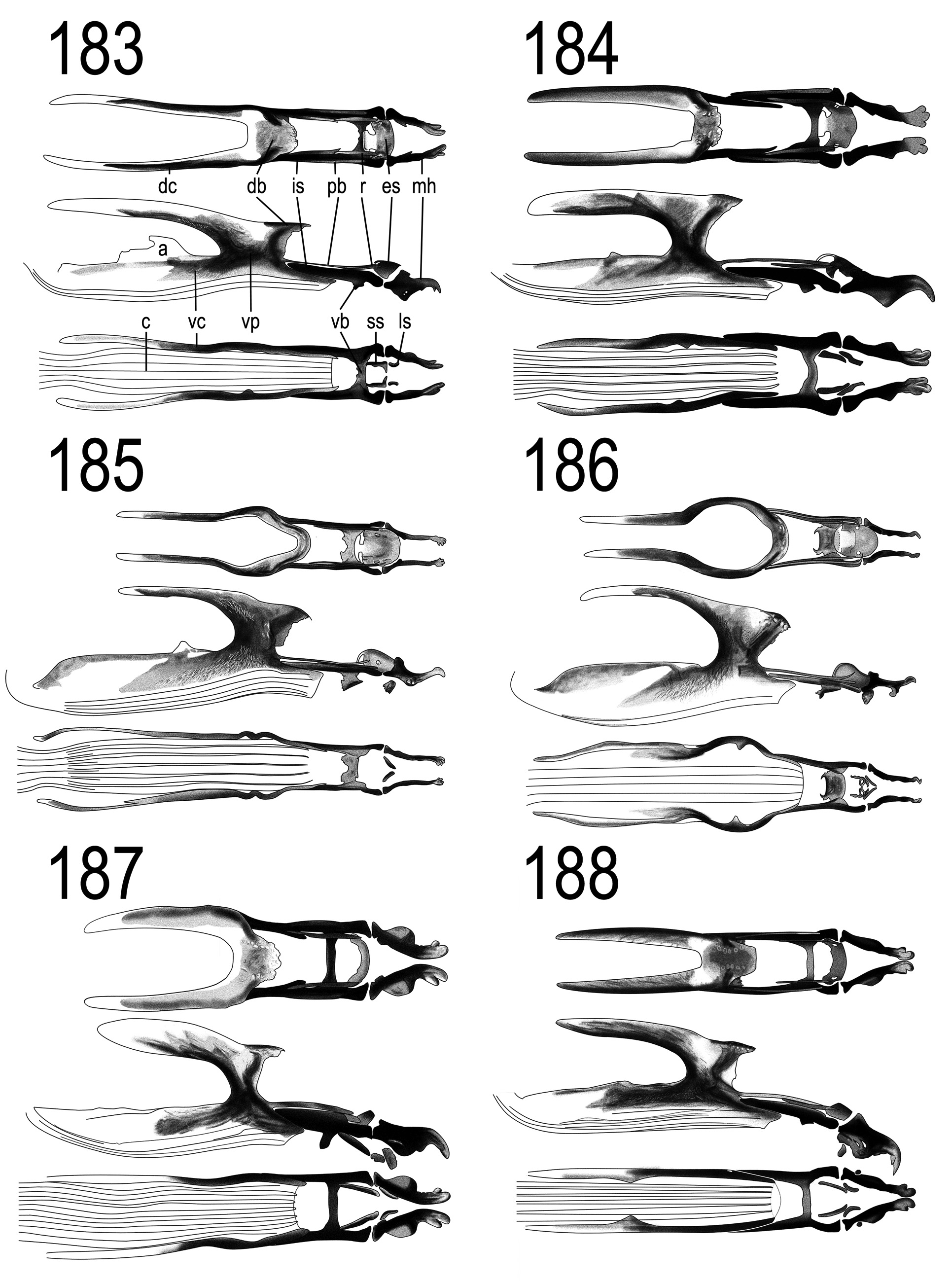
Cephaloskeleton ( Fig. 186 View FIGURES 183 – 188 ). Length 0.51–0.53 mm. Mouth hooks very slender, basal parts converging, otherwise parallel; tips simple. The epistomal sclerite longer than broad, rounded on anterior margin, convex. Subhypostomal sclerites peculiar, resembling eyeglasses in ventral view. Intermediate sclerite slender. Ventral bridge very broad, with two corners on posterior edge. Parastomal bars ascend apically. Dorsal bridge and dorsal cornua narrow in whole length. Ventral cornua broad, dorsal margin sclerotised and convex. Cibarium hyaline.
Third instar (pictures were of low quality and were not included in the present work). Length 3.35–5.37 mm. The facial mask extensive, broader than long (up to 45 scraping cirri per row). The peristomal cirri well developed, long. The ventral organ with long and slender two apical outgrowths. The thoracic segments ventrally smooth, but the ventral collar with unusually long spines. The laterals of body segments finely villous.
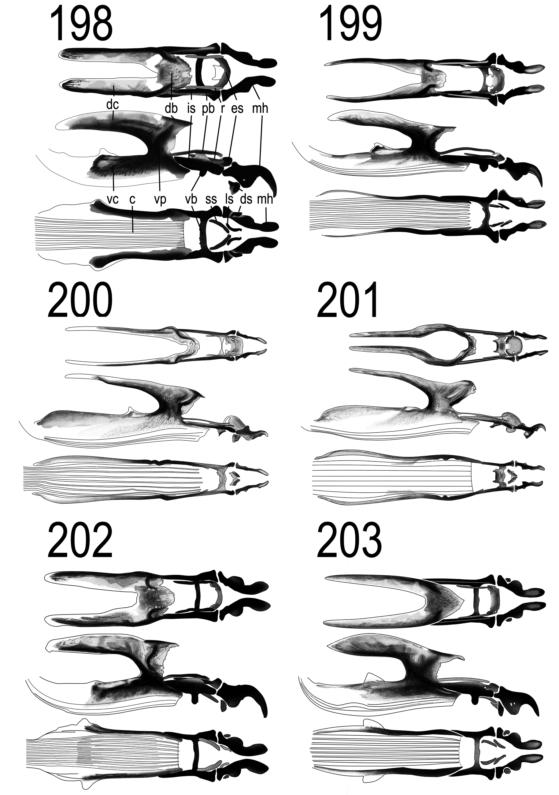
Cephaloskeleton ( Fig. 201 View FIGURES 198 – 203 ). Length 0.89 mm (n=1). Mouth hooks weak, slightly converging. Intermediate sclerite slender, ventral bridge broad with two corners on posterior edge. Epistomal sclerite rounded, as long as broad. Subhypostomal sclerites resemble eyeglasses. Parastomal bars slender, apical part ascends. Dorsal bridge and dorsal cornua narrow. Ventral cornua broad, dorsal margin sclerotised and convex. Cibarium hyaline.


No known copyright restrictions apply. See Agosti, D., Egloff, W., 2009. Taxonomic information exchange and copyright: the Plazi approach. BMC Research Notes 2009, 2:53 for further explanation.
|
Kingdom |
|
|
Phylum |
|
|
Class |
|
|
Order |
|
|
Family |
|
|
Genus |