
Eupolyphaga Chopard, 1929
|
publication ID |
https://doi.org/ 10.11646/zootaxa.4506.1.1 |
|
publication LSID |
lsid:zoobank.org:pub:6F70EE34-FCD0-4426-958E-F734994225F3 |
|
DOI |
https://doi.org/10.5281/zenodo.5978874 |
|
persistent identifier |
https://treatment.plazi.org/id/4A1B87C9-9E17-1945-FF3C-F8D1FBF1FE61 |
|
treatment provided by |
Plazi |
|
scientific name |
Eupolyphaga Chopard, 1929 |
| status |
|
Genus Eupolyphaga Chopard, 1929 View in CoL
Eupolyphaga Chopard, 1929: 261 View in CoL ; Bey-Bienko 1950: 283; Princis 1962: 53; Feng, Guo & Woo 1997: 165.
Type species: Polyphaga sinensis Walker, 1868
Diagnosis. Eupolyphaga may be confused with Polyphaga , they are sympatric distributed in north and central China. However, the former can be readily distinguished from the latter by the following characteristics: 1) male tegmina with Sc swelling, while tegmina without Sc swelling in Polyphaga ; 2) male tegmina usually maculated, rarely unicolored, CuP in cfr not whitish, while tegmina unicolored, CuP in cfr always distinctly whitish in Polyphaga ; 3) L1 with two well sclerited long processes posteriorly, while L1 with two small hyaline processes posteriorly in Polyphaga ; 4) R1L slender, sclerited, while R1L indistinct in Polyphaga ; 5) female usually small, body length at most 27.8 mm, unicolored or maculated, while female large, body length 27.0–40.0 mm, usually unicolored, pronotum with whitish margin anteriorly (except Polyphaga plancyi ).
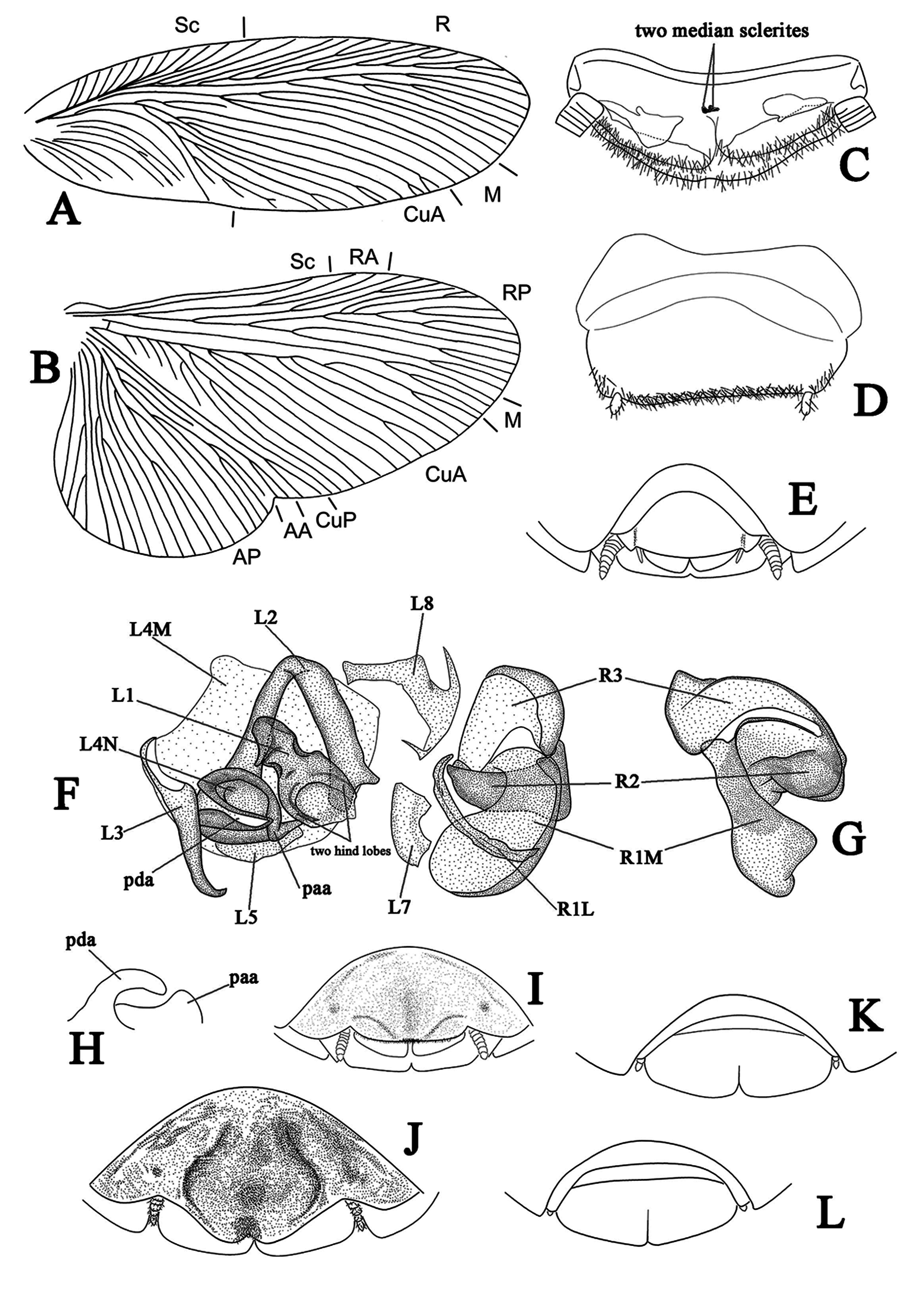
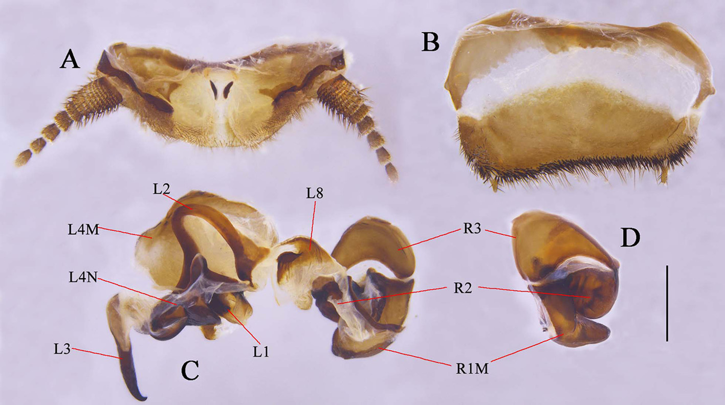
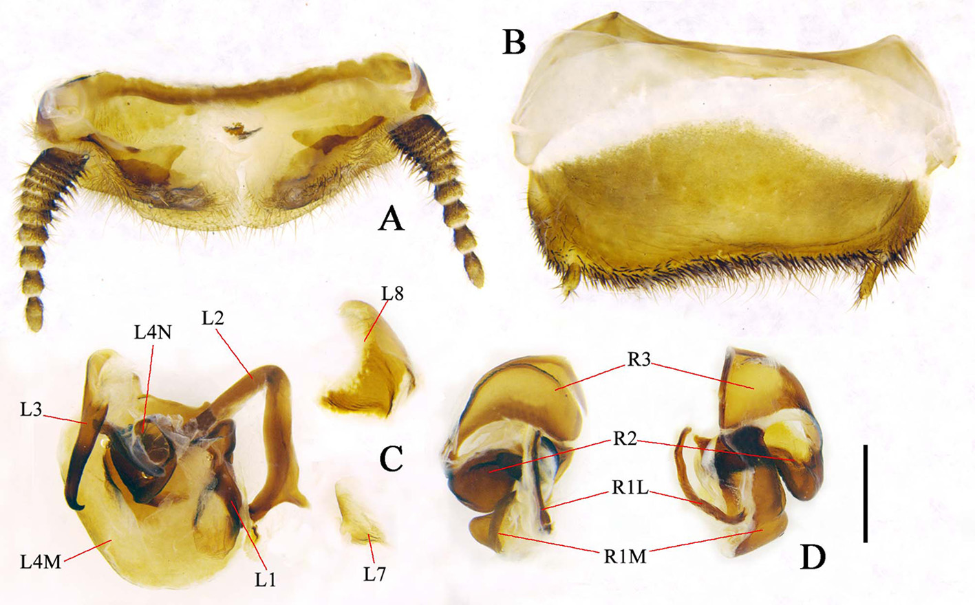
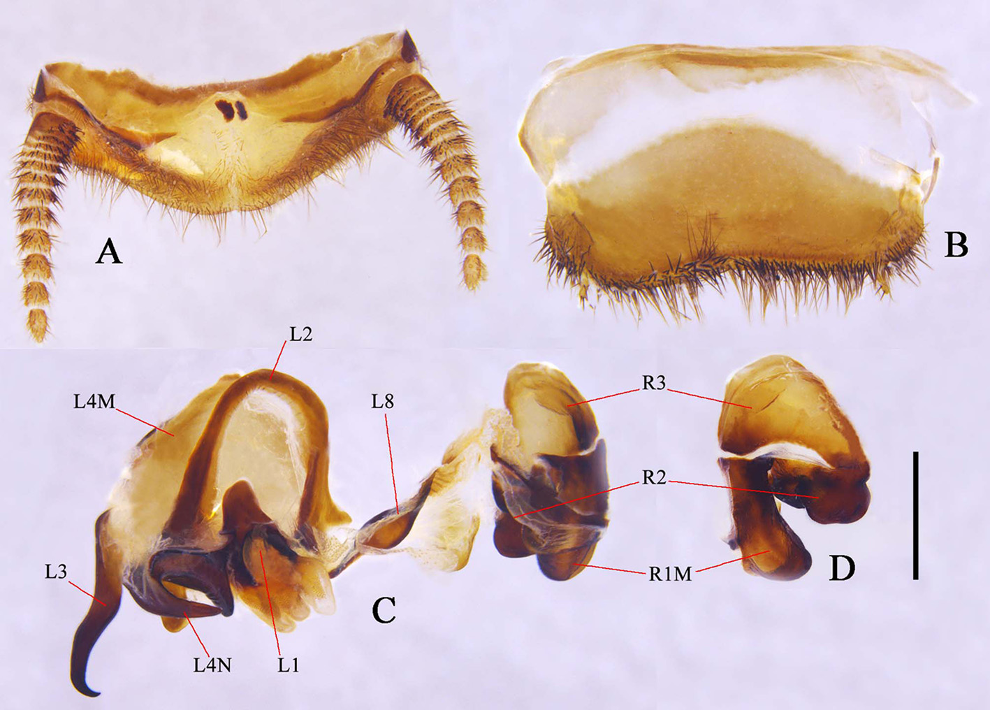
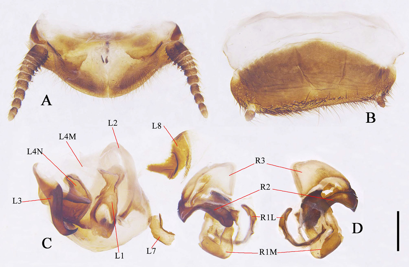
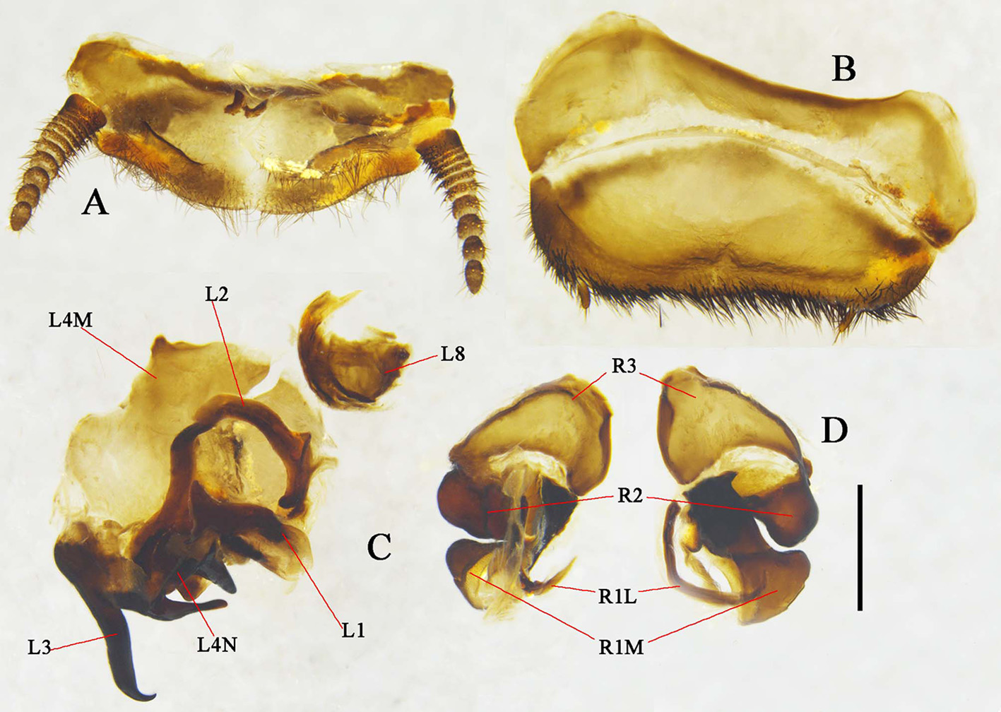
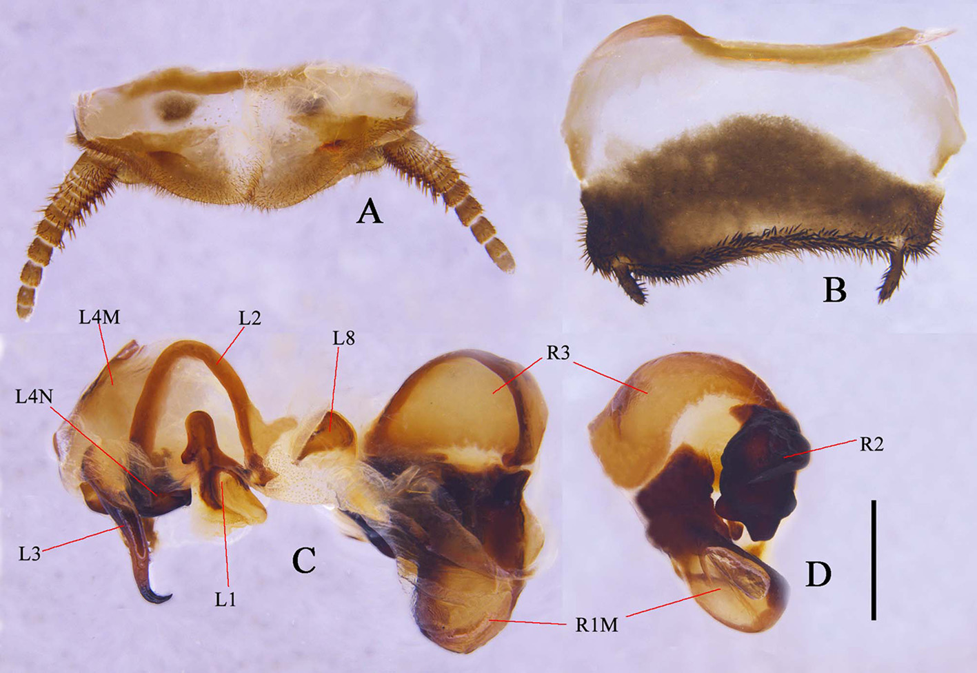
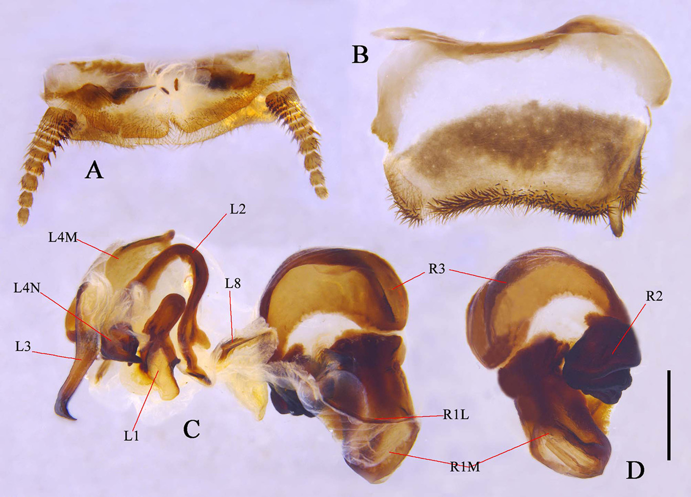
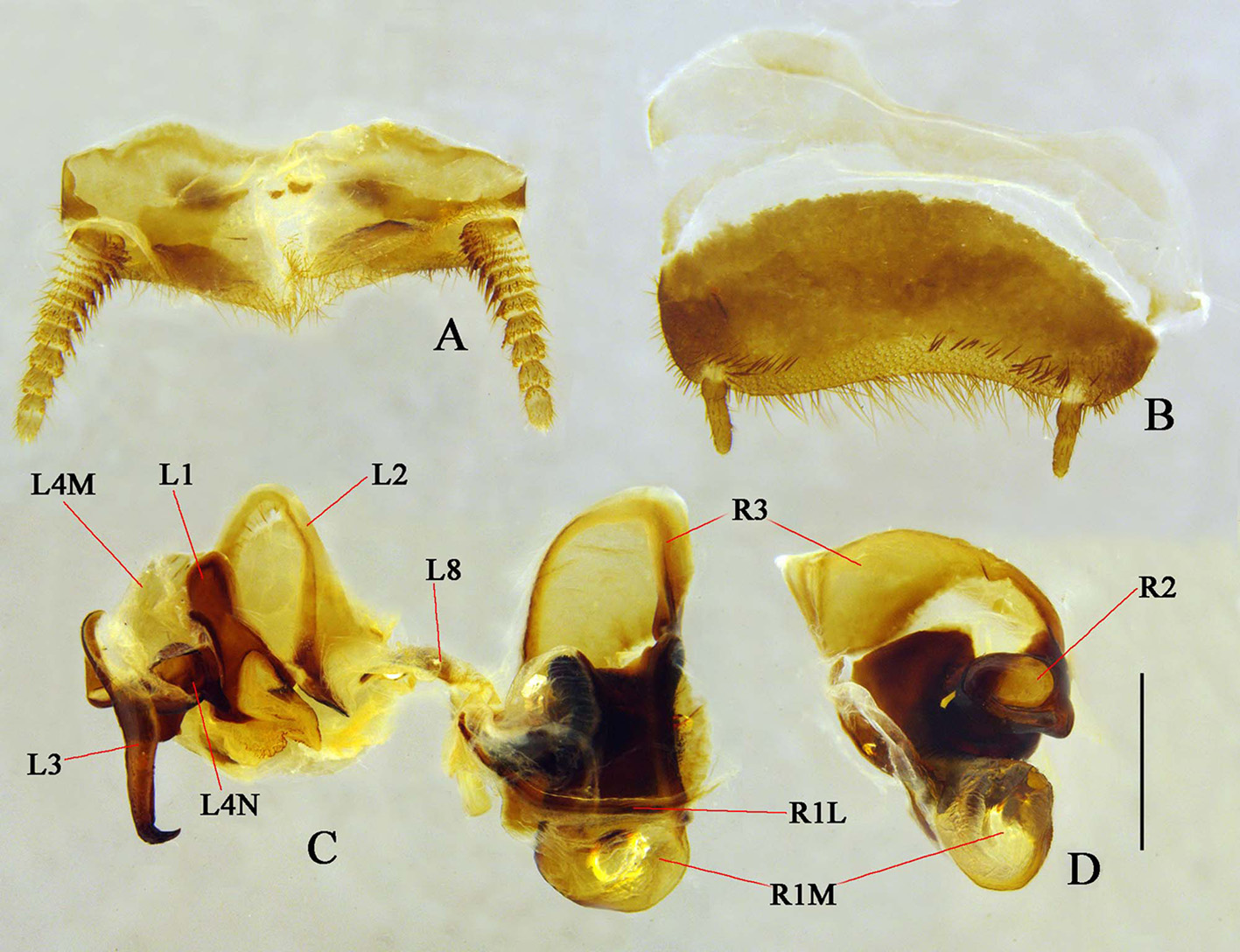
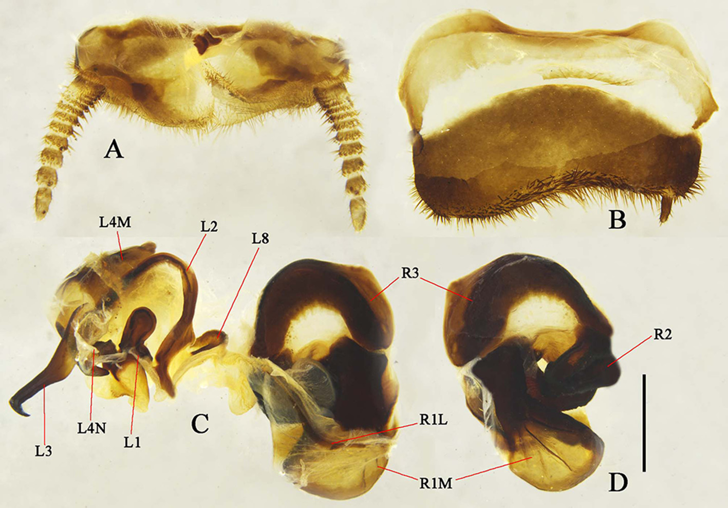
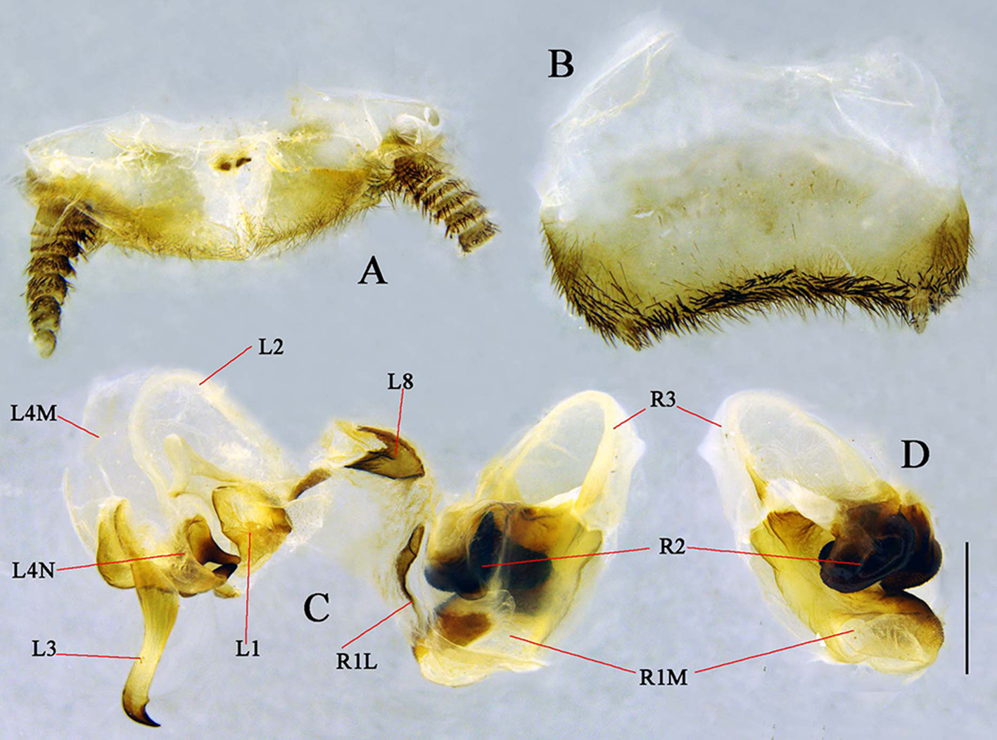
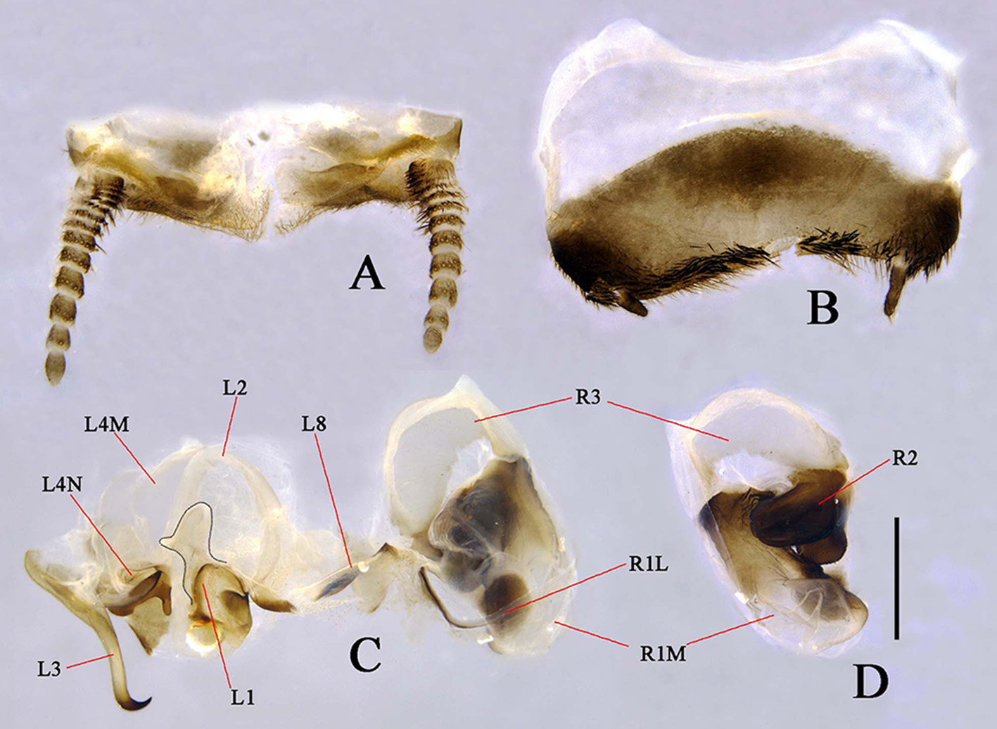
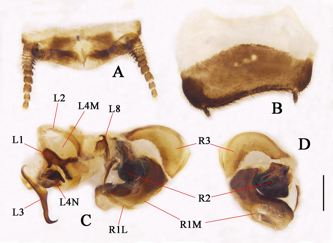
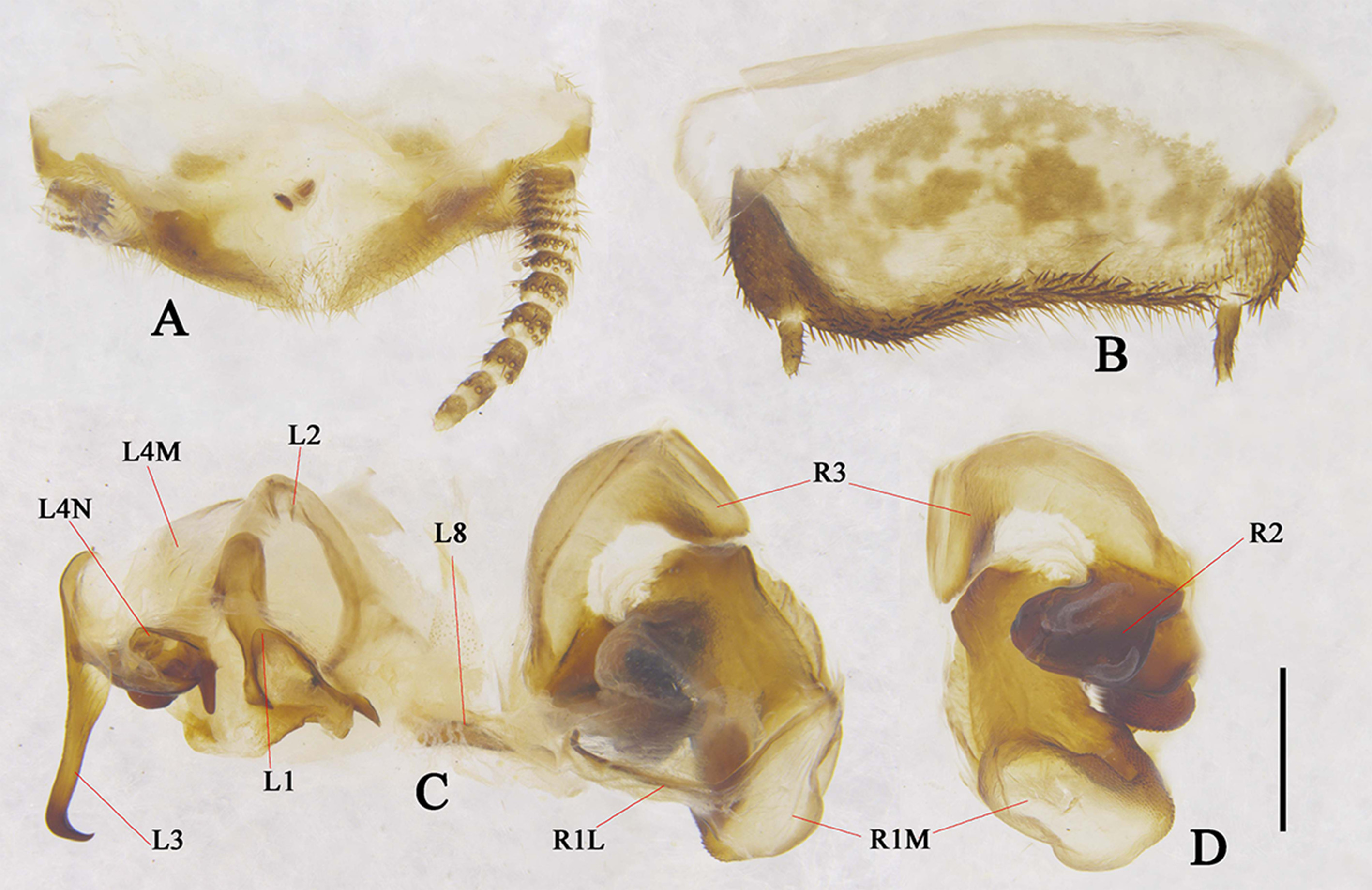
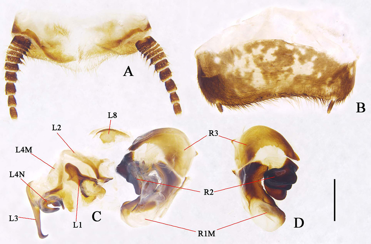
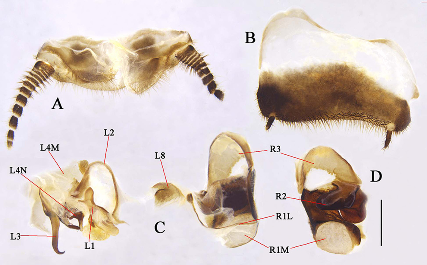
Description. Male: general: body length 12.2–29.3 mm, including tegmina 22.3–36.8 mm. Body elongate, pubescent, coloration varied, usually dull and dim. Head: eyes large, elongate, reniform, cover the lateral part of the head, about 3/4 of the head length. Interocular space narrower than or just the same as the distance between ocelli. Ocelli round, large, but not very protruded, with a ridge connecting the ocelli in between (ocelli ridge). Antennal sockets round, slightly larger than ocelli. Clypeus not surpassing dorsal border of the antennal sockets, protruded and divided into two parts: ante-clypeus usually light colored, narrow, transverse; post-clypeus bulbous, median usually with an indistinct longitudinal line. Frons usually wrinkled, below the ocelli ridge usually with two to three dimples, next to the antennal socket usually with an elongate dimple. Labrum rectangular, apex light colored and hind lateral corners rounded. Antennae unicolored, pubescent except the basal antennomeres (about 8–10 segments), scape the largest, long and robust, pedicellus similar to flagellum, the basal portion of flagellum become larger but diminish distally. Maxillary palpi with tip segment triangular. Pronotum: transverse oval, thoroughly shelters the head; usually brown, anterior margin white or yellowish white, some species unicolored. Apex arched and truncated, lateral fore margins gently oblique, usually form round angles with lateral borders. Hind margin formed blunt angles with the lateral parts. Surface covered with short pubescence, margins usually with long setae. Tegmina: length variable, in most species much exceed the end of abdomen, in Eupolyphaga everestiana just beyond the end of abdomen. Tegmina usually hyaline and spotted, the size, distribution and density of the spots varied between species, some species with unicolored tegmina. Sc swellings present. The characters of venation are as follows (taking the type species E. sinensis as example, fig. 1 A): main vein generally paralleled, Sc with about ten branches which evenly distributed; R largely occupied the lateral and distal portions, generally with two groups of branches, one group stretched straightly to the apex, the other group originated from the base and occupied the lateral portion of tegmen; M branched from about 1/3 of its length, about four branches stretch to the inner distal margin; CuA branched basally, occupied 1/4 of the tegmen area. Wings: hyaline, usually with pale maculae around the distal area. The characters of venation are as follows (taking the type species E. sinensis as example, fig. 1 B): Sc simple, long, end in the margin medially; RA bifurcate; RP with multiple branches occupied the outer apex of the wing; M simple, branched distally; CuA with straight, paralleled branches, the basal four very short; CuP straight, bifurcated distally. Legs: pubescent, front femur type C 1, spines on the tibia long, sharp, tarsal claws symmetrical, arolia present. Abdomen: Smooth, sparsely setose, usually unicolored, some species maculated. Supra-anal plate pubescent, transverse, apex protruded, two small sclerites present (the function is unknown), cerci long, pubescent ( Fig. 1 C View FIGURE 1 ). Subgenital plate transverse, hind margin usually curved or concave, densely setose, styli short, usually unequal sized ( Fig. 1 D View FIGURE 1 ). Genitalia: complex ( Fig. 1 F View FIGURE 1 ). Left phallomere: L1 round anteriorly, protruded, left usually with a small process, hind portion with two well sclerited processes (two hind lobes); L2 long, curved, right end usually forms into two processes; L3 hook like; L4N with pda and paa present ( Fig. 1 H View FIGURE 1 ), pda usually elongate, paa usually bud like, or slightly convex; L4M large, behind L2, plate like; L5 hidden behind L4N; L8 irregular, situated near the right portion of L2, large or small; L7 small, near R1M (due to it’s small, L 7 may be hidden by the membrane, not all L7 are shown in Figs. 17–37 View FIGURE 17 View FIGURE 18 View FIGURE 19 View FIGURE 20 View FIGURE 21 View FIGURE 22 View FIGURE 23 View FIGURE 24 View FIGURE 25 View FIGURE 26 View FIGURE 27 View FIGURE 28 View FIGURE 29 View FIGURE 30 View FIGURE 31 View FIGURE 32 View FIGURE 33 View FIGURE 34 View FIGURE 35 View FIGURE 36 View FIGURE 37 ). Right phallomere: R1M large, robust, hind portion usually enlarged, bulged; R1L slender, rough or thin, surrounded R1M on the left side; R2 well sclerite, transverse, irregular, median usually concave and forms into two thick chunks, the distal chunk (outer one) usually round, the basal one (inner one) usually quadrate and divided into two small sub-chunks; R3 concave, the dorsal portion large and protruded, the ventral portion narrow, in lateral-ventral view, the narrow portion connected with R2 at ventral apex ( Fig. 1 G View FIGURE 1 ).
Female. Measurements (mm). Body length: 15.2–30.2, body width: 10.6–21.5. Apterous, round, pubescent and setose, unicolored or with markings, usually brownish yellow to black. Head round, eyes wide apart, reduced, ocelli reduced into two white spots, antennae short, frons flat, clypeus convex, ante-clypeus usually whitish yellow, wide, labrum large. Dorsal surface well pubescent, margins setose, pronotum nearly triangle, apex arched, median of pronotum, mesonotum and metanotum with some irregular markings. Legs pubescent, spines long, arolia absent. Supra-anal plate pubescent, transverse, apex protruded, median emarginated, cerci very short, hide under the last terga ( Fig. 1 View FIGURE 1 K–L, the supra-anal plate are generally two types: the transverse type (with apex less protruded, strongly emarginated, Fig. 1 K View FIGURE 1 ) and protruded type (with apex distinct protruded, weakly emarginated, Fig. 1 L View FIGURE 1 )). Subgenital plate protruded, round, bulge, pubescent, apex slightly emarginated and covered with dense pubescence ( Fig. 1 J View FIGURE 1 ).
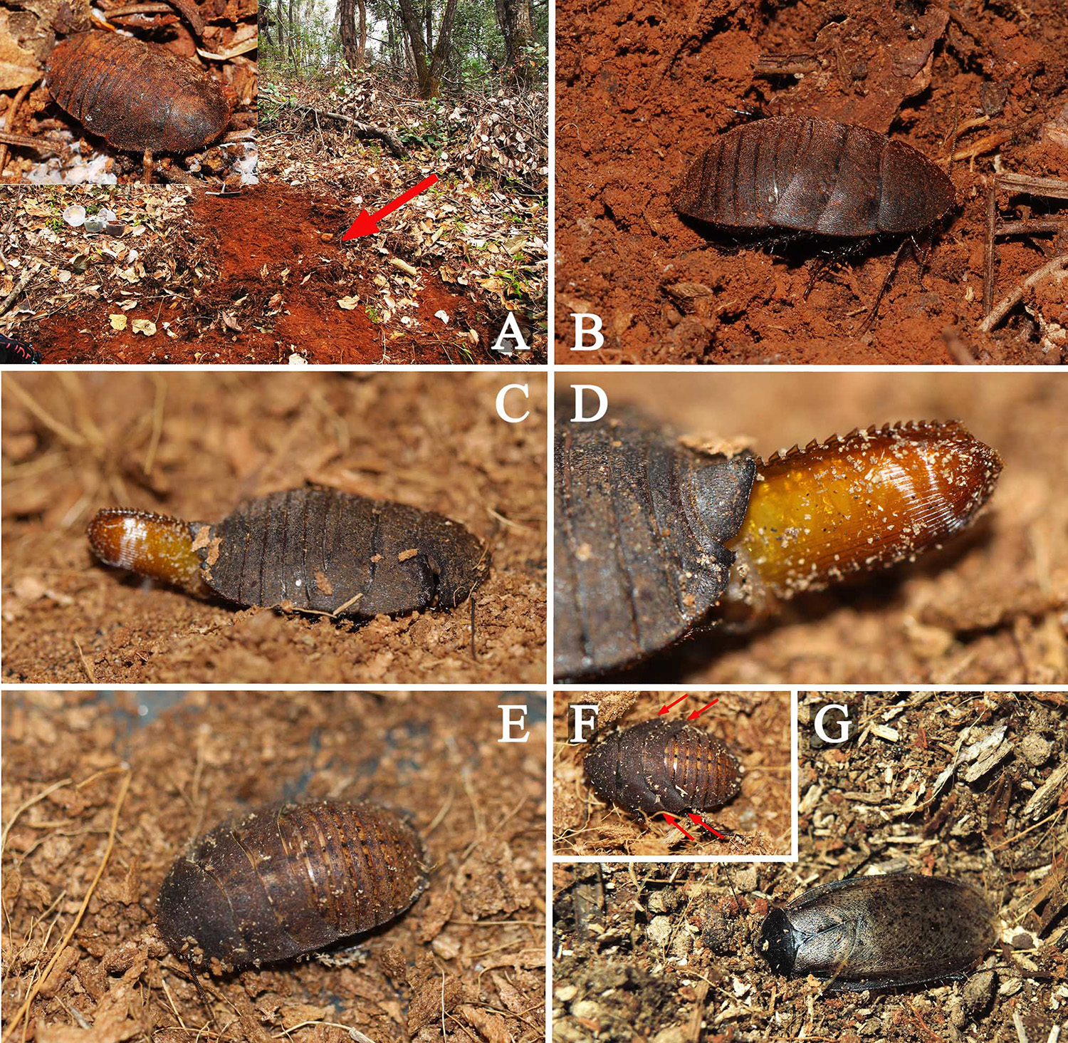
Nymph. Generally similar to the females, but may be different in marking pattern and coloration. Currently it is very difficult to identify Eupolyphaga species based on nymphs. In the last nymphal instar, it’s easy to distinguish the males from the females: 1) the male nymph with four unseparated and distinct wing buds protruded from the lateral ends of mesonotum and metanotum ( Fig. 44 F View FIGURES 44 ), while the female without wing buds; 2) the male nymph with small subgenital plate, paraprocts exposed, two styli present ( Fig. 1 E View FIGURE 1 ), while the female nymph with large subgenital plate, paraprocts exposed, styli missing ( Fig. 1 I View FIGURE 1 ). The female nymph differs from the mature female mainly by its subgenital plate, the female nymph with subgenital plate apex transverse, not bulged, two paraprocts exposed ( Fig. 1 I View FIGURE 1 ), while the mature female with large, protruded and bulged subgenital plate, paraprocts hidden ( Fig. 1 J View FIGURE 1 ).
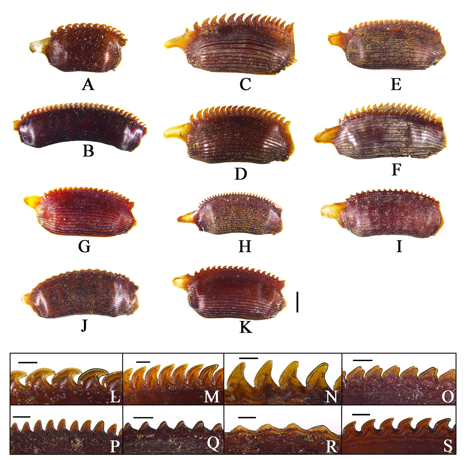
Ootheca. Elongate, surface with longitudinal line, keel serrated, the serration shape differs in different species, some species with respiratory canals well developed in the serration ( Fig. 38 View FIGURE 38 ). The size of ootheca may be varied in the same species ( Fig. 38 View FIGURE 38 A–B).
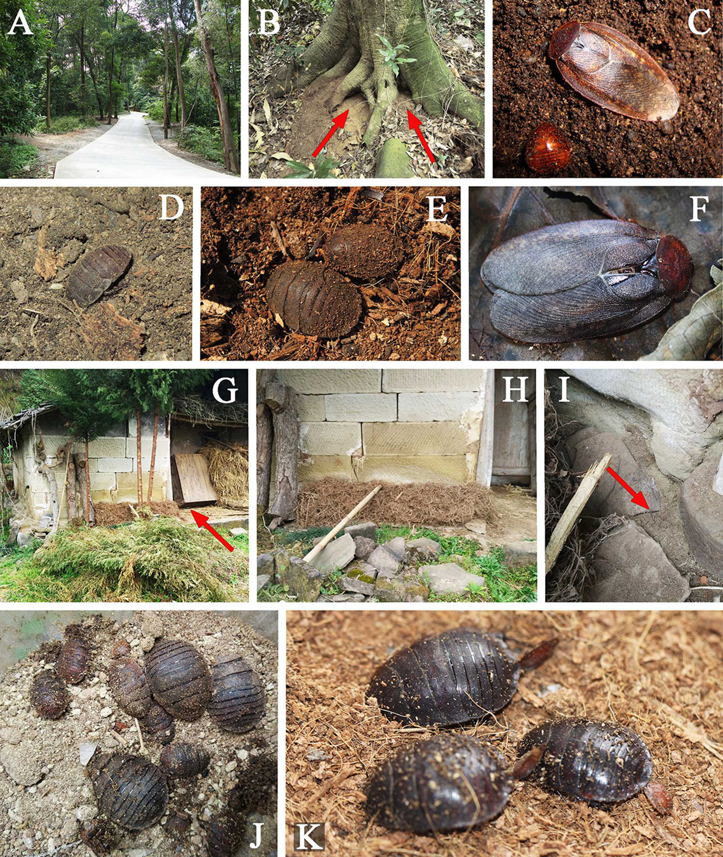
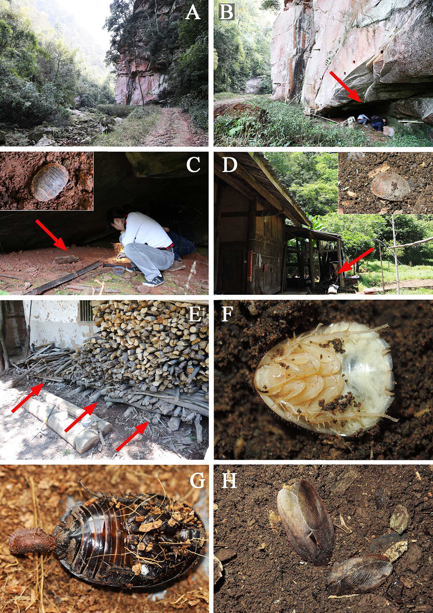
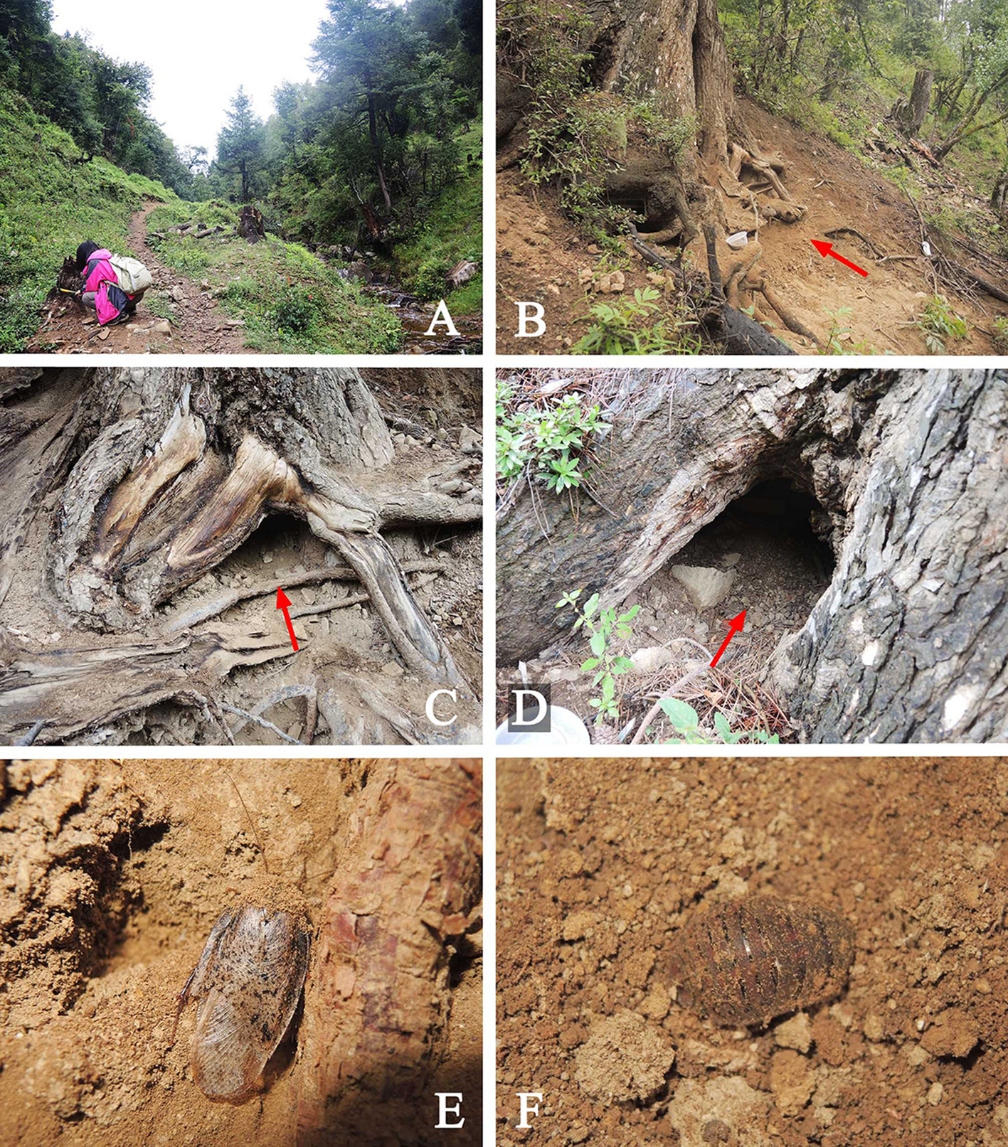
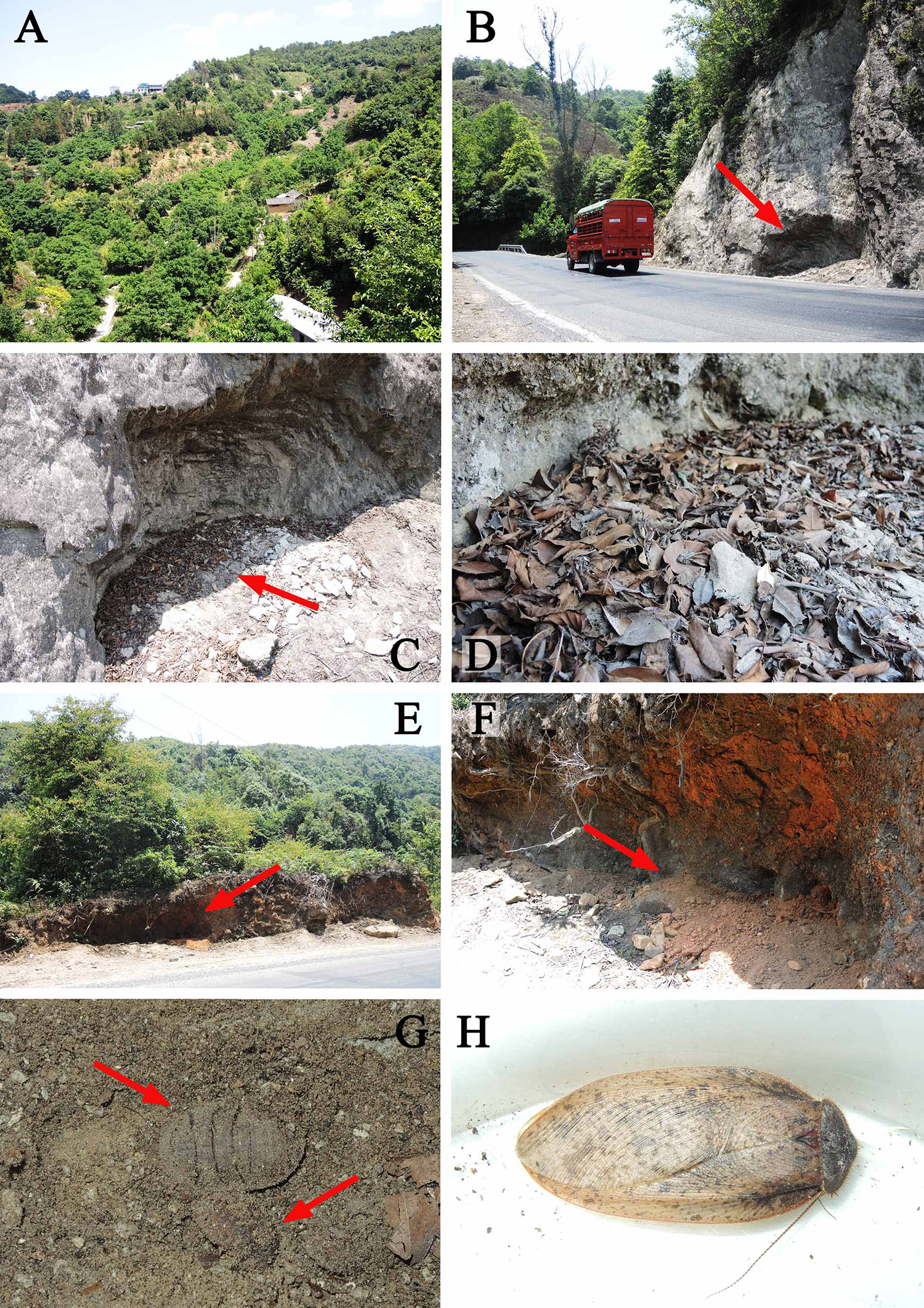
Natural history. The habitats are shown in Figs. 39–46 View FIGURES 39 View FIGURES 40 View FIGURES 41 View FIGURES 42 View FIGURES 43 View FIGURES 44 View FIGURES 45 View FIGURES 46 . Members of this genus can be found in dry lands, wet forests and human dwellings. Females and nymphs like living together in a small space, hiding inside dry and porous substrate (sand, soil dregs, wood dust, stone dregs), thus they can be found in tree holes and roots, cliff holes, old houses, and around dry excavated road sides (especially the road across the woody hill), or beneath stones. Males can be attracted by light. The rich species abundance appears in South China, especially in the wooded mountains in Southwest, though live in the wet forests, they still like to choose a small dry space to live in.
The most efficient way to collect Eupolyphaga is to dig the nymphs and females out from the substrate they hide in. Though they usually hide beneath the substrate, their abandoned exuvium and oothecae are usually exposed on the surface of the substrate; thus, to find the living individuals, one must carefully search the abandoned exuvium and oothecae in a proper place. If one collected a large mixture group of nymphs, oothecae and females, one will have the opportunity to obtain the males from rearing. Males can also be collected by light trapping, but not very often.
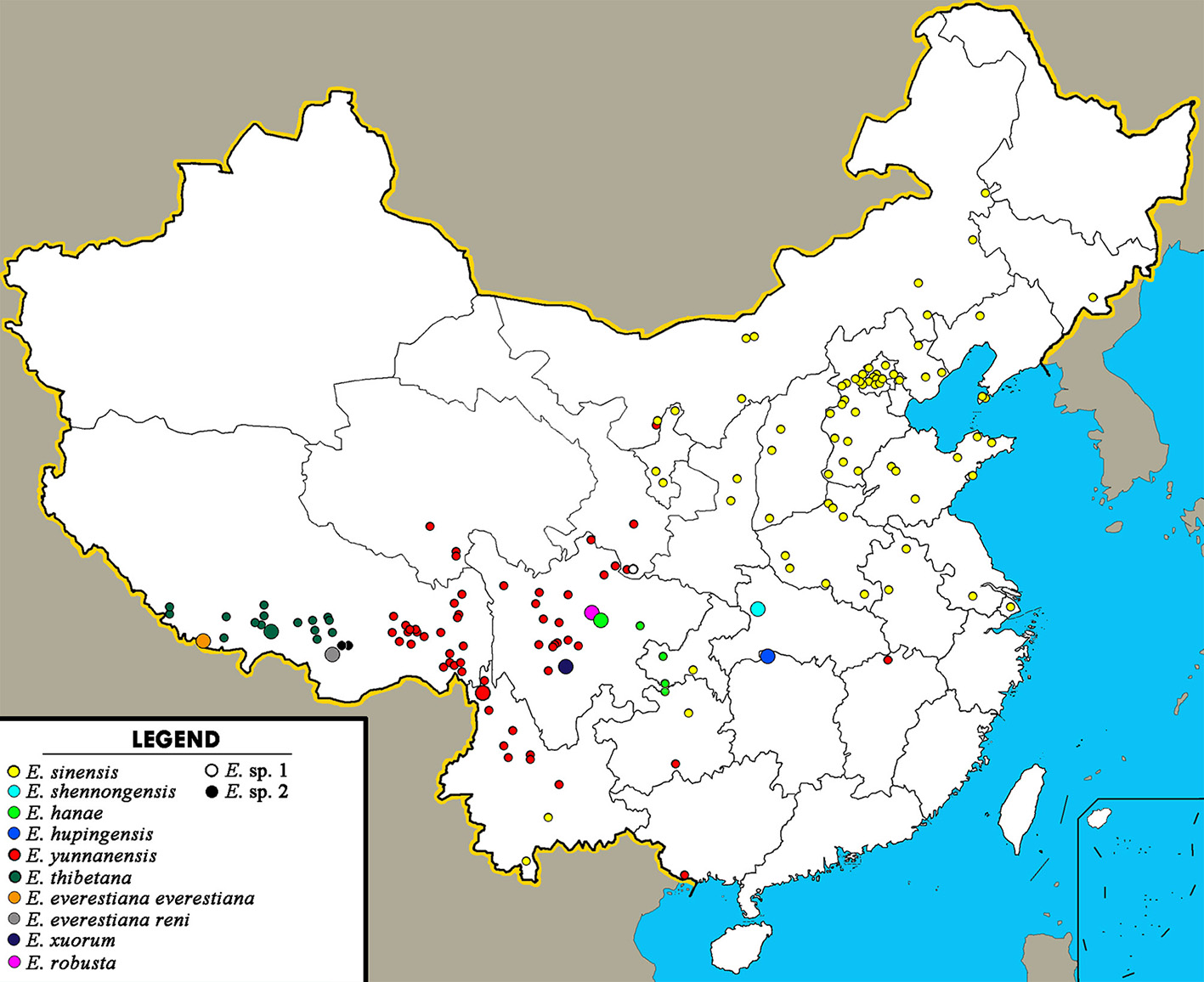
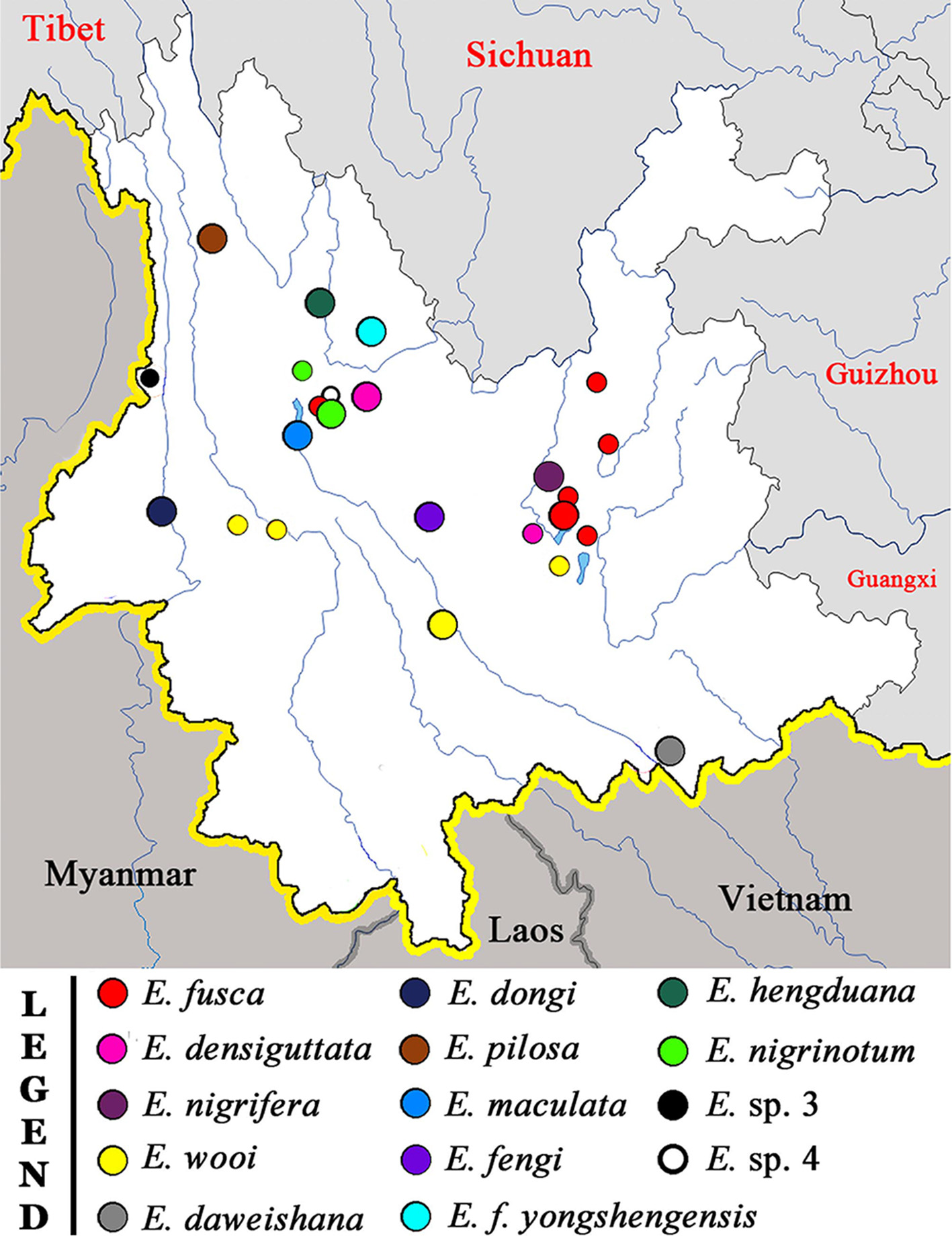
Distribution. Widely distributed in China ( Figs. 2–3 View FIGURE 2 View FIGURE 3 ), altitude ranging from tens of meters (coastal cities, e.g. Dalian City) to 5800 m (Mt. Everest). E. sinensis can also be found in South Russia.


No known copyright restrictions apply. See Agosti, D., Egloff, W., 2009. Taxonomic information exchange and copyright: the Plazi approach. BMC Research Notes 2009, 2:53 for further explanation.
|
Kingdom |
|
|
Phylum |
|
|
Class |
|
|
Order |
|
|
Family |
|
|
SubFamily |
Corydiinae |
Eupolyphaga Chopard, 1929
| Qiu, Lu, Che, Yang-Li & Wang, Zong-Qing 2018 |
Eupolyphaga
| Feng, P. - Z. & Guo, Y. - Y. & Woo, F. - C. 1997: 165 |
| Princis, K. 1962: 53 |
| Bey-Bienko, G. Y. 1950: 283 |
| Chopard, L. 1929: 261 |