
Piezodorus guildinii (Westwood)
|
publication ID |
https://doi.org/ 10.11646/zootaxa.4613.3.2 |
|
publication LSID |
lsid:zoobank.org:pub:509D6F1C-697D-48EC-AC51-E7ACC3B4BFA3 |
|
DOI |
https://doi.org/10.5281/zenodo.5611980 |
|
persistent identifier |
https://treatment.plazi.org/id/3F1A9854-FFFE-FFCC-FF2D-D2172158367F |
|
treatment provided by |
Plazi |
|
scientific name |
Piezodorus guildinii (Westwood) |
| status |
|
Piezodorus guildinii (Westwood)
(♂ syntype, Figs. 7–9 View FIGURES 7–9 )
5
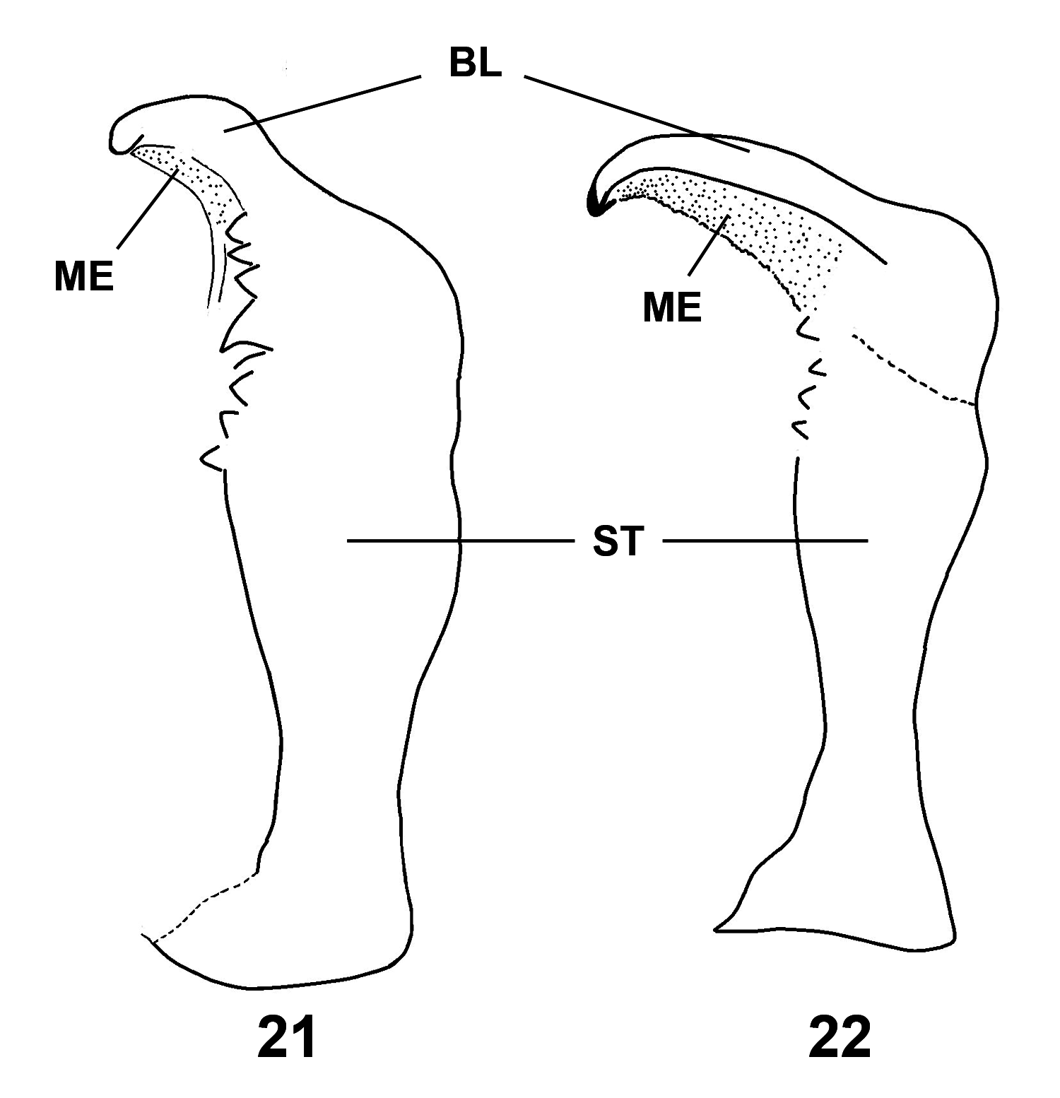
Diagnosis. Male. Pygophore longer than broad medially in dorsal view ( Fig. 10 View FIGURE 10 ), posterolateral angles tumescent (Figs. 12 and 14), medial area weakly elevated ( Fig. 14 View FIGURE 14 ), subhexagonal (hourglass) in shape (Fig. 12), sublateral area weakly elevated either side ( Fig. 14 View FIGURE 14 ); ventroposterior margin tumescent, weakly elevated and moderately reflexed, medial notch present, narrow, v-shaped, margin either side transversely longer than in P. hybneri , tumescence appearing to merge with posterolateral angle either side (Figs. 12 and 14); central opening in dorsal view with anteromedial margin lunate, broadly delimited either side by moderate triangular angulation, posterior margin with three notches, one broad medial v-shaped notch and one lateral small covered notch either side ( Fig. 10 View FIGURE 10 ). Aedeagus (Fig. 16) with three pairs of conjunctival appendages, two dorsal, one ventral, first dorsal pair projecting posteriorly, second dorsal pair projecting anteriorly, apices acute, first pair membranous, second pair membranous with slender sclerotized plate apically, first pair longer than second pair, similar to P. hybneri ; ventral pair bilobed, upper lobe broadest medially, primarily membranous, apical margin broadly rounded except for lightly sclerotized subacute medial angulation, lower lobe relatively narrow, subacute distally, less sclerotized than upper lobe. Vesica relatively short (Figs. 16–18), straight, heavily sclerotized, sometimes weakly declivent apically, reaching ca. 1/2 distance between medial margin of penial plate and lateral extensions of penial plate. Penial plate (Fig. 17) somewhat Hshaped, posterior and anterior margins curved medially, generally lightly sclerotized, lateral margins more heavily sclerotized; convex in posterior view with vesica located mediodorsally; subrectangular in dorsolateral view in basal 2/3, lateral margins gradually diverging distally, slightly converging apically, elongate and rectangular distally in dorsolateral view. Paramere (non-extracted, basal area concealed) moderately broad in basal 2/ 3 in lateral view, apex recurved ( Fig. 10 View FIGURE 10 ); paramere (extracted) with blade relatively short in lateral view, outer margin moderately sinuate, apex weakly recurved, corner broadly rounded, surface reflexed laterally; stem substraight in dorsal and lateral views ( Fig. 21 View FIGURES 21–22 ).
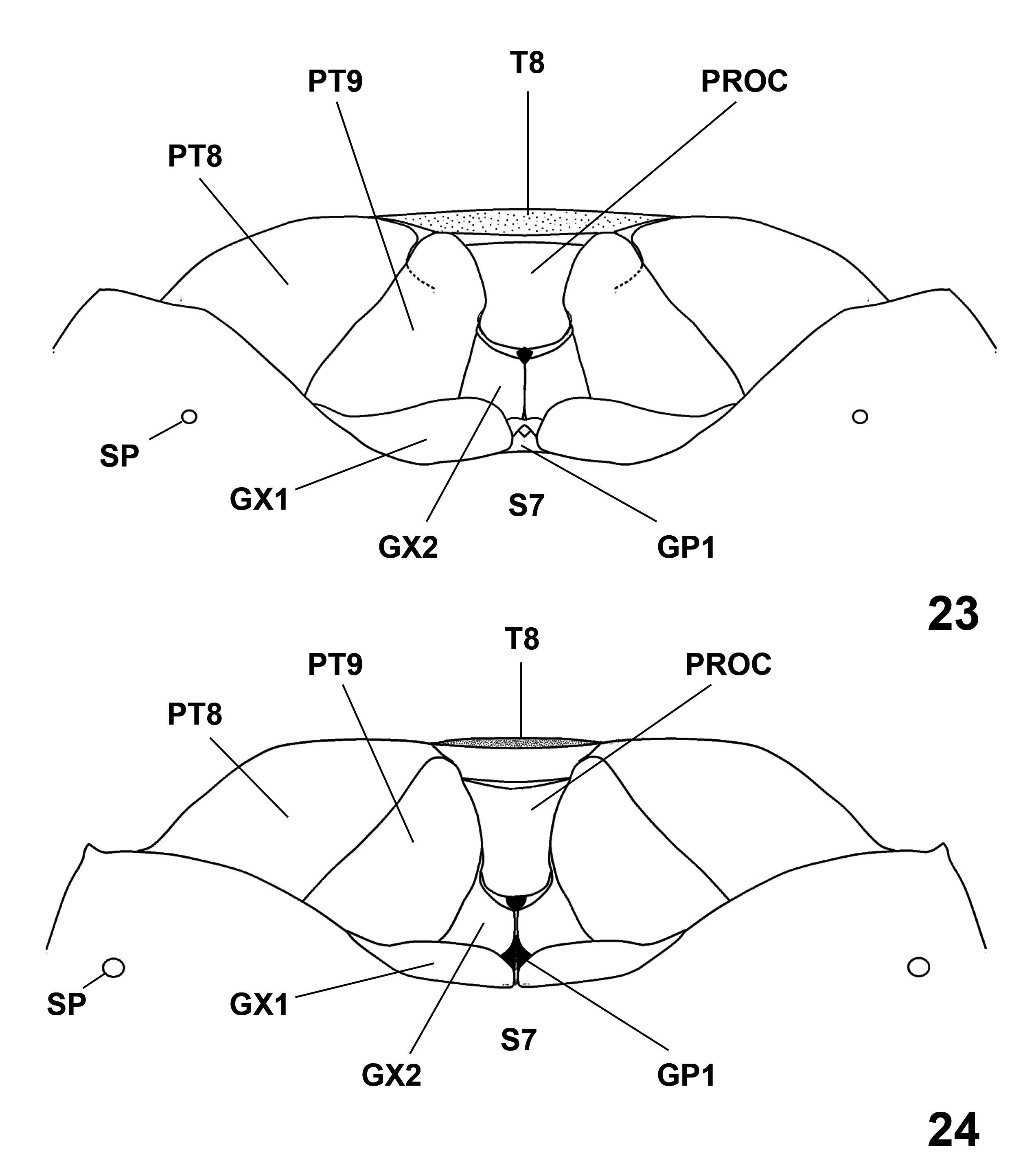
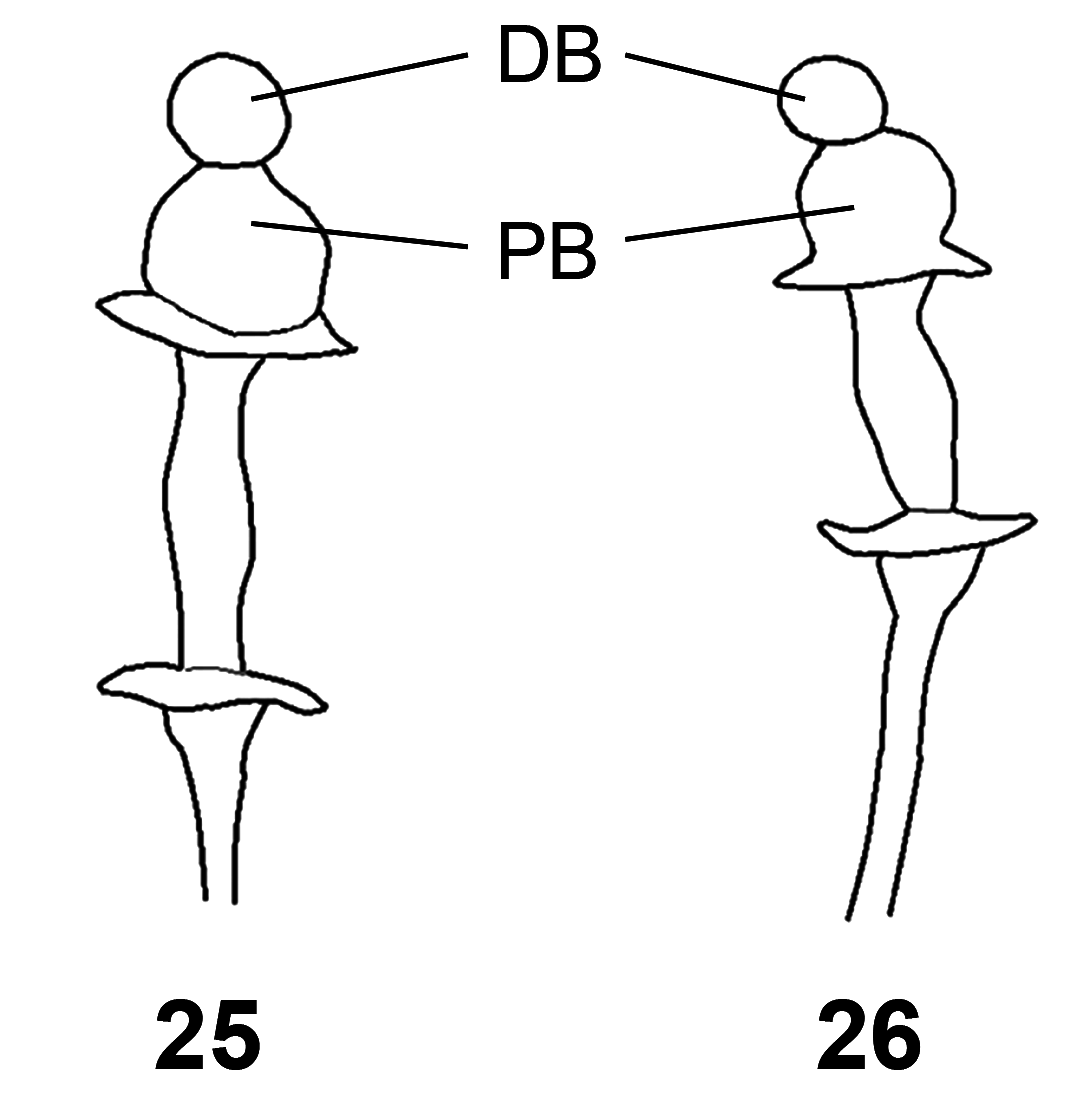
Female. Seventh abdominal sternum with posterior margin from posterolateral angle straight laterally and sublaterally, concave submedially, and weakly convex medially ( Fig. 23 View FIGURES 23–24 ). Eighth abdominal tergum ventrally moderately elevated posteriorly. First gonocoxae with posterior margin moderately exposed, weakly concave laterally, moderately convex medially. Second gonocoxae with posterior margin moderately concave. Eighth paratergite with posterior margin evenly weakly arcuate. Ninth paratergite broadly rounded distally with apex reaching ventral elevation of eighth tergum, outer margin substraight, inner margin with slight notch near distal 1/3, structure constricted at distal 1/4. Proctiger subquadrangular, clearly broader than distal 1/4 of ninth paratergite. Spermatheca ( Fig. 25 View FIGURES 25–26 ) with distal bulb moderately constricted basally, proximal bulb distinctly larger than distal bulb and in line with distal bulb.
Distribution. This species occurs from South Georgia and Argentina north through Mexico into Texas, Arkansas, and Missouri, and then east to Georgia, South Carolina, and Florida ( Bundy et al. 2018; present study). It also has been collected from several Caribbean Islands. Distribution records and numbers of specimens examined in the present study are given in Table 1 View TABLE 1 . The syntype was collected from St. Vincent Island in the Caribbean.


No known copyright restrictions apply. See Agosti, D., Egloff, W., 2009. Taxonomic information exchange and copyright: the Plazi approach. BMC Research Notes 2009, 2:53 for further explanation.