
Scydmaenus ( Parallomicrus ) inflatitibia Franz
|
publication ID |
https://doi.org/10.11646/zootaxa.5371.1.1 |
|
publication LSID |
lsid:zoobank.org:pub:D60B50D1-280B-4403-9E5B-25C0704A43A1 |
|
DOI |
https://doi.org/10.5281/zenodo.18323543 |
|
persistent identifier |
https://treatment.plazi.org/id/3E380C57-FFDB-4A73-27AC-B3DAFBF4E547 |
|
treatment provided by |
Plazi |
|
scientific name |
Scydmaenus ( Parallomicrus ) inflatitibia Franz |
| status |
|
Scydmaenus ( Parallomicrus) inflatitibia Franz View in CoL
Scydmaenus ( Allomicrus) inflatitibia Franz, 1975: 284 View in CoL .
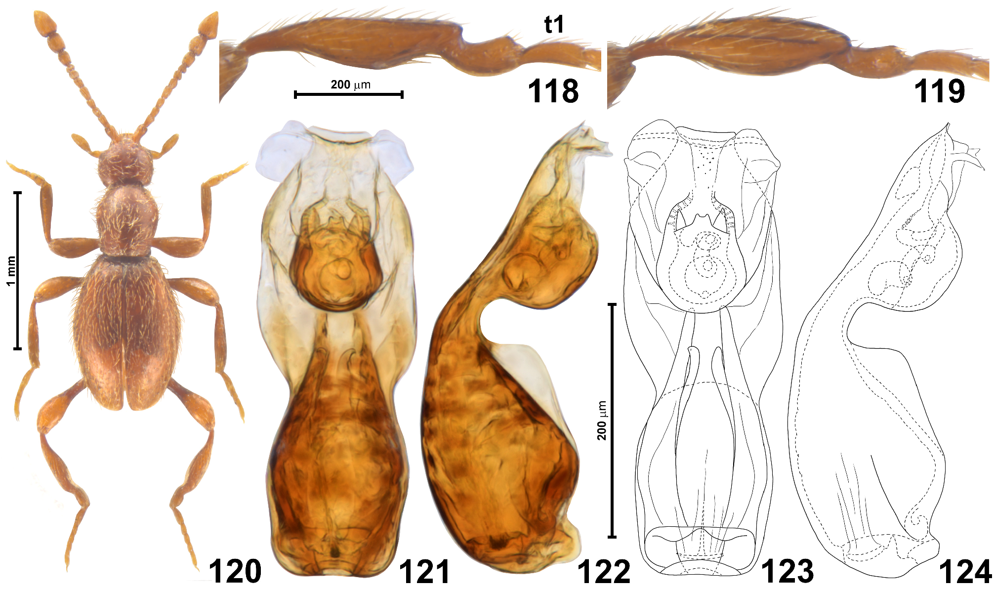
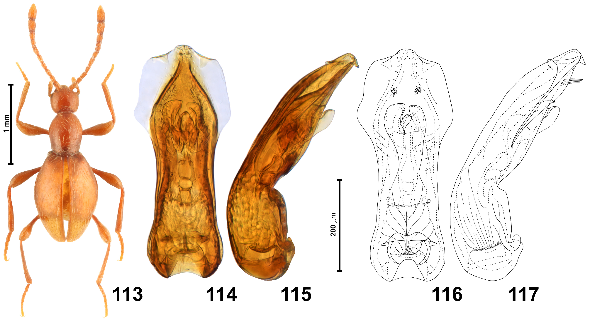
( Figs 118–124 View FIGURES 118–124 , 199 View FIGURES 189–205 )
Type material studied. Holotype ( AUSTRALIA: NEW SOUTH WALES): ♂ ( Fig. 120 View FIGURES 118–124 ), with labels illustrated in Fig. 199 View FIGURES 189–205 : “ N.S.Wales, ‘ Botany Bay / 1901. 42” [white with pink line below middle, handwritten], “ Heterognathus / A.M.Lea det.” [white, handwritten and printed], “ Scydmaenus / ( Heterognathus ) / inflatitibia / det. H.Franz m.” [white, handwritten and printed], “Typus” [red, handwritten], “Holo- / type” [white circle with red margin, printed] ( BNHM) . Paratype: ♀, “ N.S.Wales, ‘ Botany Bay / 1901. 42” [white with pink line below middle, handwritten], “Para- / type” [white circle with yellow margin] {unlabelled by Franz but listed in original description; identity uncertain, see Remarks} ( BNHM) .
Additional material studied. NEW SOUTH WALES: ♂, Acacia Plateau, J. Armstrong ( ANIC).
Revised diagnosis. In male antennomeres 3–4 each about 1.5 × as long as broad, 5 twice as long as broad, 6 1.5 × as long as broad, antennomeres 9 and 10 each 1.4 × as long as broad, and antennomere 11 less than twice as long as broad, with triangularly expanded angulate outer lateral margin ( Fig. 120 View FIGURES 118–124 , right antenna); metatibiae strongly modified ( Figs 118–120 View FIGURES 118–124 ), each medially broadened and with lateral (outer) elongate deep concavity surrounded by sharp margins and filled with setae, apical region constricted, narrowed and curved; pronotum with two pairs of antebasal pits; aedeagus in dorsal view ( Figs 121, 123 View FIGURES 118–124 ) with broad apex of median lobe, slightly narrower than half of widest (subapical) site of median lobe, flanked by weakly elongate, subtriangular weakly sclerotized lateral subapical lobes; median lobe lacking setae.
Redescription. Body in male ( Fig. 120 View FIGURES 118–124 ) slightly flattened, elongate and relatively slender, BL 1.75–1.81 mm; pigmentation uniformly light brown (including appendages); cuticle moderately glossy, covered with vestiture of setae slightly lighter than body.
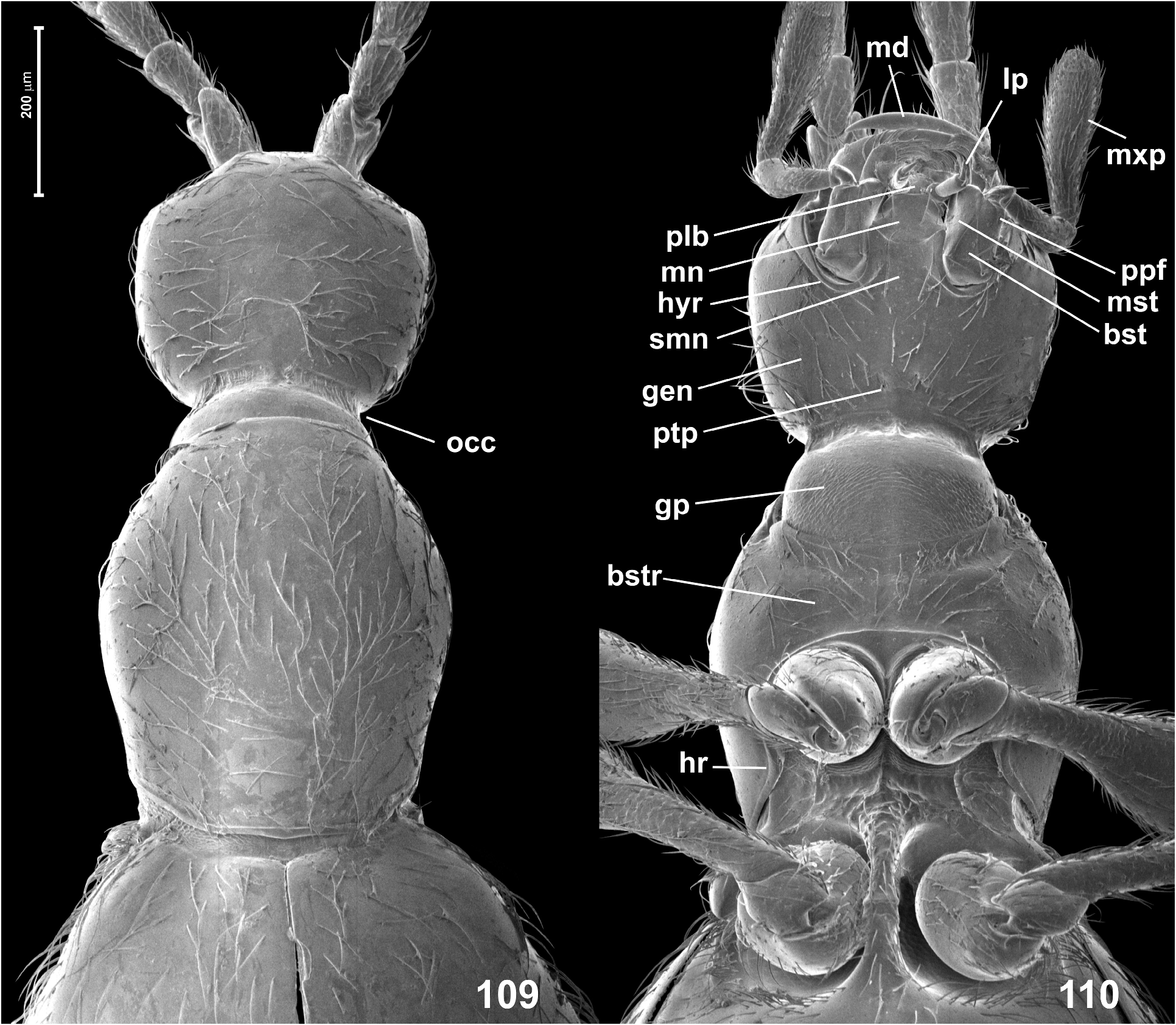
Head (Fi. 120) in dorsal view indistinctly transverse and somewhat subhexagonal, broadest at eyes, HL 0.30– 0.34 mm, HW 0.33–0.35 mm; vertex and frons confluent and weakly convex, posterior margin of vertex evenly arcuate and anteriorly convex; tempora about 2.5 × as long as length of eye in dorsal view; supraantennal tubercles indistinct; frons over antennal fossae broadly subtriangular and with rounded anterior margin. Eyes small, weakly oval, not emarginate posteriorly and oblique in relation to long axis of head. Punctures on frons and vertex fine, inconspicuous; setae (including those on tempora) short, sparse, nearly recumbent. Genae as sparsely setose as frons and vertex. Anterior (exposed) region of head capsule demarcated from neck region by short abrupt impression around occipital constriction, anterior margin of gular plate on neck region with indistinct, narrow anteriorly-directed projection. Antennae ( Fig. 120 View FIGURES 118–124 ) long and slender, AnL 1.00– 1.03 mm; three terminal antennomeres forming indistinctly delimited club; scape 2.5 × as long as broad, distinctly broadening distally; pedicel 1.8 × as long as broad; antennomeres 3–4 each about 1.5 × as long as broad, 5 twice as long as broad, 6 1.5 × as long as broad, 7 and 8 each distinctly asymmetrical and each about as long as broad, 9 1.4 × as long as broad and barrel-shaped, 10 1.4 × as long as broad and distinctly broadening distally, 11 much shorter than 9 and 10 combined, about 1.6 × as long as broad, distinctly asymmetrical, with broadly subtriangular, angulate expansion on proximal region of outer margin (visible in right antenna in Fig. 120 View FIGURES 118–124 ).
Pronotum in dorsal view ( Fig. 120 View FIGURES 118–124 ) distinctly elongate, broadest between middle and anterior third, PL 0.50 mm, PW 0.40–0.41 mm; anterior margin arcuate and laterally confluent with rounded lateral margins, so that anterior corners are not marked; posterior corners obtuse-angled and blunt; posterior margin nearly straight at middle and bent anteriorly at sides; base with very narrow and indistinct posterior marginal carina, with two pairs of distinct, small and slightly transverse pits. Pronotal disc covered with fine, shallow and unremarkable sparse punctures; setae similar to those on head, moderately dense and long, suberect. Ventrally prothorax with nearly asetose and impunctate hypomera and basisternal region only slightly longer than procoxal rests, sparsely covered with moderately long recumbent setae.
Elytra ( Figs 109 View FIGURES 109–110 , 113 View FIGURES 113–117 ) oval, broadest slightly in front of middle, EL 0.95–0.98 mm, EW 0.65–0.68 mm, EI 1.41–1.50. Humeral calli small but distinctly elevated and each mesally demarcated by shallow and transverse basal elytral impression; basal elytral foveae lacking; apices separately rounded. Elytral punctures fine and inconspicuous; setae similar to those on pronotum, moderately dense. Hind wings fully developed.
Legs ( Fig. 120 View FIGURES 118–124 ) long and slender. Metatibia ( Figs 118–120 View FIGURES 118–124 ) strongly modified, medially broadened, with strongly elongate oval cavity on lateral (outer) surface demarcated by sharp edges and filled with setae, distal 1/4 rapidly narrowed, almost constricted and distinctly curved. Protarsi with tenent setae on tarsomeres 1–3, protarsomere 1 moderately strongly broadened, weakly elongate, 2–4 each almost as long as broad, 5 about 2.5 × as long as broad; mesotarsi longer than protarsi, mesotarsomere 1 about 3 × as long as broad, tarsomeres 2–4 each weakly elongate, tarsomere 5 about 2.5 × as long as broad; metatarsi slightly longer than mesotarsi, metatarsomere 1 about 3 × as long as broad, tarsomeres 2–4 each distinctly elongate, but decreasing in length distally, tarsomere 5 about 3 × as long as broad.
Aedeagus ( Figs 121–122 View FIGURES 118–124 ) elongate but not very slender, AeL 0.38 mm, in dorsal view median lobe (excluding lateral subapical lobes) broadest in distal 1/3, narrowing both distally and proximally, but basal region again broadened, apex slightly less than half as wide as total width of median lobe, apical margin indistinctly concave; lateral subapical lobes large, each subtriangular and slightly longer than wide, projecting laterally; flagellum broadened in proximal region to form asymmetrical coils; median lobe lacking setae; ostium situated in distal third of median lobe, far from its apex.
Female. Unknown (see Remarks).
Distribution. SE Australia: CE and NE New South Wales.
Remarks. The identity of the only known paratype female is uncertain. This specimen is remarkably smaller than the holotype male (BL 1.60 mm vs. 1.75), and it may belong to a different species.
Scydmaenus inflatitibia combines some characters typical of Parallomicrus (fused metanepisterna), Choleropsis (two pairs of antebasal pronotal pits), and Corbulifer (modified metatibia in males). It remarkably differs from S. myrmecobius , especially in aedeagal features. Although the aedeagus within the large genus Scydmaenus is poorly studied, its general structure is usually stable within subgenera. For instance, it was demonstrated that despite a wide spectrum of metatibial modifications in Corbulifer , the structure of the aedeagus is very similar in all species, i.e., all of them, independently of the shape of the median lobe, share long setae and asymmetrical proximal ‘chambers’ of the flagellum ( Jałoszyński 2018). All species of Choleropsis treated in the present study also have asymmetrical proximal flagellar coils, whereas two known species of Kingius , four Australian species of Scottiscydmaenus , and two species of the newly described Ascydmaenus subgen. n. share not coils, but symmetrical expansions of the proximal flagellar region, which appear as three consecutive ‘chambers’ demarcated by constrictions. Scydmaenus ( Mascarensia) australiensis has strongly asymmetrical, broad coils, and even though the previously studied Scydmaenus ( Mascarensia) kasuganus Franz, 1976 has a clearly different shape of the median lobe, its proximal flagellar region is also strongly asymmetrical in the dorsal view ( Jałoszyński 2022). Two species of Scydmaenus belonging in the subgenus Geoscydmaenus and inhabiting Madagascar have symmetrical proximal flagellar regions ( Jałoszyński 2016b). These observations suggest that groups of closely related species have similar structures of the flagellum. However, in the Australian Parallomicrus , S. myrmecobius has symmetrical ‘chambers’, whereas in S. inflatitibia they form a loose spiral. Because external features (pronotal pits, male secondary sexual modifications) are also different in these two species, in future they may be placed in separate subgenera.

No known copyright restrictions apply. See Agosti, D., Egloff, W., 2009. Taxonomic information exchange and copyright: the Plazi approach. BMC Research Notes 2009, 2:53 for further explanation.
|
Kingdom |
|
|
Phylum |
|
|
Class |
|
|
Order |
|
|
Family |
|
|
SubFamily |
Scydmaeninae |
|
Genus |
|
|
SubGenus |
Parallomicrus |
Scydmaenus ( Parallomicrus ) inflatitibia Franz
| Jałoszyński, Paweł 2023 |
Scydmaenus ( Allomicrus ) inflatitibia
| Franz, H. 1975: 284 |