
Armatoplana kaburakii, Oya & Tsuyuki & Kajihara, 2022
|
publication ID |
https://doi.org/10.11646/zootaxa.5178.5.2 |
|
publication LSID |
lsid:zoobank.org:pub:2DEB6BCF-5DCA-4BBA-B548-FC586CA4719E |
|
DOI |
https://doi.org/10.5281/zenodo.7039807 |
|
persistent identifier |
https://treatment.plazi.org/id/3E19857F-FFD6-FF8C-8AA2-58DCB734FE5B |
|
treatment provided by |
Plazi |
|
scientific name |
Armatoplana kaburakii |
| status |
sp. nov. |
Armatoplana kaburakii sp. nov.
( Figs. 4 View FIGURE 4 and 5 View FIGURE 5 )
Etymology. The specific name is a noun in the genitive case honoring Tokio Kaburaki, who studied Japanese polyclads.
Diagnosis. Armatoplana without nuchal tentacles and common sperm duct and with prostatic vesicle located above seminal vesicle, elongated Lang’s vesicle without accessory vesicles, and male and female gonopores opening closely to each other ( Figs. 4 View FIGURE 4 and 5 View FIGURE 5 ).
Material examined. Five specimens ( one holotype and four paratypes) ( Table 1 View TABLE 1 ).
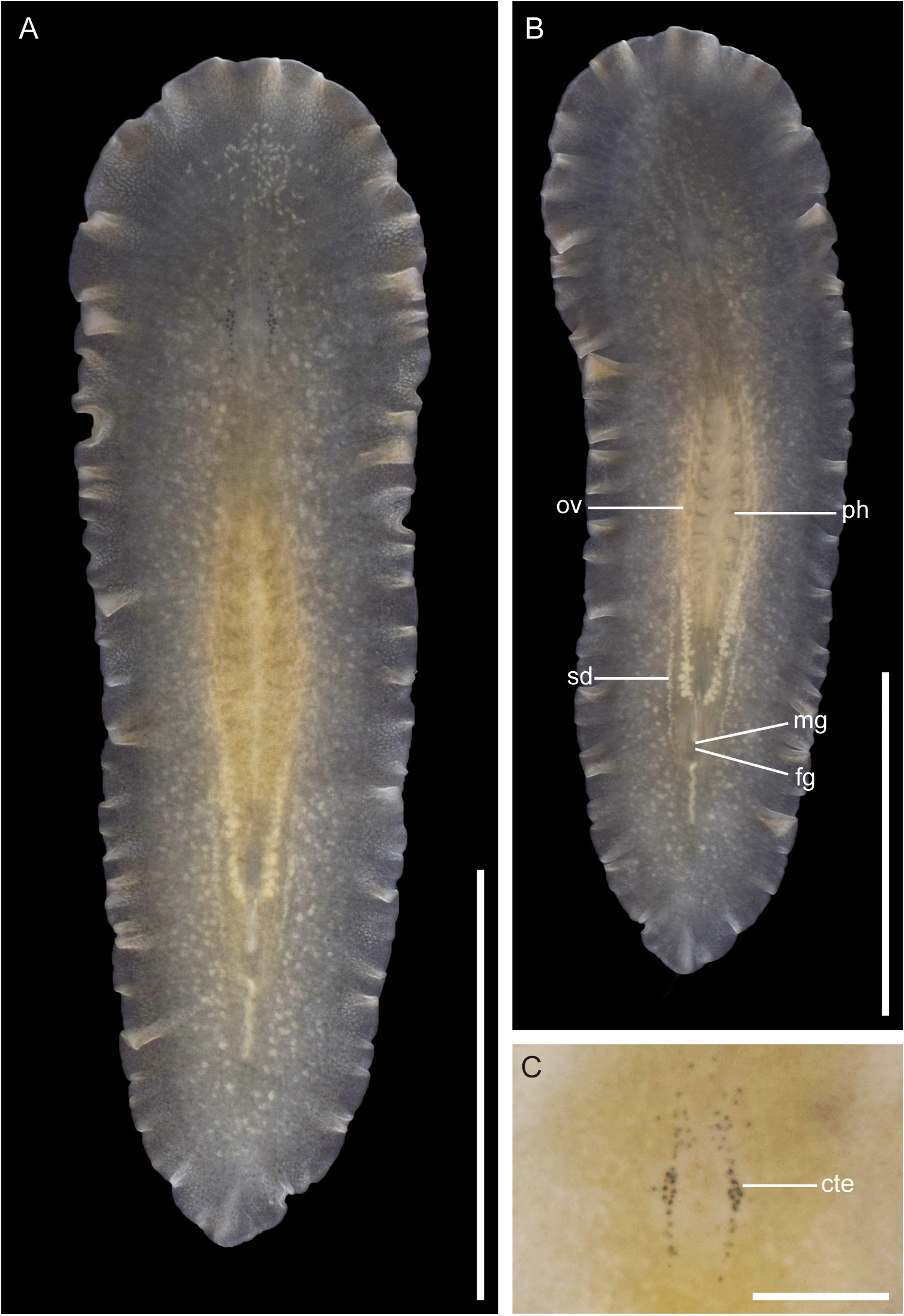
Description. Live specimens 9.0– 14 mm in length ( 13 mm in holotype), 2.5–4.2 mm in maximum width ( 3.8 mm in holotype). Body elongate oval, narrow toward posterior end ( Fig. 4A, B View FIGURE 4 ). Dorsal body tinged with light brown. Dorsal surface of body around pharynx pale yellow. Body margin translucent. General appearance of body yellowish translucent ( Fig. 4A View FIGURE 4 ). Nuchal tentacles lacking. Pair of cerebro-tentacular eye clusters, each containing 26–43 eyespots ( 30 in right cluster, 31 in left cluster in holotype), arranged near median line ( Fig. 4C View FIGURE 4 ). Eyespots located anterior to brain widely spreading but forming clear aggregation; those located posterior to brain forming almost linear aggregation. Length of anterior part of cluster almost same or longer than that of posterior one. Pharynx whitish, ruffled in shape, occupying about one-fourth to one-fifth of body length (2.0– 3.7 mm in length, 3.3 mm in holotype), located at center of body ( Fig. 4B View FIGURE 4 ). Mouth opening at slightly posterior to center of pharyngeal cavity. Intestine not anastomosed, spreading throughout body except margin. Pair of whitish sperm ducts and oviducts visible through ventral body wall ( Fig. 4B View FIGURE 4 ). Male and female gonopores close but separated; male gonopore opening at about one-fourth to one-sevenths of body length ( 2.3–3.7 mm, 3.6 mm in holotype) from posterior end; female gonopore situated 0.07–0.17 mm ( 0.07 mm in holotype) posterior to male gonopore.
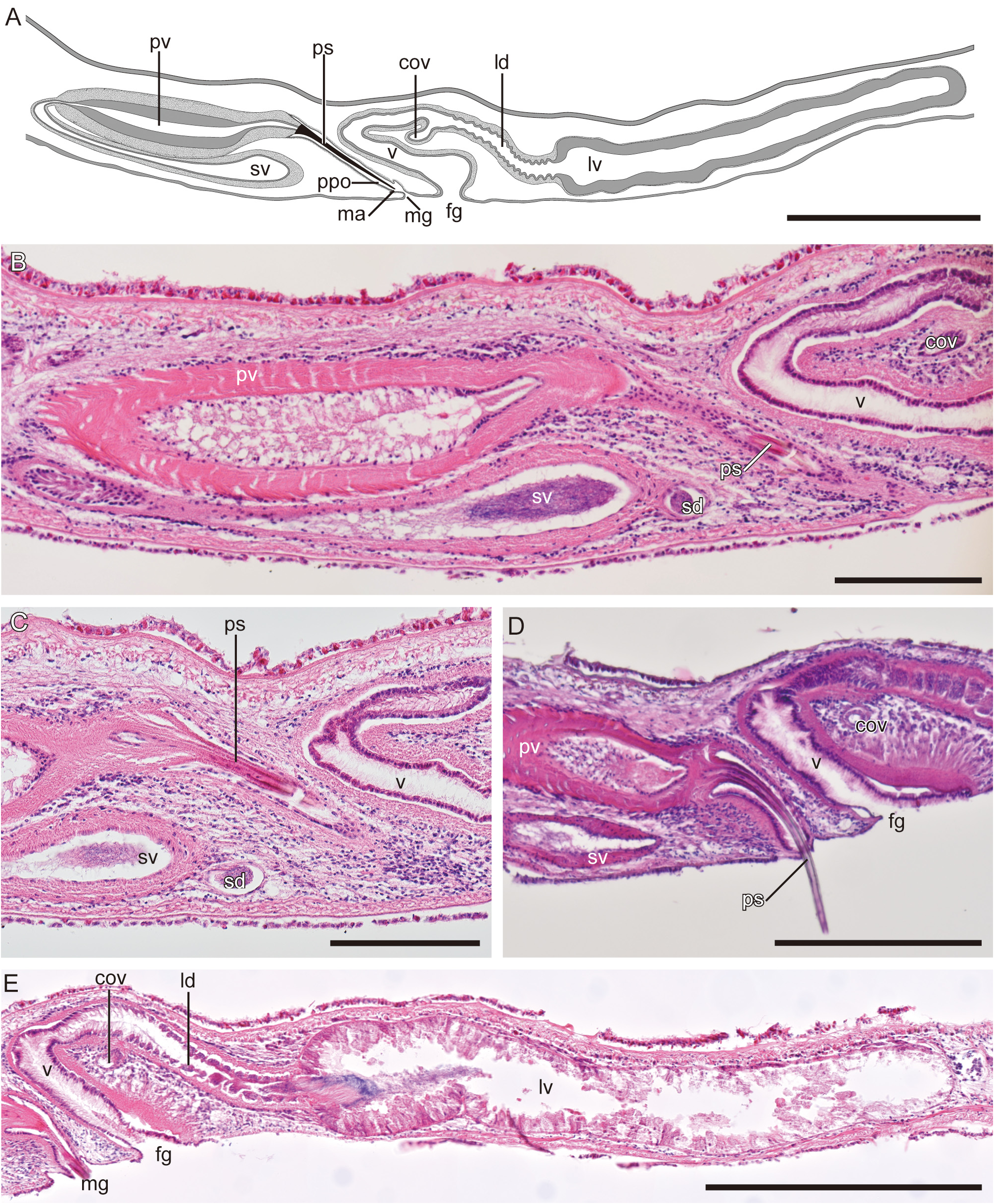
Male copulatory apparatus located posterior to pharynx, consisting of seminal vesicle, interpolated prostatic vesicle, and penis stylet ( Fig. 5A View FIGURE 5 ). Pair of sperm ducts running anteriorly, turning medially at point about one-fifth length of pharynx from posterior end, subsequently running posteriorly along both sides of pharynx and extending further posteriorly for short distance beyond level of posterior end of pharynx, then turning anteriorly ( Fig. 4B View FIGURE 4 ) and entering separately proximal end of elongated ovate seminal vesicle. Seminal vesicle directing anteriorly and having strong muscular wall ( Fig. 5B View FIGURE 5 ). Distal end of seminal vesicle slender, running dorsally before connecting to prostatic vesicle. Prostatic vesicle oval-shaped, larger than seminal vesicle, having thick muscular wall lined with smooth, thick epithelium and located dorsally above seminal vesicle ( Fig. 5A, B View FIGURE 5 ). Distal end of prostatic vesicle forming penis papilla armed with stylet ( Fig. 5C View FIGURE 5 ). Penis stylet straight or curved (straight in ICHUM 6091 [ holotype] and ICHUM 6090 [ paratype]; curved in ICHUM 6283–6285 [ paratypes]; Fig. 5A, D View FIGURE 5 ), projecting into penis pocket. Penis pocket lined with ciliated epithelium opening to male atrium ( Fig. 5A View FIGURE 5 ). Male atrium small, cone-shaped, and lined with ciliated epithelium.
Pair of oviducts forming common oviduct, latter running postero-dorsally to enter vagina. From this point, elongated Lang’s-vesicle duct (almost same length of vagina), lined with folded ciliated epithelium, running posteroventrally to connect to Lang’s vesicle ( Fig. 5E View FIGURE 5 ). Lang’s vesicle elongated, sac-shaped, and lined with columnar cells, lacking accessory vesicles ( Fig. 5E View FIGURE 5 ). Vagina curving antero-ventrally, then running postero-ventrally to exit at female gonopore. Lang’s-vesicle duct and vagina surrounded by circular muscle fibers and lined with ciliated and smooth epithelium.
Type locality. Arai-hama Beach ( 35°09′34″N, 139°36′42″E), Misaki , Kanagawa, Japan GoogleMaps .
Habitat. Intertidal to subtidal, on the surfaces of coralline algae ( Kanagawa), undersurface of stones ( Shizuoka), and among oyster beds on a mooring rope hung in the sea ( Kochi) ( Table 1 View TABLE 1 ).
Sequences of COI. The uncorrected P -distances of the partial COI sequences (712 bp) of the five specimens ( LC582945 View Materials – LC582946 View Materials and LC672056 View Materials – LC672058 View Materials ) were 0.001 –0.013 GoogleMaps .
Remarks. Armatoplana kaburakii sp. nov. resembles A. reishi ( Hyman, 1959) and A. tenuis ( Palombi, 1936) in that they share the following two characteristics: i) nuchal tentacles absent and ii) male and female gonopores opening closely to each other ( Table 3 View TABLE 3 ). Armatoplana kaburakii sp. nov. is distinguished from these two species by the shape of the Lang’s vesicle (elongated sac-shaped in A. kaburakii sp. nov.; laterally broadened in A. reishi ; spherical in A. tenuis ).
This is the first report of Armatoplana from the West Pacific along with A. albomaculata sp. nov. The uncorrected P -distances of COI (712 bp) from A. albomaculata sp. nov. were 0.180 –0.188.
No known copyright restrictions apply. See Agosti, D., Egloff, W., 2009. Taxonomic information exchange and copyright: the Plazi approach. BMC Research Notes 2009, 2:53 for further explanation.
|
Kingdom |
|
|
Phylum |
|
|
Order |
|
|
SuperFamily |
Leptoplanoidea |
|
Family |
|
|
Genus |