
Iracema caiana Triques, 1996
|
publication ID |
https://doi.org/10.1590/S1679-62252011000300001 |
|
persistent identifier |
https://treatment.plazi.org/id/3A6F87C5-1661-2E25-FED9-FDC4131DF857 |
|
treatment provided by |
Carolina |
|
scientific name |
Iracema caiana Triques, 1996 |
| status |
|
Iracema caiana Triques, 1996 View in CoL
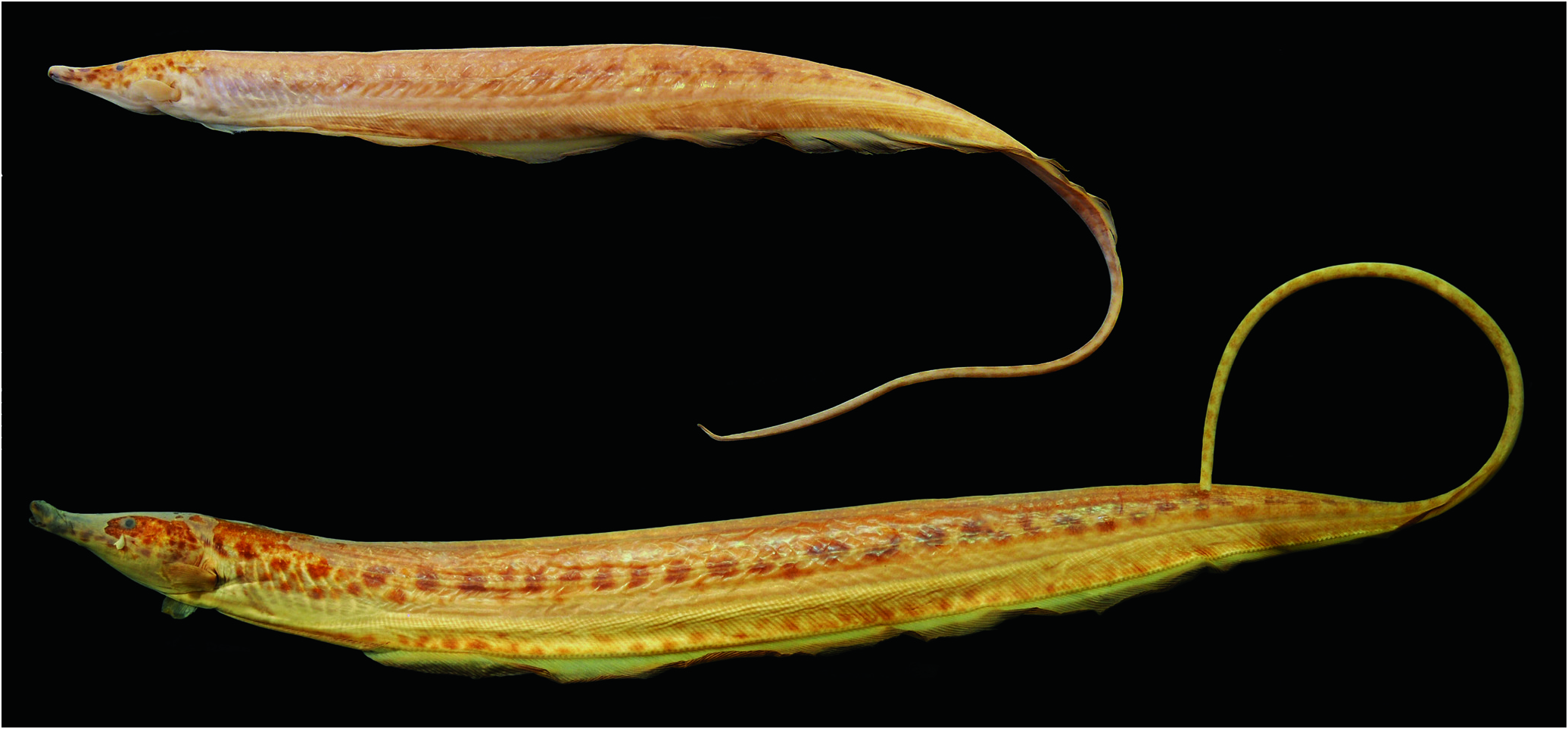
Figs. 1-2 View Fig View Fig , Table 1
Iracema caiana Triques, 1996a: 91 View in CoL [original description]. - Albert & Campos-da-Paz, 1998: 423, 425 [phylogeny and diagnosis]. - Albert, 2001: 63, 68 [phylogeny and diagnosis]. - Ferraris, 2003: 495 [listed]. -Albert & Crampton, 2005: 363 [listed]. -Crampton & Albert, 2006: 650 [listed and habitat description]. - Crampton 2011: 175 [listed]. -Winemiller & Willis, 2011: 231 [listed].
Diagnosis. The same as for genus.
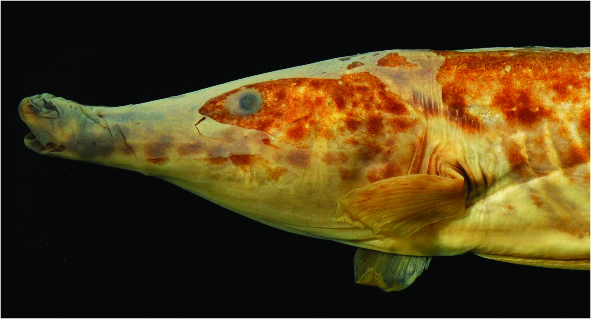
Description. Morphometrics and meristics in Table 1. Adult body size moderate for Rhamphichthyidae (maximum size 360 mm SL). Dorsal profile of snout strongly concave anterior to eye, head profile slightly convex post-orbitally. Interorbital region slightly convex. Dorsal margin of body almost straight, slightly ascending at end of body cavity. Ventral body margin slightly concave from anal-fin origin to end of anal fin. Greatest body depth at about middle of standard length. Body tapering posterior to mid body. Caudal appendage long, somewhat ovoid in cross section, its depth slightly greater than its width. Mouth subterminal, lower jaw shorter than upper jaw ( Fig. 2 View Fig ). Lower jaw included in upper jaw, rictus short and slightly upturned.Anterior and posterior nares present, without tubes. Anterior nares positioned terminally close to oral aperture, posterior nares positioned dorsally closer to tip of snout than eye. Branchial opening located posterior to opercle, its ventral margin extending to vertical with dorsal margin. No fleshy skin fold on body wall inside branchial opening. Eyes relatively large and positioned laterally. Urogenital papillae well developed, positioned more anteriorly in larger specimens. Anterior chamber of gas bladder not ossified. Posterior chamber of gas bladder apparently absent (MZUSP 49205, 345 mm SL; and Triques, 1996a: 92). Anal-fin origin at vertical with pectoral-fin insertion. Absence of thick fold of skin in proximal portion of anal-fin rays. Lateral line complete, with hourglass shape bony tubes. Lateral line with short ventral rami in anterior half of body.
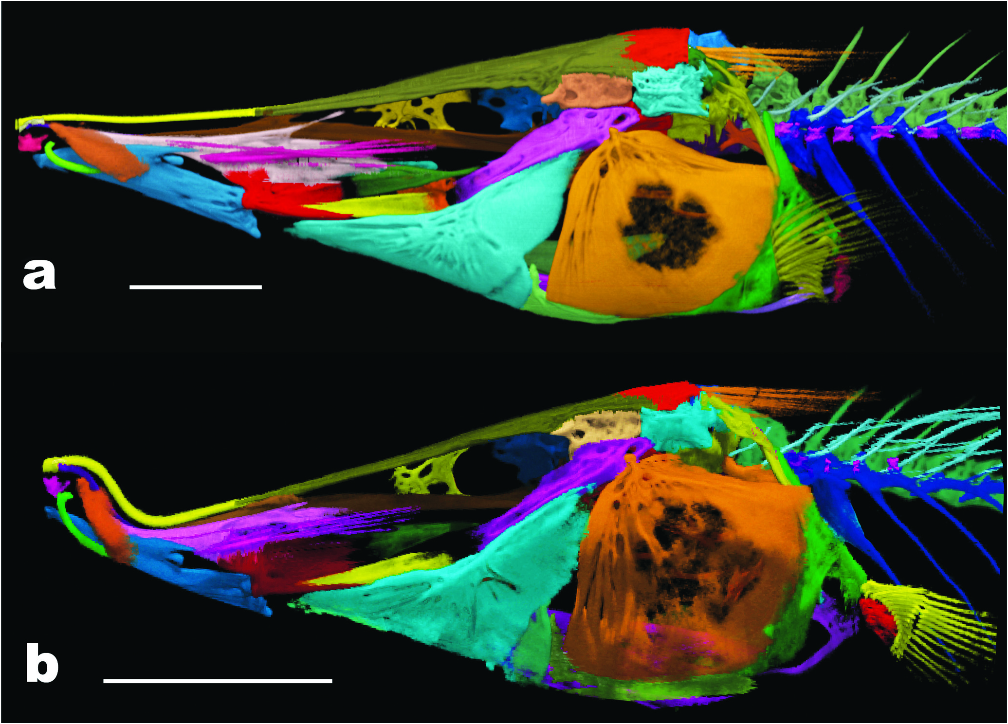
Skeletal Anatomy. Skeletal anatomy based on computed tomography (CT) scan data from two specimens of Iracema caiana ( Figs. 3-9 View Fig View Fig View Fig View Fig View Fig View Fig View Fig ).
Skull roof and neurocranium. Skull roof and neurocranium illustrated in Figure 4 View Fig . Bones on anterior portion of skull elongate, mesethmoid (met), ventral ethmoid+vomer (v), parasphenoid (pas) and frontals (fr). Mesethmoid narrow, its length greater than antorbital region of frontal; anterior tip small and rounded, flexed ventrally around ventral ethmoid+vomer; posteriorly contacting medial portion of frontal. Ventral ethmoid fused with vomer (=ventral ethmoid+vomer); somewhat crossshaped in ventral view, not fused anteriorly with mesethmoid. Lateral ethmoid not ossified and thus not visible on scan. Frontals covering more than half skull length.Antorbital process of frontals absent, lateral margin almost straight in dorsal region of orbit. Posterolateral region of frontals extending medially to orbit, contacting pterosphenoid (pts); posterior portion with imbricate suture with parietal (pa). Cranial fontanels large, frontals forming bridge in middle portion. Parietals forming lateral margins of fontanels contacting each other posteriorly, supporting supratemporal laterosensory canal. Small bony ridge on parietal at point of contact with extrascapular. Parietal ridge with inflection at point of contact with thin intermuscular bones of epaxial musculature. Occipital region of braincase consisting of supraoccipital (soc), basioccipital (bo), exoccipital (exo) and epioccipital (epo) bones. Supraoccipital contacting parietal anteriorly and epioccipital ventrolaterally. Epioccipital forming posterodorsal corner of brain case, contacting exoccipitals ventrally and pterotics anteriorly. Exoccipital contacting basioccipital ventrally, pterotics anterodorsally and supraoccipital posterodorsally. Posterodorsal portion of exoccipital together with posteroventral portion of supraoccipital forming condyle articulating skull with supraneural. Exoccipital forming most of foramen magnum, formed ventrally by basioccipital. Basioccipital forming ventral wall of lagenar otolith chamber, covered dorsally by exoccipital. Anterior portion of braincase (sphenoid and otic regions) consisting of orbitosphenoid (obs), pterosphenoid, prootic (pro), sphenotic (spo) and pterotics (pto) bones. Pterotic forming lateral portion of skull supporting semicircular canals of otic capsule. Pterotic contacting prootic and exoccipital ventrally, epioccipital posterodorsally, and exoccipital posteroventrally. Sphenotic contacting frontal dorsally and prootic ventrally. Parasphenoid extending to about three quarters of total length of neurocranium; anterior margin positioned dorsal to ventral ethmoid+vomer medially; posterior margin contacting basioccipital and prootic posteriorly; lateral margin extending dorsally to horizontal line with trigeminal foramen of prootic; ventral margin almost straight in lateral profile. Orbitosphenoid tapering anteriorly, contacting frontals dorsally and parasphenoid ventrally. Orbitosphenoid not contacting pterosphenoid, probably connected by cartilage (as in Rhamphichthys ). Orbitosphenoid and pterosphenoid with porous, reticulated surfaces. Pterosphenoid contacting sphenotic posteriorly and parasphenoid ventrally. Sphenotic somewhat rectangular with anterodorsal process contacting frontal. Prootic, sphenotic and anteroventral portion of pterotic articulating with hyomandibula. Prootic contacting basioccipital, exoccipital, pterotics, sphenotics, and parasphenoid. Dorsolateral margin of sphenotic straight, anterior margin ventral to frontal. Prootic ossifying around numerous foramina for nerves and blood vessels. Prootic encapsulating anteriormost pair of otoliths.
Cephalic sensory canals. Small and tubular ossifications surrounding sensory canals of cephalic sensory canal system. Weakly ossified canals of preopercular-mandibular sensory canals; posteriorly contacting preopercle. Infraorbital series represented by five or six long bony tubes; series curved dorsoposteriorly at about third infraorbital. Two or three ossifications of supraorbital sensory canal anteriorly, canal entering frontal posteriorly. One or two ossifications in supratemporal sensory canal overlying parietals, with variable and asymmetrical appearance. Antorbital (ant) laminar; without canal ( Fig. 5 View Fig ).
Suspensorium and oral jaws. Suspensorium consisting of hyomandibula (h), symplectic (sym), quadrate (q), metapterygoid (mpt) and endopterygoid (enp) bones ( Fig. 5 View Fig ). Long axis of hyomandibula highly oblique, almost parallel to main axis of neurocranium ( Fig. 3 View Fig ). Dorsal (proximal) head of hyomandibula narrow and robustly ossified, with two articulating surfaces. Ventral (distal) limb of hyomandibula narrow and thinly ossified, with slender shelf on its anterior margin at middle of bone. Ventral limb of hyomandibula articulating with metapterygoid anterodorsally, and symplectic anteroventrally (almost certainly by cartilage as in other rhamphichthyoids). Hyomandibula with single large foramen on dorsomedial surface, two foramina on lateral surface, and one on posterodorsal margin, all through which pass cranial nerves. Posterior lateral line nerve with separate lateral foramen located on posterodorsal margin of hyomandibula. Large foramen on lateral surface of dorsal portion of hyomandibula (laterosensory, Trigeminal, and Facial nerve components). Preopercular-mandibular (also laterosensory, Trigeminal, and Facial nerve components) foramen located on lateral surface of ventral hyomandibula about 2/3 distance to its tip. Metapterygoid narrow and triangular, with anterior dorsal process contacting endopterygoid. Posterior margin of metapterygoid directly abutting hyomandibula, anteroventral margin contacting symplectic. Symplectic triangular and narrow, about same size as metapterygoid. Anterior portion of symplectic contacting medial surface of quadrate between ascending blade and posteroventral process of quadrate. Quadrate contacting anguloarticular (ang) anteriorly and endopterygoid dorsally, with interdigitated contact margin. Quadrate anterodorsal process somewhat squared, not rounded. Endopterygoid elongate anteriorly, edentulous. Ascending process of endopterygoid long and thin, directed obliquely backward towards frontal and orbitosphenoid, not contacting these bones. Ascending endopterygoid process located about midlength of bone. Autopalatine not ossified and thus not visible in scan. Several isolated filamentous bones in adductor mandibulae (ib), located lateral to endopterygoid, extending from posterodorsal portion of dentary (d) obliquely to articulation of hyomandibula with neurocranium. Intermuscular ossifications absent in levator operculi, dilator operculi and protractor hyoidei.
Upper jaw consisting of maxilla (m) and premaxilla (p). Maxilla elongate, sickle shape, its dorsal margin concave and ventral margin convex; anterior margin rounded; anterior maxillary process absent; descending process of maxilla narrow, tapering distally. Premaxilla gracile, with small ascending process. Bones of lower jaw consist in dentary, anguloarticular, retroarticular (rar), and coronomeckelian (cm) bones. Dorsal portion of dentary straight; posterior margin deeply forked; anteroventral portion with small hook-shaped process directed posteroventrally.Anguloarticular in close contact with dentary anteriorly and extensively contacting quadrate posteriorly. Anguloarticular condyle socket positioned along posterodorsal margin of lower jaw. Anguloarticular contributing more than retroarticular to joint with quadrate. Retroarticular triangular in shape, anterior margin pointed, dorsoposterior portion contacting quadrate ( Fig. 5A View Fig ). Coronomeckelian bone elongate and teardrop shaped, located at dorsoposterior portion of dentary posterior concavity. Teeth absent in both jaws.
Opercular series. Opercular series consists of preopercle (pop), interopercle (iop), subopercle (sop), and opercle (op; Fig. 6 View Fig ). Preopercle long, anterior margin pointed; without concavity (as in Steatogenys Boulenger and Hypopygus Hoedeman ). Dorsal margin concave, most concave in medial portion; anteroventral margin straight, posteroventral margin expanded. Posterodorsal portion of preopercle closely associated with posteroventral margin of hyomandibula. Preopercular-mandibular sensory canal fused with surface of preopercle, with five laterally oriented canal rami leading to pores. Subopercle thin, ventral surface convex and dorsal surface concave. Anterodorsal arm of subopercle long, reaching about same height of dorsoposterior portion of interopercle. Interopercle thin, weakly ossified, triangular in shape, anterior portion in close contact to preopercle. Posterior margin of interopercle convex in contact with subopercle. Opercle broad and fan shaped, dorsal margin concave posteriorly and with small crest anteriorly. Anterior margin of opercle almost straight, surface covered with series of vertical ridges and grooves. Central surface of opercle with reticulated texture. Opercle ventral margin rounded and convex, contacting subopercle anteroventrally.
Branchial basket. Gill arches illustrated in Figure 7 View Fig . This description includes skeletal support of gills and associated elements, including basihyals (bh), urohyal (uh) and branchiostegals rays (br). Bones of gill arches poorly ossified (or demineralized during preservation), showing some degree of reticulated texture. Dorsal elements of branchial skeleton consisting of four epibranchials (bb), pharyngobranchials (poorly ossified?) and pharyngobranchial toothplate (tp). Epibranchial three with anterior uncinate process and epibranchial four forked posteriorly.Ventral elements of branchial skeleton consisting of five ceratobranchials and three basibranchials. Basihyal fused with basibranchial 1, forming long, rod shaped hollow bone (its core not ossified). Dorsal surface of basihyal rounded, not concave. Basibranchials two and three weakly ossified. Ventral hypohyal (vhh) located anterior to dorsal hypohyal (dhh), these located over anterior ceratohyal (ach).Anterior ceratohyal narrow medially. Posterior ceratohyal (pch) rounded, half ovoid, with distinct notch in dorsal margin. Hypobranchials apparently not ossified. Urohyal fusiform in ventral view; inverted T shape in cross section; with medial ventral slit along long axis, head large with lateral ridges, and anterior portion flat. Posterior portion of urohyal hyperossified forming elongate blade, reaching vertical with third ceratobranchial (cb). Interhyal small and located dorsoposteriorly to posterior ceratohyal. Four branchiostegal rays. First (anterior) branchiostegal ray filamentous, second thin, larger than first, both contacting anterior ceratohyal. Third and fourth branchiostegals lamellar, third contacting limit between anterior and posterior ceratohyal, fourth contacting posterior ceratohyal.
Weberian Apparatus. Anterior vertebrae and Weberian Apparatus illustrated in Figure 8 View Fig . Neural arches (na) of third and fourth vertebrae and supraneural (sn) meet together forming roof of neural canal. Supraneural adjacent to supraoccipital and neural arch of third vertebra adjacent to exoccipital. Bones of Weberian complex forming socket for dorsal contact between occipital bones of skull and axial skeleton (in Gymnorhamphichthys only supraoccipital articulates with neural complex; Mago-Leccia, 1994: 40). Scaphium (s) barrel-shaped and located anterior to, and slightly ventral to third neural arch. Intercalarium (i) small and round, positioned posterior to scaphium and anterior to tripus (t). Tripus positioned obliquely and descending posteriorly between scaphium and ventral portion of fourth centrum. Tripus narrow anteriorly, broad in median region and tapering posteriorly. First centrum (c1) anteroposteriorly compressed and disk-like shape. Second centrum (c2) larger and with prominent parapophysis behind ossified Baudelot’s ligament (bl), extending posterolaterally and fused with fourth parapophysis (pp4) and pleural rib complex. Third centrum located posterior to other components of Weberian complex. Fourth vertebra with fourth parapophysis and pleural rib complex fused to second parapophysis anteriorly. Os suspensorium (os) fused with ventral portion of fourth vertebrae, which extends anteriorly, tapering to reach ventral portion of second centrum. Fifth centrum bearing fifth rib (r5), slightly expanded along antero-posterior axis in midregion. Fifth vertebra bears first neural spine with spinous morphology, located in anterior portion of vertebra, and oriented obliquely posterodorsally. Subsequent ribs widest close to articulation with vertebrae; tapering distally. Anterior chamber of gas bladder not enclosed in bony capsule as in Rhamphichthys ( Mago-Leccia, 1994: 40) .
Vertebral column. Eighteen precaudal vertebrae (n=2; x-rayed specimens) including vertebrae of Weberian apparatus; 84 vertebrae to end of anal fin (n=1; x-rayed specimen). Neural and hemal spines positioned anteriorly on centrum in anterior vertebrae, transitioning to posterior position on centrum at about vertebra 62. Intermuscular bones (observed in CT scans) composed by dorsal series of myorhabdoi, epicentral bones and series of filamentous bones of anterior epaxial muscles. Epicentral bones in contact with dorsoposterior portion of ribs, rod shaped and somewhat sinusoidal, directed towards posterodorsal portion of body. First two epicentral bones not contacting ribs. Anterior portion of first epicentral bones located slightly anteriorly to vertical with scaphium; second slightly anterior to contact between neural arches three and four, third contacting fourth rib. Dorsal series of myorhabdoi obliquely positioned, anterior and posterior ends branched multiply filamentous bones in anterior dorsal muscles positioned posterior to epioccipital simple or sometimes branched.
We were not able to count precisely number of displaced hemal spines, but radiographs show at least more than six in MZUSP 49205, 345 mm SL.Displaced hemal spines all of similar size; absence of enlarged anterior displaced hemal spine ( Albert, 2001: fig. 35c-d; posteroventral abdominal bone of Hilton et al., 2007: fig. 16b).
Anal fin and supports. Anal-fin rays range from 240-257 rays (n=4). All rays posterior to rays 23-42 branched, no rays branched more than once. Branched portions of rays always adnate from branching origin to distal end of fin. Three analfin pterygiophores between adjacent hemal spines.
Pectoral fin and girdle. Pectoral fin with 16-17 rays (n=4), anomalous specimen with 12 fin rays on left side (MZUSP 49205, 235 mm SL; as noted by Triques 1996: 92). Pectoral fin lanceolate, dorsal rays longer than ventral rays, distal fin margin slightly rounded. One or two dorsalmost rays unbranched.
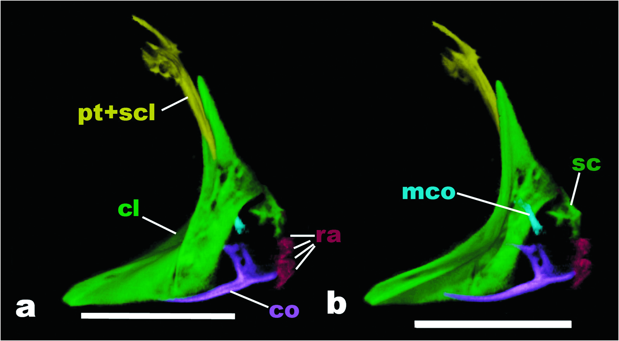
Dermal elements of pectoral girdle consisting of posttemporal (pt), supracleithrum (scl) and cleithrum (cl; Fig. 9 View Fig ); postcleithrae apparently absent. Posttemporal bone fused with supracleithrum (pt+scl), bearing large foramen for passage of posterior lateral line nerve. Dorsal portion of posttemporal + supracleithrum bones positioned over exoccipital and epioccipital. Cleithrum large, dorsal and ventral portions of similar size.
Chondral components of pectoral girdle consist of scapula (sc), coracoid (co) and mesocoracoid (mco; Fig. 9 View Fig ). Scapula not well ossified; axe-blade shape. Coracoid with long, thin and curved ventral process directed anteriorly. Dorsal process of coracoid hatchet-shaped with hook-shaped process on dorsal surface. Mesocoracoid present, rod-like hourglass shaped structure, obliquely oriented anterodorsally to posteroventrally. Support for pectoral fin rays composed of four radials (ra; MZUSP 49205, 345 mm SL). Baudelot’s ligament ossified, branching anteriorly and posteriorly; extending from posteroventral portion of basioccipital and posterolateral portion of exoccipital to cleithrum; with “><“ shape in lateral view.
Scales. Absence of scales on anterior two-thirds of body. Scales covering entire caudal appendage. Absence of scales in anterior portion of lateral line. Lateral line ossicles embedded in thick layer of skin. Scales above lateral line in posterior portion of body ovoid in shape, elongate on horizontal axis, about twice as long as deep ( Fig. 10 View Fig ).
Electric organs. No information available regarding electric organ discharges (EOD) in Iracema caiana . Hypaxial electric organ extending anteriorly to mental region. Four rows of electrocytes present above posterior end of anal fin.
40-50 km above its mouth with the rio Negro , approximately 01º05’S 61º35’W, T. Roberts, 19 Nov 1968. MZUSP 49205 View Materials , 3 paratypes, 208-345 mm SL, collected with the holotype GoogleMaps .
Color in Alcohol. Background color pale yellow, dorsolateral and lateral surfaces of head and body darker. Head almost unpigmented ventrally except for dark chromatophores on anterior portion of lower jaw. Dorsal portion of head and temporal region strongly pigmented, forming dark brown speckles from snout to nape region; speckles of about eye size; most concentrated dorsolaterally and absent on dorsal midline of head. Lateral region of body yellow with dark reddish and diffuse chromatophores. Anal-fin pterygiophore region lighter than other parts of body. Scattered dark brown speckles over anal-fin pterygiophores. About 20 to 30 dark brown diffuse blotches on lateral surface of body. Caudal appendage pale yellow, except for diffuse dark brown bands not extending to dorsal or ventral midline. Distal third of caudal appendage pale yellow, not darkly pigmented. Pectoralfin membranes mostly hyaline with scattered chromatophores at distal end; anal-fin membrane hyaline except for dark brown band covering entire extent of distal two-thirds of rays. Iridescent coloration throughout entire lateral portion of body.
Distribution. Iracema caiana is known only from the type locality on the rio Jauaperi, a tributary of rio Negro in the Amazon basin. Despite a recent survey of several museum collections with large holdings of rhamphichthyid specimens, no additional material of Iracema caiana was identified. This makes Iracema the rarest species of Rhamphichthyidae and one of the rarest of Gymnotiformes ( Nogueira et al., 2010) . This limited distribution does not seem the result of a sample bias, since the rio Negro basin is relatively well surveyed ( Goulding et al., 1988; Chao, 2001).
Material examined. Iracema caiana . Brazil. MZUSP 8952 View Materials , holotype, 360 mm SL, Amazonas, rio Jauaperi , sand beach about


| T |
Tavera, Department of Geology and Geophysics |
No known copyright restrictions apply. See Agosti, D., Egloff, W., 2009. Taxonomic information exchange and copyright: the Plazi approach. BMC Research Notes 2009, 2:53 for further explanation.
|
Kingdom |
|
|
Phylum |
|
|
Class |
|
|
Order |
|
|
Family |
|
|
Genus |
Iracema caiana Triques, 1996
| Carvalho, Tiago P. & Albert, James S. 2011 |
Iracema caiana
| Triques 1996: 91 |