
Aspidoscopulia tetrasymmetrica, Tabachnick, Konstantin R., Menshenina, Larisa L., Pisera, Andrzej & Ehrlich, Hermann, 2011
|
publication ID |
https://doi.org/ 10.5281/zenodo.203661 |
|
DOI |
https://doi.org/10.5281/zenodo.5612026 |
|
persistent identifier |
https://treatment.plazi.org/id/345787A2-ED71-FF95-3EF7-56F3120DFE72 |
|
treatment provided by |
Plazi |
|
scientific name |
Aspidoscopulia tetrasymmetrica |
| status |
sp. nov. |
Aspidoscopulia tetrasymmetrica sp. n.
( Figures 1–4 View FIGURE 1 View FIGURE 2 View FIGURE 3 View FIGURE 4 ; Tables 1 View TABLE 1 , 3 View TABLE 3 )
Holotype. ZINRAS 11133 ( Figs. 2–4 View FIGURE 2 View FIGURE 3 View FIGURE 4 ), Philippines Sea, Komahasi underwater mount ( Fig. 1 View FIGURE 1 ): R.V. ‘Academic Oparin’ - 13, stn. 56, 23.04.1991, 28°4.8’ N 134°38.97’ E, 705 m.
Paratypes. IORAS 5/2/sp403; sp405; sp488, same data as the holotype.
Etymology. The species is named after its 4-rayed symmetry found in lateral branches arising from the main stem.
Diagnosis. Aspidoscopulia with four rayed symmetry when seen from the top, with large anchorate clavules and microscleres of oxyoidal type.
Description. Morphology: The holotype is represented by a plexiform unit 200 mm in length and 90 mm in diameter composed of branching-anastomosing tubes 10–16 mm in diameter ( Fig. 2 View FIGURE 2 A). It has an anisotomous type of branching, its lateral branches have the same diameter as the main one and undergo further dichotomous branching and anastomosing. Obvious 4-rayed symmetry is observed from the apex of the main stem ( Fig. 2 View FIGURE 2 C). The mode of branching is always associated with the formation of carina. Some lateral oscula are overgrown by a secondary framework ( Fig. 2 View FIGURE 2 D). The lateral branches arise at right angles to the central axis, each branch is situated in alternate order, the angle observed in the transverse section with the neighboring lateral branches is regular - about 60° or 120°. So, if the first and second branches form an angle of 60°, the second and third ones will form one with 120°, the third and the forth with 60° again, and so on, in such a way that four alternate rows of lateral branches are observed on the main stem. Hence the sponge displays 4-rayed symmetry observed when seen from above. The central tube has the same diameter as most lateral branches but it has thicker walls (about 4 mm) at base, while in the upper parts walls have the same thickness as lateral branches (about 1 mm). The lateral branches are only rarely covered by a secondary dictyonal skeleton, similar to that of the wall. Usually the lateral branches anastomose to their neighbors forming intercavaedia. Dictyonal framework: The skeleton contains mostly the primary type or farreoid framework (with rectangular, sometimes square meshes; Fig 2 View FIGURE 2 E). The secondary skeleton is represented in some upper parts of the branches on the outer (dermal) side and is well developed at base of the main stem on both dermal and atrial sides of the wall ( Fig. 2 View FIGURE 2 F, Fig. 3 View FIGURE 3 A, B), and in the overgrown oscular constructions. Most tubes have walls consisting of 3 layers of primary skeleton with smooth beams 30–60 μm in diameter which form meshes about 80x 80 μm or 80x 300 μm. The free rays are rough and about 300 μm long. The secondary skeleton in the upper parts of the body consists of beams 30–80 μm in diameter, usually with triangular meshes about 200 μm in diameter. The secondary skeleton at base has remarkable secondary silica deposition so that the primary skeleton is not visible ( Fig. 3 View FIGURE 3 E–G). The beams are 60–80 μm in diameter, the lumen about 80 μm in diameter. Small hexactines ( Fig. 3 View FIGURE 3 D) are always connected by fusion with dictyonal skeleton and with each other and have smooth rays 30–70/2–7 μm in dimensions.
Free spicules: Dermalia and atrialia are pentactines ( Fig. 4 View FIGURE 4 A, B) with unpaired ray directed inside the body and with tangential rays slightly bent inside the body in their distal parts. Dermal pentactines are usually slightly larger than atrial ones, with less rough rays and with less clavate outer ends. The tangential rays of dermal pentactines are 122–296 μm long, the unpaired rays are 141–296 μm long, with diameter 5–18 μm. The tangential rays of atrial pentactines are 59–248 μm long, the unpaired rays are 44–289 μm long with diameter 3–18 μm. Clavules are of two types with discoidal-clavate ( Fig. 3 View FIGURE 3 J, Fig. 4 View FIGURE 4 C, D, H, I) and anchorate heads (rarely with 5, usually with 8–9 teeth) ( Fig. 3 View FIGURE 3 K, L, Fig. 4 View FIGURE 4 I–G), the latter often have several long, curved spines situated close to the head. Their shafts are slightly rough, the end directed inside the body is rough-spiny, usually lanceolate in shape. The discoidal (pileate) clavules are 222–337 μm long, their heads are 7–19 μm long and 19–35 μm in diameter, the diameter of the shaft is about 2 μm in the middle. The anchorate clavules are 244–629 μm long, their heads are 15–44 μm long and 35–78 μm in diameter, the diameter of the shaft is about 3 μm in the middle. The aspidoscopules ( Fig. 3 View FIGURE 3 M–N, Fig. 4 View FIGURE 4 J– O) have 8- usually10–14 terminal spines with conically pointed outer ends and short-spiny surface. Their shafts are similar in shape to those of the anchorate clavules. The aspidoscopules are 178–344 μm long, with 33–59 μm long heads and the tuft 37–70 μm in diameter. The shaft diameter is about 3 μm in the middle. There are also abnormal forms of anchorate scopules ( Fig. 3 View FIGURE 3 N, Fig. 4 View FIGURE 4 G) and aspidoscopules ( Fig. 4 View FIGURE 4 N– O) with some teeth bent distally in the former and spines bent proximally in the latter. Anchorate clavules are located mostly in the dermal layer while aspidoscopules in the atrial one, discoidal clavules are found in both layers. Uncinates ( Fig. 4 View FIGURE 4 Q) are 700–3 000/7–30 μm.
ZINRAS 11133 (Holotype) IORAS 5/2/sp403
n avg min max std n avg min max std Microscleres: Oxyhexasters with 2-usually-4-rarely up to 7 secondary rays are 61–108 μm in diameter with primary rosette 25–65 μm in diameter ( Fig. 3 View FIGURE 3 H–I, Fig. 4 View FIGURE 4 R). Rarely it is possible to find their derivatives: oxystauractines, hemioxyhexasters and spicules with some outer ends of onychoidal-discoidal types ( Fig. 4 View FIGURE 4 S–V).
Remarks. The paratypes of Aspidoscopulia tetrasymmetrica sp. n. are small plexiform units that may be fragments of the holotype. The newly described species has extremely large rather specific anchorate clavules and microscleres of only oxyoidal types. These two characters allow to distinguish it from A. furcillata ( Lévi, 1990) , the only species known in the genus so far.
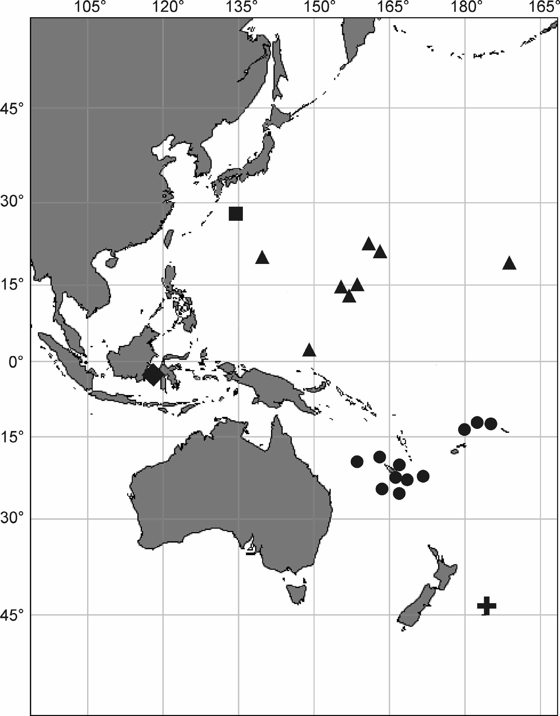
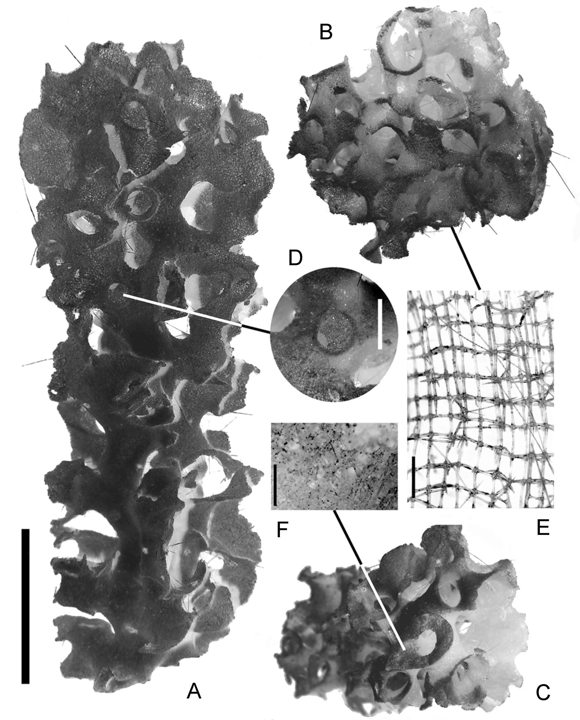
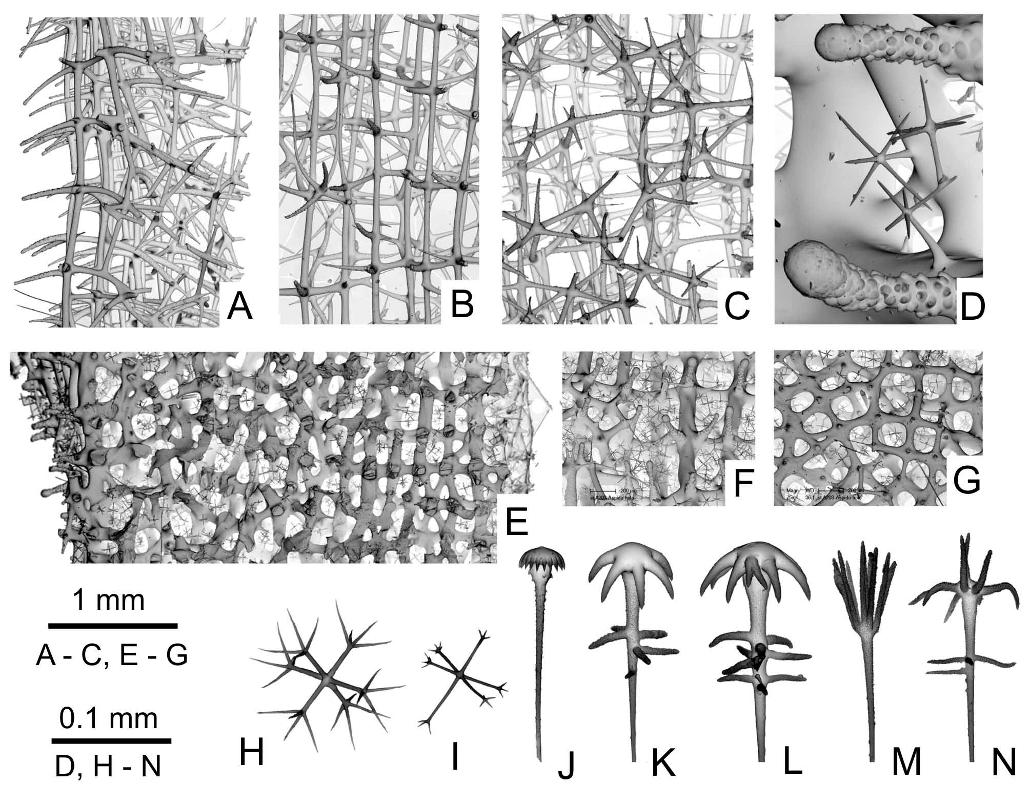
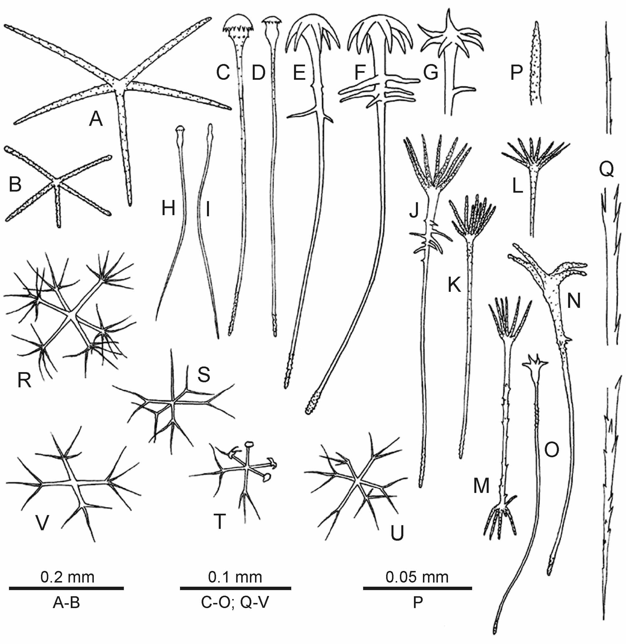
TABLE 1. Spicule dimensions of Aspidoscopulia tetrasymmetrica sp. n. (in μm). L—length, D—diameter, d—diameter of the primary rosette.
| L dermal pentactine tangential ray L dermal pentactine unpaired ray L atrial pentactine tangential ray L atrial pentactine unpaired ray L pileate clavule L pileate clavule head | 25 25 25 25 25 25 | 200 122 296 227 141 296 137 59 248 132 44 289 284 226 318 13 7 19 | 40 41 48 67 26 3 | 15 15 | 277 14 | 222 11 | 337 19 | 27 3 |
|---|---|---|---|---|---|---|---|---|
| d pileate clavule head L anchorate clavule L anchorate clavule d anchorate clavule head | 25 25 25 25 | 27 22 33 330 244 544 30 15 44 57 35 67 | 4 80 7 8 | 15 15 15 15 | 29 412 34 60 | 26 344 26 41 | 35 503 44 70 | 3 50 5 8 |
| L aspidoscopule L aspidoscopule head | 25 25 | 260 178 296 45 33 59 | 28 7 | 14 15 | 288 46 | 226 37 | 333 52 | 31 5 |
| d aspidoscopule head D oxyhexaster d oxyhexaster | 25 25 25 | 52 37 70 88 61 108 41 25 50 | 7 10 7 | 15 15 15 | 55 90 46 | 41 72 32 | 67 108 65 | 8 10 9 |
| continued. | ||||||||
| L dermal pentactine tangential ray L dermal pentactine unpaired ray L atrial pentactine tangential ray | IORAS 5/2/sp405 n avg | min | max | std | ||||
| L atrial pentactine unpaired ray L pileate clavule L pileate clavule head d pileate clavule head | 15 278 15 0 12 15 28 | 211 7 19 | 307 15 33 | 24 2 3 | ||||
| L anchorate clavule L anchorate clavule | 14 477 15 34 | 381 26 | 629 44 | 68 5 | ||||
| d anchorate clavule head L aspidoscopule L aspidoscopule head d aspidoscopule head | 15 66 5 280 6 36 6 44 | 61 241 26 37 | 78 344 41 48 | 4 43 5 4 | ||||
| D oxyhexaster d oxyhexaster | 15 88 15 40 | 79 32 | 101 54 | 6 7 |
No known copyright restrictions apply. See Agosti, D., Egloff, W., 2009. Taxonomic information exchange and copyright: the Plazi approach. BMC Research Notes 2009, 2:53 for further explanation.