
Papuaneon, Maddison, 2016, Maddison, 2016
|
publication ID |
https://doi.org/10.11646/zootaxa.5150.1.8 |
|
publication LSID |
lsid:zoobank.org:pub:1E186CB7-BDB2-46BF-913B-12E11C2A5BEB |
|
DOI |
https://doi.org/10.5281/zenodo.6611752 |
|
persistent identifier |
https://treatment.plazi.org/id/332D87BB-C323-731E-94D8-FF6765E6FA24 |
|
treatment provided by |
Plazi |
|
scientific name |
Papuaneon |
| status |
|
Key View in CoL View at ENA : Females
1. Lateral receptacles of the spermathecae extend further forward than the median receptacles and are further apart than the atria and surrounding sclerotised areas........................................................................ 2
– Lateral receptacles of the spermathecae extend no more than a little further forward than the median receptacles and are not further apart than the atria.............................................................................. 3
2. Strong lateral stripes on the dorsal abdomen; a moustache-shaped marking in the middle of the dorsal abdomen; palp mid brown with off-white metatarsus; thick connection between spermathecal receptacles............... P. eurobodalla View in CoL ( Figs 20–31 View FIGURES 20–26 View FIGURES 27–31 )
– Plain, stippled dorsal abdomen; palps mid brown with off-white tips; abdomen oval with a distinctive pattern of stripes; narrow connections between spermathecal receptacles........................................... P. ewingar View in CoL ( Figs 32–39 View FIGURES 32–39 )
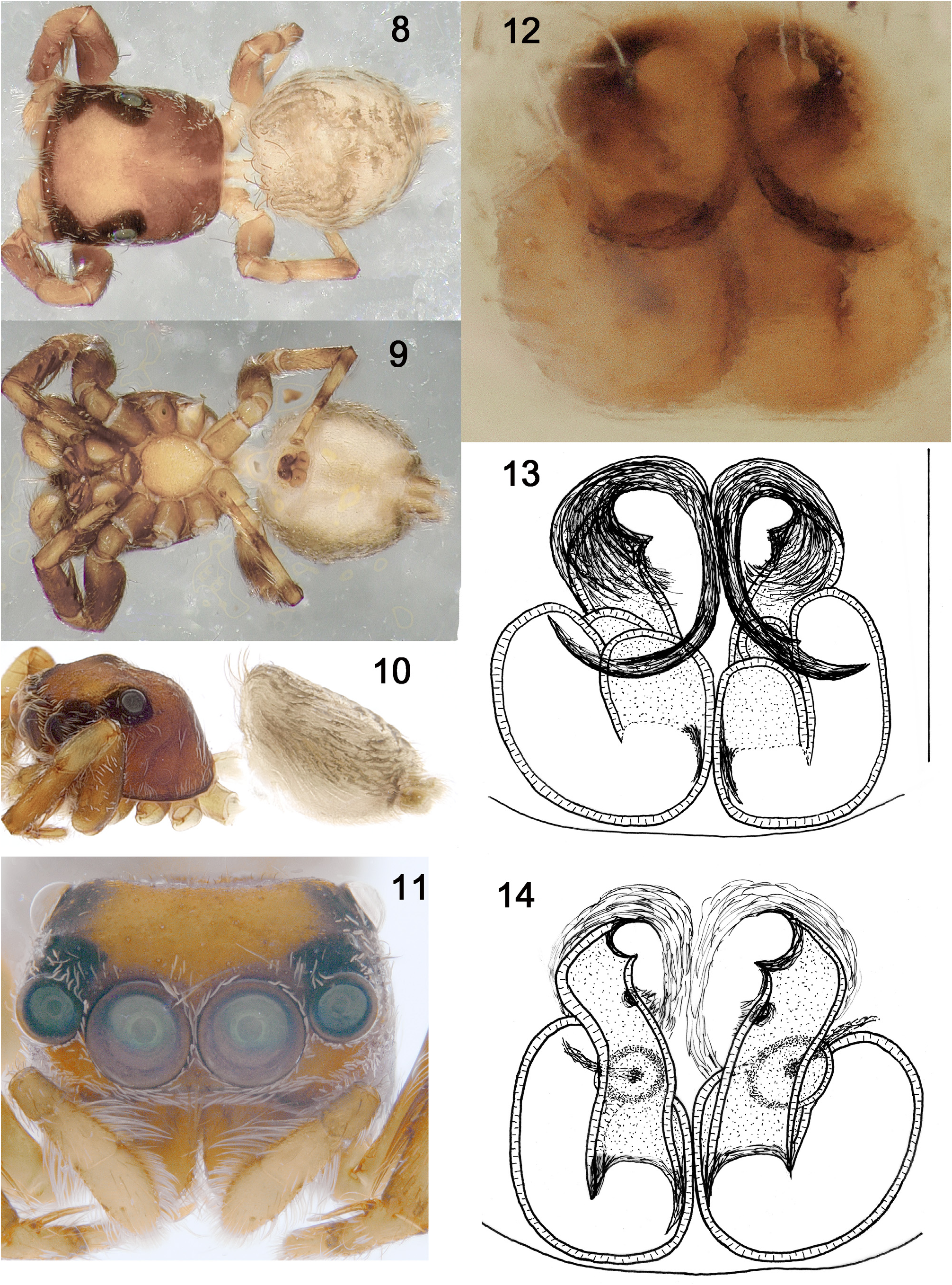
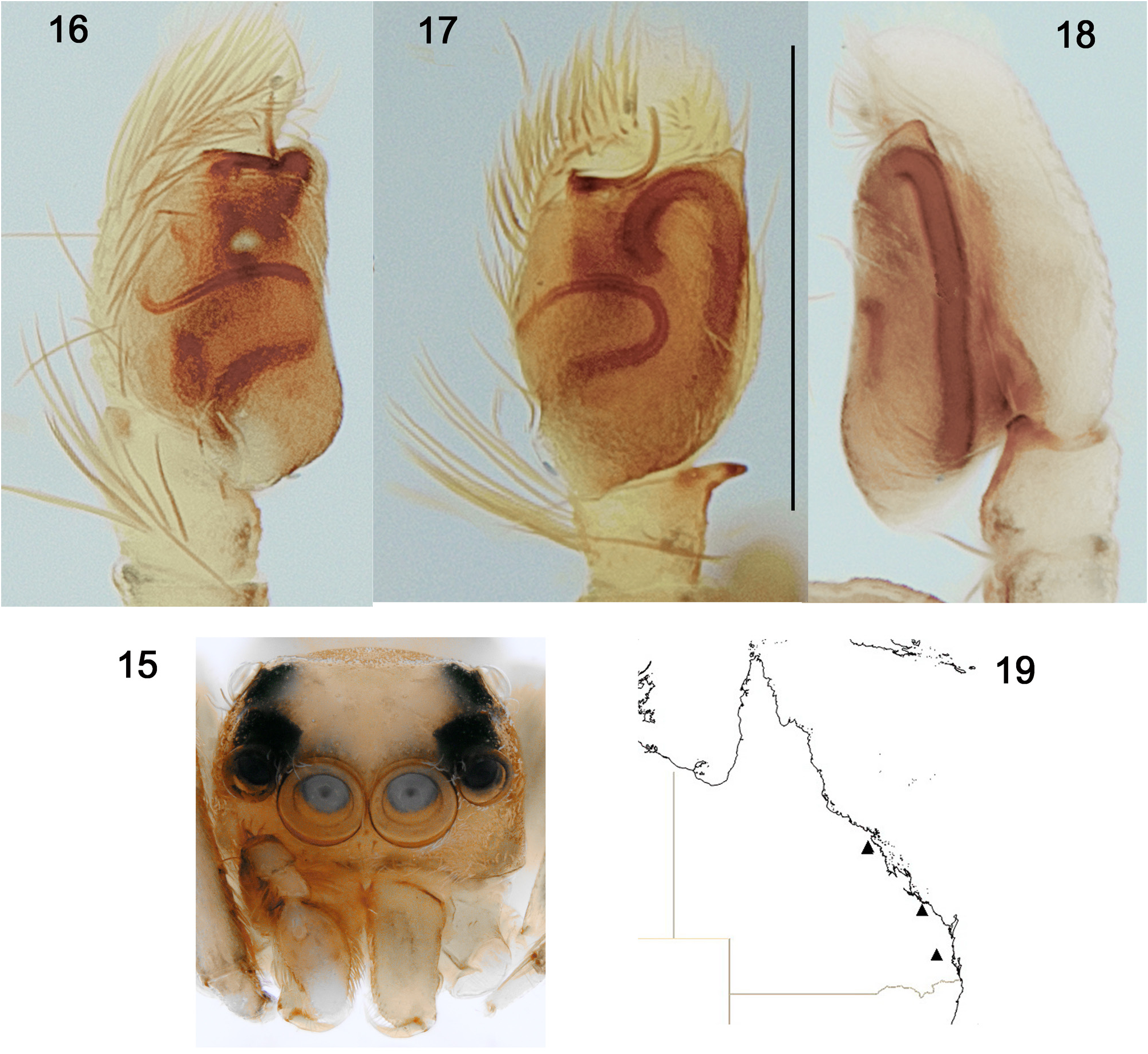
3. Lateral and medial spermathecal receptacles almost the same size............................ P. eungella View in CoL ( Figs 8–19 View FIGURES 8–14 View FIGURES 15–19 )
– Medial spermathecal receptacle much smaller than lateral receptacle............................................ 4
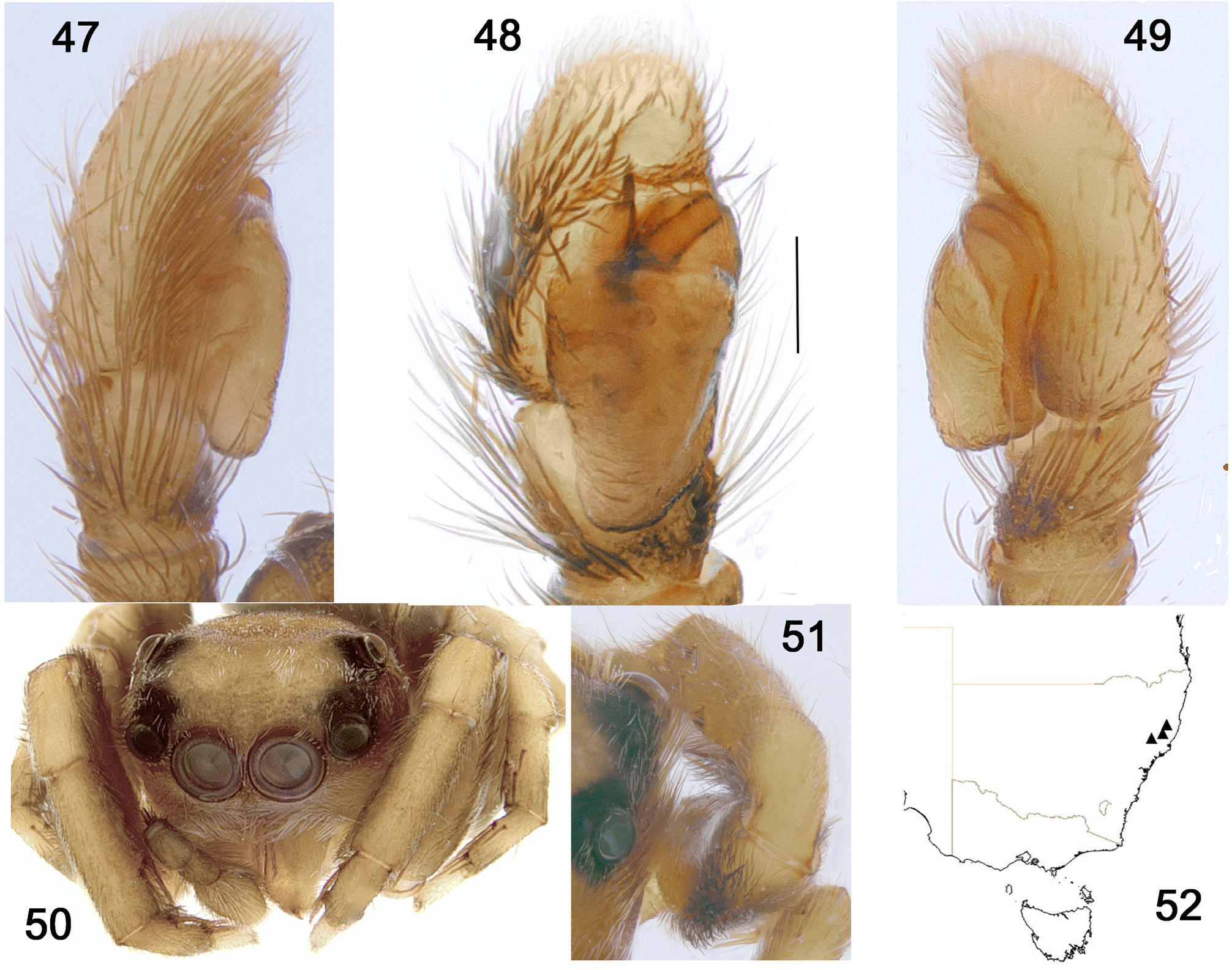
4. Atria round and further apart than the median receptacles..................................... P. tapin View in CoL ( Figs 40–52 View FIGURES 40–46 View FIGURES 47–52 )
– Atria including copulatory openings close together, directly anterior to the medial spermathecal receptacle and much larger than the medial receptacle …................................................................................ 5
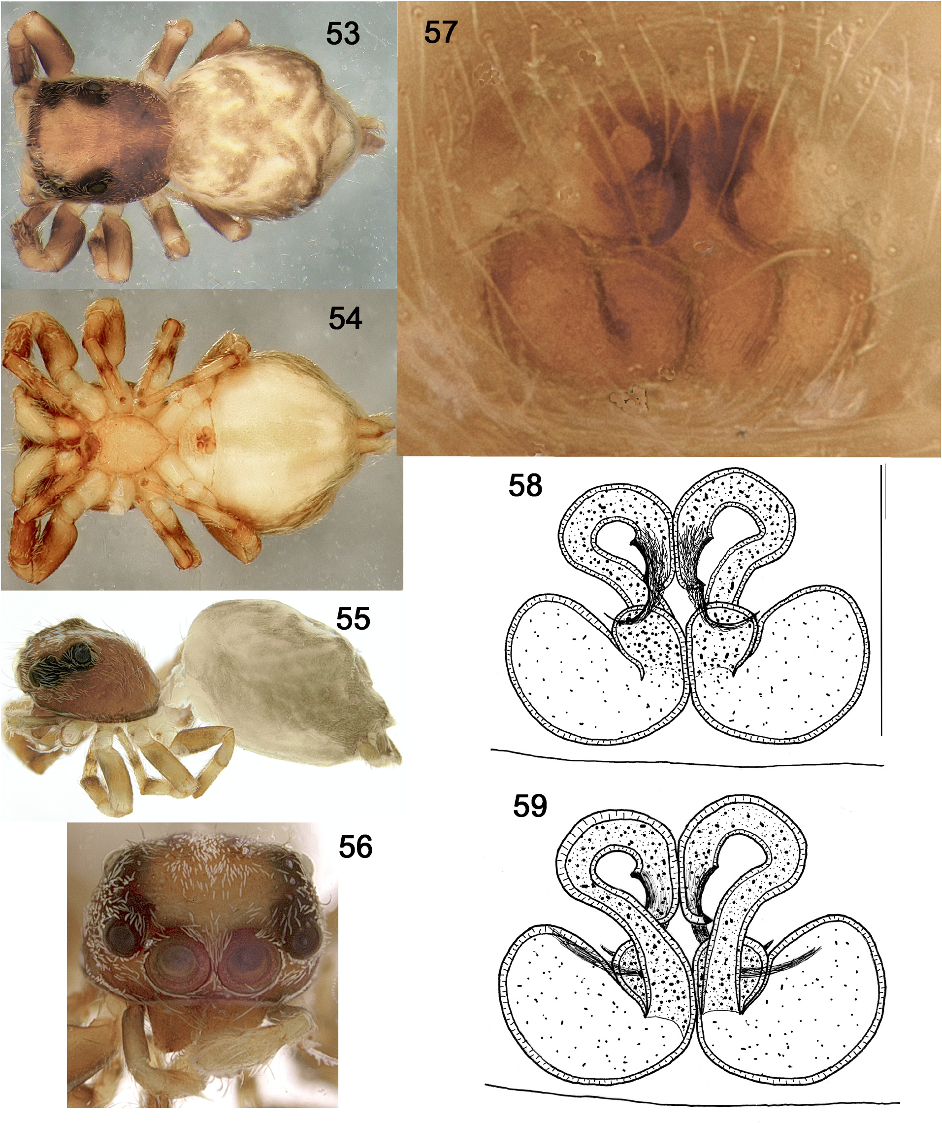
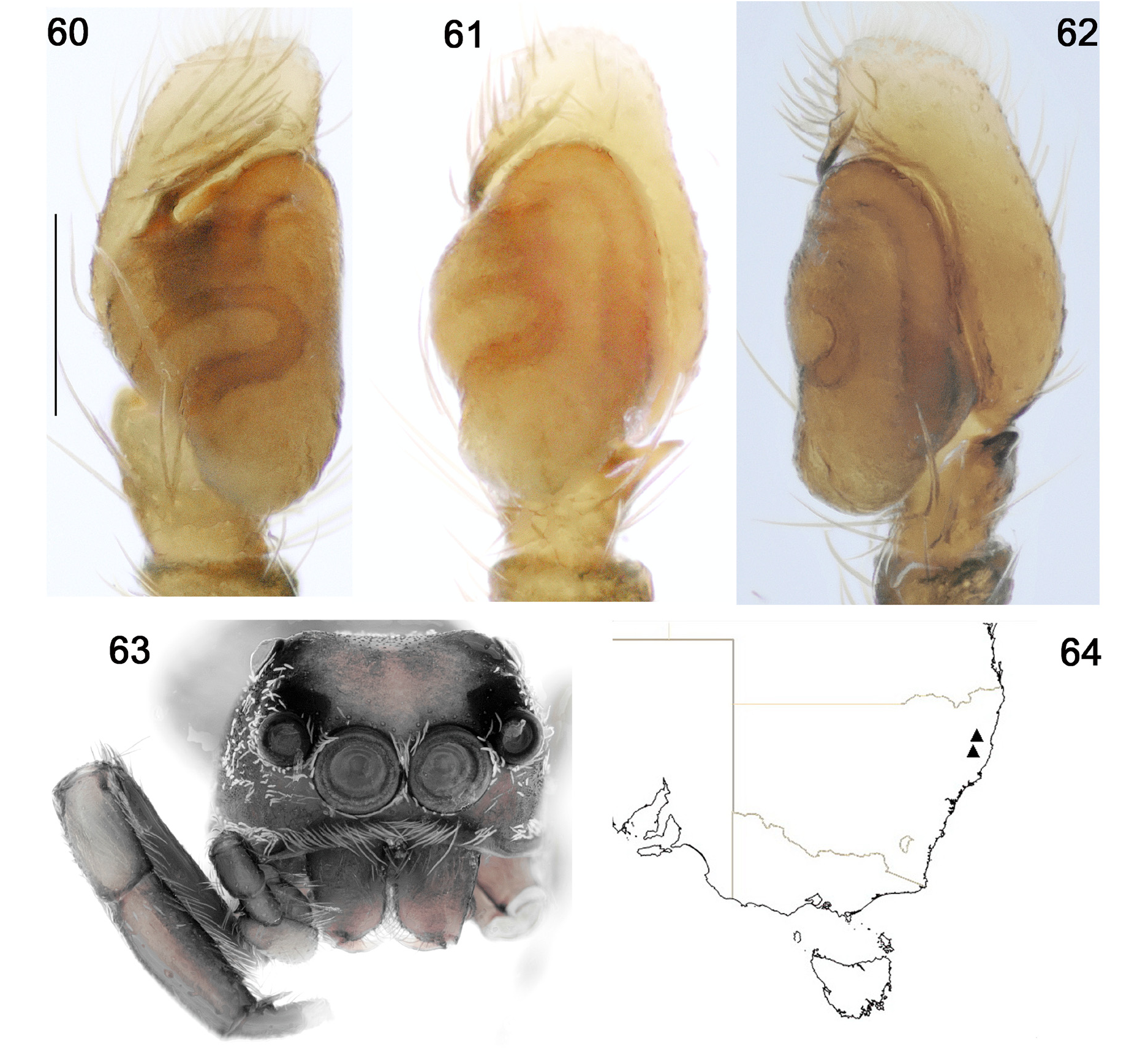
5. Abdomen with large lateral bulges, transverse cleft between spermathecal receptacles; palp white with white fringing...... ….......................................................................... … P. werrikimbe View in CoL ( Figs 53–64 View FIGURES 53–59 View FIGURES 60–64 )
– Abdomen with normal shaped abdomen, longitudinal cleft between spermathecal receptacles; palp brown, metatarsus white with white, feather-like fringing......................................................... P. tualapa View in CoL ( Figs 3–7 View FIGURES 3–7 )
Key: Males
1. Bulb of embolus hidden behind a tegular shelf, tibial apophysis short, pointed and aimed directly away from the tibia..... 2
– bulb of embolus clearly distal to the tegular shelf, tibial apophysis not short, pointed and aimed directly away from the tibia (either almost absent or pointing distally); with a sparse, grey clypeal fringe...................................... 4
2. Embolus curving ventrally away from the face of the tegulum before twisting in a three-quarter circle and then moving gently in a distal direction................................................................... P. tualapa View in CoL ( Figs 3–7 View FIGURES 3–7 )
– Embolus moving parallel to the distal edge of the tegulum.................................................... 3
3. Abdomen without distinct, posterior lateral bulges; large posterior tegular lobe, tapering; with a postero-distal shoulder on tegulum; finely built embolus moving parallel to the distal edge of the tegulum before curving a quarter circle away in a distal direction.......................................................................... P. eungella View in CoL ( Figs 8–19 View FIGURES 8–14 View FIGURES 15–19 )
– Abdomen with distinct posterior lateral bulges; large posterior tegular lobe, broad and rounded; strongly built embolus moving parallel to the distal edge of the tegulum before curving slightly away in a distal direction....... P. w errikimbe ( Figs 53–64 View FIGURES 53–59 View FIGURES 60–64 )
4. Embolus short, straight and pointed....................................................... P. tapin View in CoL ( Figs 40–52 View FIGURES 40–46 View FIGURES 47–52 )
– Embolus moving parallel to the distal edge of the tegulum before curving away in a distal direction.................................................................................................. P. eurobodalla View in CoL ( Figs 20–31 View FIGURES 20–26 View FIGURES 27–31 )


No known copyright restrictions apply. See Agosti, D., Egloff, W., 2009. Taxonomic information exchange and copyright: the Plazi approach. BMC Research Notes 2009, 2:53 for further explanation.
|
Kingdom |
|
|
Phylum |
|
|
Class |
|
|
Order |
|
|
Family |